

How an Impacted Canine Is Surgically Exposed and Guided Into Place
Key Takeaways
- Upper canines are the second most commonly impacted teeth after wisdom teeth and travel farther through the jaw than any other tooth, which is why they so often lose their way.
- Exposure surgery does not move the tooth; it uncovers the crown and bonds a bracket and fine chain so the orthodontist can apply light force over months.
- A Cochrane systematic review comparing open and closed exposure for palatal canines found no clear difference in success, gum appearance, or patient comfort.
- Removing the baby canine early, roughly between ages 10 and 13, can sometimes let a straying adult canine correct its own path and avoid surgery.
- Soft-tissue healing takes about one to two weeks, but active traction ranges from a few months to over a year depending on how far the tooth must travel.
- A canine that has not moved after several months of steady force may be fused to bone, and the plan should include an agreed fallback before surgery begins.
Impacted canine exposure surgery is a short oral surgery in which a surgeon lifts or removes the gum (and sometimes a thin layer of bone) covering a canine tooth that failed to erupt, then bonds a small bracket or chain to it so an orthodontist can gently pull the tooth into the arch over months. It is usually done under local anesthesia, often with sedation, and is combined with braces.
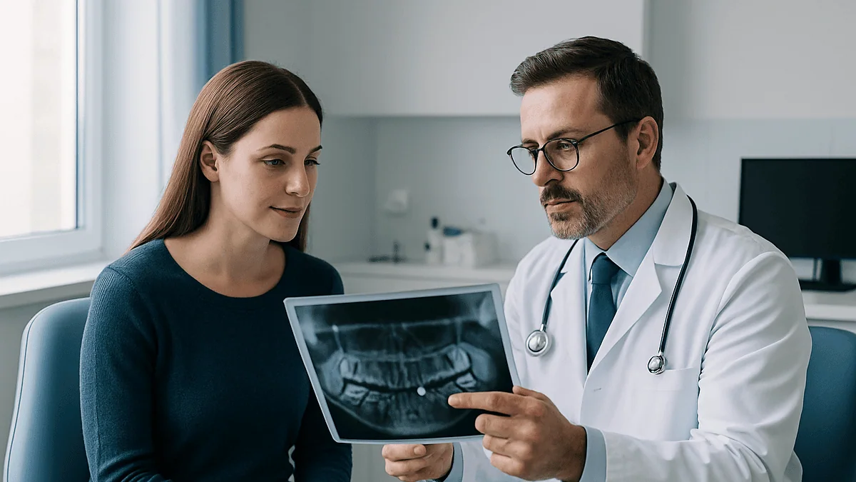
It usually starts with an ordinary orthodontic check. A 13-year-old opens wide, the dentist counts teeth, and pauses: the pointed upper canine that should have come through by now is nowhere to be seen. A baby tooth still sits in its spot, wobbly and overdue. An X-ray shows the adult canine parked high in the roof of the mouth, tilted like a car that took the wrong exit.
For many families, that image is the first time they hear the phrase impacted canine exposure surgery. The words sound dramatic. The reality is a well-rehearsed, two-part plan: a surgeon uncovers the hidden tooth, and an orthodontist coaxes it, millimeter by millimeter, into the gap that has been waiting for it.
This explainer walks through what happens, why canines in particular get stuck, how the two main surgical approaches differ, what recovery tends to look like, and which questions are worth asking before anyone picks up a scalpel.
What is an impacted canine, and why does it matter?
A tooth is described as impacted when it cannot erupt into its normal position because something blocks its path: another tooth, bone, thick gum tissue, or simply too little room in the jaw. Wisdom teeth are the most familiar example, but the upper canines come next in line, according to patient information from Cleveland Clinic.
Canines have a hard job. They form high in the jaw, close to the floor of the nose, and travel farther than any other tooth to reach the gumline. They are also among the last front teeth to arrive, typically around the early teens, which means they must squeeze into whatever space the neighbors have left. When the arch is crowded, or a nearby lateral incisor has a small or missing root to guide it, the canine can drift toward the palate or, less often, toward the lip.
Why not simply ignore a tooth that is not causing pain? Three reasons come up repeatedly in the clinical literature. First, the retained baby canine eventually loosens and falls out, leaving a gap. Second, an unerupted canine can press on the roots of adjacent teeth and slowly dissolve them, a process called root resorption. Third, the canine is a cornerstone tooth: it turns the corner of the smile and carries heavy chewing forces, so a missing one is hard to replace convincingly.
An impacted canine is often silent. It is usually spotted on routine dental X-rays or when a dentist notices the baby tooth is overstaying its welcome. MedlinePlus lists gum swelling, a bad taste, or difficulty opening the mouth as possible signs of an impacted tooth, but stresses that many cause no symptoms at all and are found on imaging (MedlinePlus).
How impacted canine exposure surgery works, step by step
Picture the canine as a tooth wearing a blanket of gum and bone. Exposure surgery peels back enough of that blanket to reach the crown, then gives the orthodontist a handle to pull on.
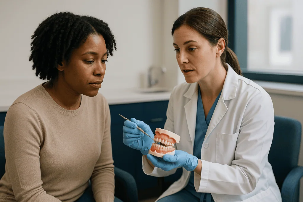
The appointment itself follows a predictable sequence. After numbing, the surgeon makes a small incision in the gum over the tooth’s location, which was mapped in advance on imaging. If bone covers the crown, a thin layer is removed with fine instruments, taking care to leave the enamel and the neighboring roots untouched. The surgeon then clears away the soft tissue sac, called the dental follicle, that surrounds an unerupted tooth.
Next comes the attachment. Once the crown is dry, a tiny orthodontic bracket or button is bonded to it with the same adhesive used for braces. A fine gold-colored chain is usually linked to that bracket. Gold is chosen because it resists corrosion and stays soft enough to bend, and the chain is what the orthodontist will later shorten link by link.
What happens to the gum afterward depends on the technique. In a closed exposure, the flap is stitched back over the tooth with only the chain poking out through the gum. In an open exposure, a window of tissue is left away so the crown stays visible, sometimes protected by a small dressing for a week or so.
The whole procedure commonly takes less than an hour, and most people go home the same day, as is typical for minor oral surgery described by the NHS. Nothing is pulled on that day. The tooth begins its journey only after the site has settled, when the orthodontist attaches the chain to the braces.
Open vs closed exposure canine surgery: what the two techniques involve
Two surgical philosophies exist, and both have loyal followers. The table below summarizes the practical differences.
| Feature | Closed exposure | Open exposure |
|---|---|---|
| What the surgeon does | Uncovers crown, bonds chain, stitches gum back over tooth | Uncovers crown and leaves a window in the gum so the tooth stays visible |
| What you see afterward | Only a chain emerging through the gum | Part of the tooth crown, sometimes under a dressing at first |
| When traction starts | Soon after healing, via the chain | Tooth may erupt partly on its own before or during traction |
| If the bracket comes off | Usually needs a second minor surgery to rebond | Often rebonded in the orthodontic chair |
| Typical use | Deeply positioned teeth, especially toward the lip | Palatal teeth closer to the surface |
Which is better? The honest answer is that high-quality evidence has not crowned a winner. A Cochrane systematic review comparing open and closed exposure for canines displaced toward the palate found no clear difference in whether the tooth was successfully brought into the arch, in how the gum looked afterward, or in how patients rated the experience, while noting that the underlying trials were small. Clinical preference, the tooth’s exact position, and the surgeon’s experience tend to drive the choice.
What matters more than the label is planning. A tooth angled steeply toward the palate needs a different pulling direction than one lying flat behind the incisors, and the surgeon and orthodontist should agree on that vector before the incision is made.
Who is usually offered exposure, and who is asked to wait
Not every hidden canine goes straight to surgery. The decision hinges on age, how far the tooth has strayed, and whether there is room to receive it.
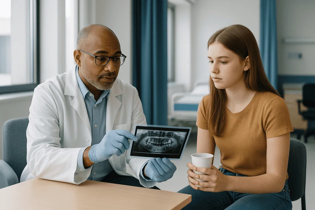
Exposure and traction are most often proposed for adolescents and young adults whose canine is clearly off course but whose root is still developing or recently completed. Younger bone is more forgiving, and the tooth’s own eruptive drive can help. Adults can and do have the procedure, but the tooth may move more slowly and the risk that it has fused to bone rises with age, a point discussed further below.
Some children are asked to wait, in a good way. If a palatally displaced canine is detected early, often around ages 10 to 13 when the tooth should be approaching the surface, removing the baby canine can sometimes give the adult tooth room to correct its own path. This interceptive approach is a recognized first step in orthodontic guidance, and it avoids surgery altogether for the children in whom it works. Follow-up imaging a year or so later shows whether the tooth has improved or whether exposure is now needed.
Others are steered away from exposure. Signs that a canine is unlikely to respond include a tooth lying almost horizontally, a crown positioned well beyond the midline, a fully mature root in an older adult, or radiographic evidence that the tooth is already fused to surrounding bone. In those cases the team may recommend leaving the tooth and monitoring it, or removing it and closing or restoring the space.
Medical factors count too. Bleeding disorders, medications that affect bone healing, or conditions that make sedation riskier will shape both the timing and the setting of surgery. The orthodontist and surgeon weigh all of this together; the plan is theirs to make with you, not a formula.
Before the operation: imaging, planning, and getting the braces ready
Good exposure surgery is mostly decided before the day itself. The first task is locating the tooth in three dimensions. Ordinary dental X-rays show that a canine is impacted but can be ambiguous about whether it sits toward the palate or toward the lip. Two films taken from different angles help; increasingly, a cone-beam CT scan, a low-dose three-dimensional dental scan, is used to map the crown, the root, and its relationship to neighboring roots. That scan also reveals early root resorption on the incisors, which can change the urgency.
Space is the second question. A canine cannot be pulled into a gap that does not exist. Braces are therefore usually fitted before surgery to open and hold room in the arch, often with a firm wire that can act as an anchor for traction later. Some orthodontists prefer to complete this preparation several months ahead so that the tooth can be moved as soon as the surgical site heals. Orthodontic treatment overall, of which this is one phase, commonly runs 18 to 24 months according to NHS guidance, though impacted teeth can lengthen that.
The surgical consultation covers practical points: which anesthesia option suits you, what to eat beforehand if sedation is planned, who will drive you home, and how long to expect off school or work. Bring a list of medicines and supplements, because some affect bleeding or healing and the surgeon may want to coordinate with whoever prescribes them. Never stop a prescribed medicine on your own initiative.
Ask, too, how the surgeon and orthodontist communicate. The best plans are joint ones, where the orthodontist has specified where the attachment should be bonded and in which direction the chain should exit so that traction pulls the tooth along the safest path.
Anesthesia and the day of surgery: what it actually feels like
Most canine exposures are performed under local anesthesia, sometimes with sedation given by mouth or through a vein to take the edge off anxiety, according to guidance on oral surgery from Mayo Clinic. General anesthesia, in which you are fully asleep, is reserved for younger children, for very deeply placed teeth, or for people who cannot tolerate awake treatment. The choice sits with the surgical and anesthesia team and is tailored to age, health, and the complexity of the case; no option is universally better.
Under local anesthesia you will feel pressure and vibration but not sharp pain. The sound of instruments near the palate can be startling, and many people find the injection itself the least pleasant moment. Sedation blurs the memory of the procedure and often leaves people surprised that it is already over.
Expect to be in the chair for roughly 30 to 60 minutes for a straightforward case. Palatal teeth sometimes take longer because the roof of the mouth is thicker and access is awkward. Once the attachment is bonded and the site is closed or dressed, the surgeon checks the chain is secure, usually tying its free end to the braces or a nearby bracket so it does not dangle.
Before you leave you should receive written aftercare instructions and a contact number. If you had sedation, you will need an adult to take you home and stay with you, and you should not drive, operate machinery, or sign important documents for the rest of the day. The numbness fades over a few hours; a mild ache and a feeling of fullness in the palate are common as it does.
Gold chain impacted tooth traction: how the canine is guided into place
The surgery gets the headlines, but the slow part is orthodontic. Once the gum has healed, typically within one to two weeks, the orthodontist connects the chain to the archwire or to a small spring and applies a light, steady force. Light is the operative word. Teeth move because sustained pressure remodels the bone around the root: cells on the compressed side dissolve bone, and cells on the stretched side lay down new bone. Too much force does not speed this up; it can damage the root or cause the tooth to stop moving.
At visits every several weeks, the orthodontist shortens the chain by a link or two, or adjusts the spring, and the tooth inches toward the arch. Direction changes along the way. A palatal canine is first pulled down and away from the incisor roots, then across toward its final position, so that it clears the neighboring roots rather than grinding into them.
Once the crown breaks through the gum, the temporary attachment is replaced by a standard bracket, and the tooth is aligned like any other. The last stage often involves rotating the tooth and adjusting the gum contour so the canine matches its partner on the other side.
Patients notice the chain most in the early weeks: a foreign feeling on the tongue, occasional snagging on food, and mild tenderness after each adjustment. Wax from the orthodontist can cover sharp ends. A chain that comes loose from the braces is a phone call, not an emergency; one that seems to have detached from the tooth itself needs prompt review, because rebonding may require a return to the surgeon, especially after a closed exposure.
Impacted canine surgery recovery: the first days and weeks
Recovery from exposure resembles recovery from any minor oral surgery, with a twist: you are also wearing braces and a chain.
In the first 24 hours, expect oozing that tints saliva pink, some swelling of the cheek or palate, and an ache as the local anesthetic wears off. Biting gently on gauze for the first hour, keeping the head raised, and applying an ice pack to the outside of the face in short intervals help with bleeding and swelling, as Mayo Clinic advises for comparable oral procedures (Mayo Clinic). Your surgeon will discuss pain relief; over-the-counter options are usually enough, and prescription pain medicine is uncommon for this procedure. Follow the specific instructions you were given rather than general advice.
Days two and three are typically the peak for swelling and soreness, after which things improve steadily. Soft, cool foods are easier: yogurt, scrambled eggs, smoothies without straws, mashed vegetables. Avoid very hot drinks, spicy foods, and anything crunchy that could catch the chain or disturb the stitches. Rinsing gently with warm salt water after meals from the second day helps keep the site clean; vigorous swishing or spitting can dislodge the clot.
Stitches are usually dissolvable and fall away over one to two weeks. If a dressing was placed over an open exposure, it is removed at a review visit around the same time. Most teenagers return to school within one to three days; contact sports are generally paused for about a week or until the surgeon clears them.
Brushing continues around, not over, the surgical site, then resumes fully as tenderness allows. Good hygiene matters more than ever, because braces plus a healing wound make plaque control harder and gum inflammation can slow tooth movement.
How long does the whole process take?
People often ask for a single number, and there is none that applies to everyone. What can be described are typical ranges, each dependent on how far the tooth has to travel.
The surgery is a single visit of under an hour. Soft-tissue healing takes one to two weeks. Traction, the phase where the tooth is actively pulled, is where variation is widest: a canine sitting just beneath the gum near its correct spot may appear within a few months, whereas one lying high in the palate can take a year or more to reach the arch. Once the tooth is in the line of the other teeth, several further months of fine alignment usually follow.
Set against the whole orthodontic course, which NHS information puts at commonly 18 to 24 months for braces in general (NHS), an impacted canine often stretches treatment toward the longer end and sometimes beyond it. Orthodontists usually give a range at the outset and revise it as they see how the tooth responds in the first few months of traction; a tooth that moves briskly early is a good sign, while one that has not budged after a few months of consistent force prompts a re-evaluation for ankylosis, meaning fusion of the root to the bone.
Age influences pace. Bone in adolescents remodels faster than adult bone, which is one reason earlier identification tends to mean shorter treatment. Missed appointments, broken brackets, and poor hygiene each add weeks; keeping every visit is the single most controllable factor.
Think of it as a season rather than a date on the calendar, and ask your orthodontist to explain which milestones will tell you the plan is on track.
Risks and complications, described plainly
Exposure and traction are routine, but any surgery carries risk, and it is fair to ask about the specific ones.
Common and minor: bleeding that settles within a day, swelling and bruising, temporary difficulty eating, and infection of the surgical site, which presents as increasing pain, swelling, a bad taste, or fever a few days after surgery. Infection is uncommon and usually responds to cleaning and, if needed, treatment prescribed by the surgeon.
Bracket or chain detachment is the most frequent technical hitch. It happens when the bond fails or when the chain is snagged. After an open exposure it is often fixable in the orthodontic chair; after a closed exposure it may mean a second small procedure.
Less common but more consequential: damage to the roots of the neighboring incisors during surgery or traction, particularly if the canine lies against them; loss of gum height or an uneven gumline around the canine once it erupts, which may need minor gum surgery to correct; and injury to the pulp of the moved tooth, occasionally leading to root-canal treatment later. A palatal wound can be uncomfortable to keep clean and may heal more slowly than one on the cheek side.
The outcome that changes the plan entirely is ankylosis. If the canine has fused to bone, no amount of traction will move it, and the choice becomes leaving it in place, removing it, or, in select cases, surgically loosening or transplanting it. Age, a very displaced position, and long delay before treatment raise this risk. Mayo Clinic’s general guidance on impacted teeth notes that outcomes are individual and depend on the tooth’s position (Mayo Clinic). Your team should tell you how they will spot problems early and what the fallback is.
Palatally impacted canine treatment alternatives: what else can be done
Exposure and traction is the most common route, but it is not the only one, and a good consultation lays out the others without pushing you toward any.
Watchful monitoring means leaving the tooth where it is, keeping the baby canine or the gap, and taking periodic X-rays to check for cyst formation or root resorption of neighbors. This suits some adults with a deeply buried, symptom-free tooth whose removal would be more invasive than its presence. The trade-off is lifelong surveillance and an eventual decision about the space.
Extraction of the impacted canine and orthodontic space closure moves the first premolar forward to stand in for the canine, reshaping it to look the part. It avoids traction but sacrifices the tooth and can be less convincing in appearance and function, particularly where the smile is wide.
Extraction with later replacement holds the gap open, often with the baby canine or a temporary tooth, until the jaw has finished growing, and then restores the space with an implant or a bridge. It defers the definitive fix and adds a second treatment episode in adulthood.
Autotransplantation, moving the canine surgically out of the bone and replanting it in its correct socket in one operation, offers speed when the root is still developing. It is technically demanding, is used selectively, and carries a risk that the replanted tooth’s root will later dissolve.
Interceptive baby-tooth removal, discussed earlier, is the alternative for younger children caught early enough. Cleveland Clinic’s overview of impacted teeth summarizes that treatment ranges from monitoring to surgical exposure to removal depending on the tooth and the person (Cleveland Clinic). Each path has a different balance of time, surgery, and long-term maintenance; the right one is a conversation, not a verdict.
Teens versus adults: does age change the plan?
The biology of tooth movement does not stop at 18, but it changes tempo. Adolescent jawbone is metabolically busy, remodels quickly, and contains a canine whose root may still be finishing, which gives the tooth some eruptive momentum of its own. Adult bone is denser, cell turnover is slower, and the canine has long since abandoned any attempt to erupt. Both can be treated; the expectations differ.
For teenagers, the plan is usually straightforward exposure and traction integrated into comprehensive braces. The main challenges are compliance with hygiene, keeping the chain intact, and coordinating surgery with school. Parents often ask whether the procedure is frightening for a young person. Most describe pressure and noise rather than pain, and sedation options exist for anxious patients; comfort planning is a legitimate topic for the consultation.
For adults, the pre-treatment assessment carries more weight. The team looks harder for signs of ankylosis, checks the health of the gum and bone around the neighbors, and asks whether existing dental work such as crowns or a bridge changes the goal. Adults also tend to have firmer views on appearance during treatment, which can shift preference toward clear aligners for the alignment phase where that is feasible, though traction itself generally relies on fixed attachments.
Timelines lengthen with age, and the chance that a tooth will refuse to move increases. That is not a reason to rule out treatment; it is a reason to define, in advance, how many months of non-movement would trigger a change of plan. Mayo Clinic’s information on braces notes that orthodontic treatment is effective at any age when the gums and bone are healthy (Mayo Clinic). Healthy foundations, not a birthday, are the real requirement.
What people often get wrong about impacted canine surgery
Myths gather around any procedure that involves the words surgery and teeth. Here are the ones clinicians hear most, with what the evidence actually supports.
“The surgeon pulls the tooth into place during the operation.” No. Surgery only uncovers the tooth and attaches a handle. Movement happens over months under gentle orthodontic force. A tooth yanked into position in an afternoon would lose its blood supply and its bone support.
“If it doesn’t hurt, it doesn’t need treatment.” Most impacted canines are painless. The concern is silent damage: resorption of neighboring roots and, rarely, cyst formation around the buried crown, both of which show up on imaging long before symptoms.
“Open exposure is always faster” or “closed is always kinder.” Systematic review evidence has not shown one technique to be clearly superior in success, appearance, or patient comfort. The choice is driven by the tooth’s position and the team’s judgment.
“Once the tooth is through, treatment is over.” Breaking the gumline is a milestone, not the finish. Rotation, alignment, and settling of the gum contour usually take several more months.
“Adults can’t have this done.” Adults are treated regularly. The risk of a tooth being fused to bone is higher and the pace is slower, which is why adult planning includes a fallback.
“The gold chain is permanent.” It is temporary hardware, removed once the crown is accessible and replaced with a normal bracket.
“Pulling the baby tooth always fixes it.” Interceptive extraction helps some children when done early enough; it is a reasonable first step, not a guarantee, and follow-up imaging decides the next move.
Questions to ask your care team before impacted canine exposure surgery
A twenty-minute consultation goes further when you arrive with questions. These are the ones that tend to change decisions.
- Where exactly is the tooth, and have you seen it in three dimensions? Is it toward the palate or the lip, and how close is it to the incisor roots?
- Is there already any sign of root resorption on the neighboring teeth, and does that make timing more urgent?
- For a younger child: is removing the baby canine and waiting a reasonable first step, and how would we know if it has worked?
- Which exposure technique do you recommend for this tooth, and why? What would make you choose the other?
- Have the surgeon and orthodontist agreed on where the attachment will go and the direction of pull?
- What anesthesia options are appropriate here, and what does each involve for recovery on the day?
- Roughly how many months of traction do you anticipate, and what milestones will tell us we are on track?
- If the tooth has not moved after a set period, what is the plan? Would we consider stopping, extracting, or another approach?
- What are the specific risks for this tooth, including to the gum contour and to the teeth next to it?
- If the bracket or chain comes off, who do I call, and does it mean another surgery?
- How should I manage brushing around the site and the braces in the first weeks?
- What is the total expected length of orthodontic treatment with this tooth included, compared with without it?
Write down the answers. A plan with clear checkpoints and a stated fallback is a plan you can trust, whichever technique the team ultimately chooses.
When to call your doctor
Most recovery after canine exposure is uneventful, and mild soreness, slight oozing, and swelling that peaks around the second or third day are expected. Some signs, though, warrant a prompt call to the surgeon or orthodontist rather than waiting for the next scheduled visit.
Contact your care team the same day if you notice bleeding that does not slow with 20 to 30 minutes of firm gauze pressure, swelling that keeps increasing after the third day or spreads toward the eye or neck, pain that worsens rather than improves after day three, a fever, pus or a persistent foul taste from the site, or numbness of the lip, cheek, or palate that has not resolved by the following day. Difficulty swallowing or breathing, or swelling that makes it hard to open the mouth, needs emergency care.
Call the orthodontist if the chain detaches from the tooth or the braces, a wire or link is cutting into the tongue or palate and wax does not help, or the dressing over an open exposure falls off early. None of these are dangerous, but early attention keeps the plan moving and avoids a repeat surgery where it can be prevented.
In the longer term, get in touch if the canine has visibly stopped moving over several appointments, if the gum around the erupting tooth looks swollen, receding, or unusually red, or if a neighboring front tooth becomes loose or tender. These can signal ankylosis, gum inflammation, or root involvement, each of which the team will want to assess with an examination and imaging.
Guidance on impacted teeth from MedlinePlus and Cleveland Clinic echoes the same principle: expected discomfort fades; anything escalating deserves a professional look. Every decision about next steps belongs with the treating team who know your tooth’s position and history.
Frequently asked questions
What does impacted canine exposure surgery involve?
It is a minor oral surgery in which the surgeon opens the gum over an unerupted canine, removes any thin bone covering the crown, and bonds a small bracket with a fine chain to the tooth. The gum is then either stitched back over the tooth with the chain emerging or left open so the crown is visible. The orthodontist later uses the chain to guide the tooth into the arch over months.
Is open or closed exposure better for a canine in the palate?
Current evidence does not favor one over the other. A Cochrane systematic review comparing open and closed techniques for palatally displaced canines found no clear difference in whether the tooth reached the arch, how the gum looked afterward, or how patients rated the experience. Surgeons choose based on the tooth’s depth and position, their own experience, and how easily a detached bracket could be replaced.
How painful is impacted canine surgery recovery?
Most people describe soreness rather than severe pain, peaking around the second or third day and settling within a week. Swelling, mild bleeding, and difficulty eating hard foods are typical early on. Over-the-counter pain relief recommended by the surgeon is usually sufficient. Palatal wounds can feel more awkward than cheek-side ones because the tongue keeps finding them. Pain that worsens after day three warrants a call to the surgeon.
What is the gold chain on an impacted tooth for?
The chain is a temporary handle. One end is bonded to the hidden canine during surgery and the other is attached to the braces once the gum has healed. At each orthodontic visit the chain is shortened link by link, applying a light, steady pull that gradually draws the tooth toward its place. Once the crown emerges, the chain is removed and replaced with a standard bracket.
How long does it take to pull an impacted canine down?
There is no single figure. A canine lying just below the gum near its correct spot may appear within a few months, while one high in the palate can take a year or longer. Add several months of fine alignment after the tooth emerges. Overall orthodontic treatment commonly runs 18 to 24 months, and an impacted canine often pushes it toward the longer end, according to NHS guidance.
Can adults have impacted canine exposure surgery?
Yes. Adults are treated regularly when the gums and bone are healthy. The differences are pace and risk: adult bone remodels more slowly, so traction takes longer, and the chance that the tooth has fused to bone is higher. For that reason adult plans usually specify in advance how many months without movement would prompt a switch to another option such as extraction and replacement.
What happens if the impacted canine will not move?
A tooth that stays put despite months of steady force may be ankylosed, meaning its root has fused to the surrounding bone. Imaging and a clinical check help confirm this. Options then include leaving the tooth and monitoring it, removing it and closing the space with orthodontics, holding the space for a later implant or bridge, or in select cases surgically repositioning the tooth. The treating team decides with you.
Can removing the baby canine fix a palatally impacted canine without surgery?
Sometimes. If a palatally displaced canine is detected early, roughly between ages 10 and 13, extracting the retained baby canine can allow the adult tooth to correct its path on its own. This interceptive step is a recognized first option, but it does not work for every child, so follow-up X-rays about a year later determine whether exposure and traction are still needed.
What are the risks of impacted canine exposure surgery?
Common, minor risks include bleeding, swelling, temporary difficulty eating, and infection of the site. The bracket or chain can detach, sometimes requiring a second minor procedure. Less common but more significant risks include damage to neighboring incisor roots, an uneven or receded gumline around the erupted canine, injury to the tooth’s pulp, and failure of the tooth to move because it is fused to bone.
When should I call the surgeon after canine exposure?
Call the same day for bleeding that does not stop with firm gauze pressure, swelling that keeps increasing after the third day, worsening pain, fever, pus or a foul taste, or numbness lasting beyond the next day. Seek emergency care for difficulty breathing or swallowing. Contact the orthodontist if the chain detaches or a wire is cutting into the tongue or palate.
References
This article is for general information only and is not a substitute for professional medical advice. Please consult a qualified doctor about your individual situation.

More from the Blog

How Long to Stay in Istanbul After Orthognathic Surgery Before You Travel Home
Most surgical teams ask patients who have had orthognathic (corrective jaw) surgery far from home to stay near the hospital through at least the…

Who Treats Halitosis, a Dentist or an ENT? How the Cause Decides the Referral
Most bad breath begins in the mouth, so a dentist is usually the right first appointment: gum disease, tongue coating, decay and dry mouth…

What to Bring to an Oral Health and Systemic Diseases Assessment: Reports, Tests, Medications
Bring an up-to-date list of every medicine and supplement you take, recent lab results such as HbA1c or INR if you have diabetes or…

Before Gummy Smile Treatment: Questions About Cause, Technique and Long-Term Stability
Before gummy smile treatment, the most useful questions concern cause, technique and durability. A gummy smile can come from a short or overactive upper…

Who Needs Scaling and Root Planing (SRP)? Pocket Depths That Call for a Deep Cleaning
Scaling and root planing is usually recommended for people whose gum pockets measure 4 millimeters or deeper, especially when the gums bleed on probing,…
TMJ Splint Therapy: How a Night Guard Is Made, Fitted and Adjusted Over Time
TMJ splint therapy uses a custom-made oral appliance, usually a hard acrylic night guard, to reduce jaw muscle strain and protect the teeth in…