

How Carotid Angiography Maps the Arteries to the Brain Through a Small Catheter

Key Takeaways
- Carotid angiography photographs the neck arteries by injecting contrast directly through a catheter placed via the groin or wrist, which is why it remains the reference standard for measuring a narrowing.
- The catheter tip usually sits in the neck, not the brain; the arteries inside the skull are imaged by watching the dye flow onward.
- The warm flush behind the eye during each contrast injection is expected and fades within seconds; the sharpest pain of the day is typically the local anesthetic.
- A stenosis percentage compares the open channel with a normal segment of the same artery, so seventy percent narrowing means roughly thirty percent of the diameter remains.
- Ultrasound and CT are the usual first tests; catheter angiography is reserved for disagreements between scans, unusual findings, or planning a stent.
- A completely occluded carotid is usually not treated with surgery or a stent, because there is no longer a channel for plaque fragments to travel through.
Carotid angiography is an imaging test in which a thin, flexible tube called a catheter is guided through an artery in the groin or wrist up toward the neck, iodine-based contrast dye is injected, and rapid X-ray pictures show the carotid arteries filling. It is usually done under local anesthetic with light sedation, takes about one to three hours, and is followed by several hours of rest while the puncture site seals.
The ultrasound report said “moderate to severe narrowing,” and then a second scan disagreed with the first. Now a vascular specialist has suggested a catheter angiogram, and the phrase that lodged in your mind on the drive home was “a tube threaded up toward the brain.” That sentence is accurate and also, in a way, misleading: the tube stays in the artery, the artery is a plumbing system, and the person doing the threading watches every millimeter on a screen.
Understanding how carotid angiography is done takes most of the menace out of it. It is the oldest and most detailed way of photographing the two arteries in the front of the neck that carry roughly three-quarters of the brain’s blood supply, and it is still the reference standard against which ultrasound and CT are judged. It is also a real procedure with real, if uncommon, risks, which is exactly why it is no longer the first test anyone orders.
What follows is the honest version: what happens, what it feels like, what the pictures mean, and where the decisions genuinely sit.
What carotid angiography shows that other scans cannot
Two carotid arteries run up either side of the windpipe, one under each side of the jaw where you can feel a pulse. Each splits into an internal branch that supplies the brain and an external branch that supplies the face and scalp. The fork, called the bifurcation, is where fatty deposits known as plaque most often build up, and plaque that cracks or sheds fragments is one of the major causes of ischemic stroke, the type caused by a blocked vessel. Cleveland Clinic’s overview of carotid artery disease describes this mechanism plainly: debris travels with the blood flow until it lodges in a vessel too small to pass it.
Catheter angiography photographs that fork from the inside. Contrast, a liquid that blocks X-rays, is injected directly into the artery, and a series of images captures it flowing through. Because the dye is delivered exactly where the pictures are taken, the vessel edge is crisp, small branch arteries show clearly, and the radiologist can watch timing: how fast blood moves, whether it stalls behind a narrowing, and whether the arteries inside the skull fill normally. That last point matters. A plan for the neck depends partly on what the brain’s own vessels look like.
Ultrasound infers narrowing from how fast blood is moving. CT and MR angiography build a three-dimensional model from a dye injected into a vein. All three are excellent and, for most people, sufficient. The catheter test remains the tie-breaker when those pictures conflict, when calcium in the vessel wall confuses the CT, or when a treatment such as a stent is already being considered and the team wants the definitive map before committing. MedlinePlus describes cerebral angiography in these terms: a test ordered when other imaging does not give enough information.
How carotid angiography is done, step by step
You lie on a narrow table under a C-shaped X-ray arm that can swing around your head and neck. A nurse places an intravenous line, attaches a heart monitor and blood pressure cuff, and, in most centers, gives light sedation so you feel calm but can follow instructions. General anesthesia is rarely needed for a diagnostic study; the team wants you awake enough to hold still and hold your breath on request.
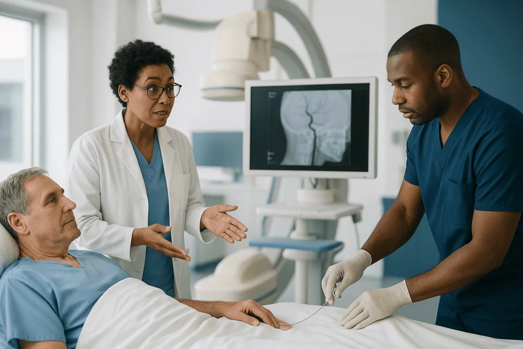
The access point is either the femoral artery at the top of the thigh or the radial artery at the wrist. After cleaning and numbing the skin with local anesthetic, the physician punctures the artery with a needle, slides in a soft guidewire, and then a short plastic tube called a sheath, which acts as a door that stays open through the whole procedure. Everything else passes through the sheath.
Through it goes the catheter, a hollow tube roughly the width of cooked spaghetti. Under fluoroscopy, which is continuous low-dose X-ray shown live on a screen, it is steered along the aorta, the body’s main artery, and its tip is parked at the origin of each carotid in turn. Contrast is injected by a power pump for a second or two while the machine fires a burst of images. A typical study photographs both carotids from two or more angles and often the vertebral arteries at the back of the neck, then the vessels inside the head.
The catheter and sheath come out. The artery is closed either by ten to twenty minutes of firm hand pressure or by a small closure device, a plug or stitch placed from inside. MedlinePlus and Johns Hopkins both put the total time in the room at roughly one to three hours, most of it setup and imaging rather than the catheter work itself.
Getting ready: the days before the test
The pre-procedure phone call is long for a reason. The team is screening for the handful of things that change the plan: a previous reaction to contrast dye, kidney disease, diabetes, pregnancy, and every medicine you take, including supplements and over-the-counter pain relievers. Blood-thinning drugs are the central question. Some are continued because stopping them is riskier than a puncture site that oozes; others are paused for a defined window. The decision belongs to the prescribing clinician, and the safest thing you can do is give a complete, honest list rather than guess which items matter.
Blood tests usually check kidney function and clotting. Iodine contrast is filtered by the kidneys, and Johns Hopkins notes that people with reduced kidney function may need extra fluids or a modified plan; it is one of the reasons a CT or MR study is sometimes chosen instead.
Fasting instructions vary, but a common pattern is nothing to eat for several hours beforehand while clear fluids are allowed until closer to the appointment. Follow the written instructions you are given rather than a general rule, because sedation practices differ. Drink normally the day before unless told otherwise; being well hydrated helps the kidneys handle the dye.
Practical preparation is mostly about the hours afterward. You will not be allowed to drive home after sedation, so arrange a ride. Wear loose clothing that is easy to remove, leave jewelry at home, and if the groin route is planned, expect a small area of hair to be trimmed. Bring reading glasses and something to pass a few flat hours. Most people spend far longer resting afterward than they do on the procedure table.
Is a carotid angiogram painful? What it feels like in the room
The truthful answer is that the sharpest moment is the local anesthetic, a sting and pressure lasting a few seconds, and after that most people describe pressure rather than pain. Arteries have very few pain nerves along their inner lining. You will not feel the catheter travel through your chest or neck, and people are often surprised when told it is already in position.

The contrast injection is the sensation everyone remembers. When dye enters the carotid, many people feel a wave of heat behind the eye and across one side of the face, sometimes with a metallic taste or a brief flash of light in the vision. MedlinePlus lists this warm flush as a normal, expected effect; it fades within seconds. The technologist warns you before each run so it never arrives as a surprise.
Noise and stillness are the other challenges. The X-ray arm whirs and repositions, and you will be asked to hold your breath and keep your head perfectly still during each image burst, because even a swallow blurs the picture. Sedation helps with the boredom and the anxiety without making you unable to cooperate. Tell the team if you are claustrophobic or have a bad back; the table is hard and lying flat for a long stretch can be uncomfortable in a way that has nothing to do with the arteries.
Pain afterward is usually confined to the puncture site: a bruise-like soreness for a few days, more noticeable at the wrist than the groin because there is less tissue to cushion it. Severe, escalating pain at the site is not typical and is one of the signs covered in the section on when to call.
How radiologists measure a blockage on the pictures
Stenosis is the medical word for narrowing, and the percentage on your report is a ratio, not a measure of how much plaque is present. The radiologist finds the tightest point in the internal carotid, measures the width of the channel that still carries blood, and compares it with the width of a healthy segment of the same artery farther up. A seventy percent stenosis means the channel is about thirty percent of the normal diameter. Cleveland Clinic describes this grading in its patient material because the number drives nearly every downstream decision.
Catheter angiography is treated as the most accurate way to make that measurement because the dye outlines the true lumen, the open channel, without being fooled by calcium. On CT, dense calcium can bloom and make a narrowing look worse than it is. On ultrasound, the number is estimated from blood velocity, which rises as the channel tightens, a bit like water speeding up through a pinched hose. Both are good; both can disagree with each other, which is often how someone ends up on the angiography table.
The pictures answer other questions too. Is the narrowing short and focal or a long, irregular stretch? Is the surface of the plaque smooth or ulcerated, with crevices where clot can form? Is there a tortuous loop in the artery that would make placing a stent awkward? Has the artery beyond the plaque shrunk from years of low flow? And do the vessels inside the skull show damage from earlier small strokes?
Reports often use bands: mild, moderate, severe, and near-occlusion or complete occlusion, meaning the artery has closed. Ask which band applies and which measurement method produced it. The same artery can sit in two different bands on two different scans.
Who is usually offered a carotid angiogram, and who is asked to wait
Nobody arrives at catheter angiography as a first test. The typical path starts with a carotid duplex ultrasound, often after a transient ischemic attack (a TIA, sometimes called a mini-stroke, in which stroke symptoms resolve within a day), after a stroke, or after a physician hears a whooshing sound called a bruit over the neck. If the ultrasound suggests significant narrowing, CT or MR angiography usually comes next.
The catheter study is reserved for particular situations. The commonest is disagreement: ultrasound says severe, CT says moderate, and the difference decides whether surgery is even on the table. A second is planning. When a team is weighing carotid stenting, a catheter study maps the exact anatomy the stent would have to negotiate, and in practice the diagnostic run is often the first stage of the same session. A third is a rare or unusual finding, such as a tear in the artery wall, called a dissection, or a suspected inflammatory disease of the vessels. Mayo Clinic’s material on carotid stenting describes angiography as the step that confirms and measures the blockage before treatment.
People are commonly asked to wait, or steered toward a different test, for reasons that are about safety rather than urgency. Reduced kidney function raises the risk from iodine contrast. An uncontrolled bleeding disorder or a blood thinner that cannot be safely paused makes the puncture riskier. A recent large stroke sometimes means waiting for the brain to settle. Pregnancy prompts a discussion about radiation. An acute infection at the planned puncture site is a reason to postpone.
Someone with mild narrowing and no symptoms is usually not offered angiography at all, because the result would not change management. That is a good outcome, not a dismissal.
Carotid angiogram vs ultrasound, CT and MR angiography
Each test trades something for something. Ultrasound is safe and repeatable but operator-dependent and blind to the arteries inside the skull. CT angiography is fast and widely available but uses contrast and radiation and struggles with heavy calcium. MR angiography avoids both radiation and iodine but is slow, noisy, and unsuitable for some implants. Catheter angiography gives the sharpest picture and the option to treat in the same sitting, at the cost of an arterial puncture and a small risk of stroke.
| Test | How it sees the artery | Contrast and radiation | Strength | Main limitation |
|---|---|---|---|---|
| Duplex ultrasound | Sound waves; narrowing estimated from blood speed | None | Safe, repeatable, no needles | Operator-dependent; cannot see inside the skull |
| CT angiography | 3D X-ray model after dye into a vein | Iodine contrast; X-ray dose | Fast; shows neck and brain vessels | Calcium can exaggerate narrowing |
| MR angiography | Magnetic imaging, with or without dye | Usually no iodine; no X-rays | No radiation; good soft-tissue detail | Slow; not for some implants; may overestimate |
| Catheter angiography | Dye injected directly into the artery | Iodine contrast; X-ray dose | Reference-standard detail; treatment possible | Invasive; small stroke and bleeding risk |
The sequence most teams follow reflects that ladder: start with the test that cannot hurt, escalate only when the answer would change what happens next. Johns Hopkins frames cerebral arteriography the same way, as a study chosen when less invasive imaging has not settled the question. If you have been offered the catheter test, it is reasonable to ask which of the earlier rungs was inconclusive and why.
The risks, honestly stated: contrast, radiation, bleeding and stroke
The most common problems are at the puncture site. A bruise is nearly universal. A hematoma, a firm collection of blood under the skin, is less common and usually resolves over weeks. Rarely the artery wall fails to seal and forms a pseudoaneurysm, a pocket of blood that pulses, or a connection to the neighboring vein; both are checked for with ultrasound and sometimes need a minor repair. Wrist access has fewer bleeding complications but a small chance the radial artery narrows or closes afterward, which most people never notice because the hand has a second supply.
Contrast reactions range from hives to, very rarely, a severe allergic response; the team has treatment at hand and screens for prior reactions. Contrast can also temporarily strain the kidneys, which is why kidney function is checked beforehand and why people with existing kidney disease are handled differently. Johns Hopkins lists these among the recognized risks of cerebral arteriography.
The risk that gives the test its reputation is stroke. A catheter manipulated near a plaque can dislodge a fragment, or a small clot can form on the catheter itself. MedlinePlus describes this as a rare but serious complication of cerebral angiography, and it is the reason the test is no longer used for screening. The risk is not zero and is higher in people whose arteries are heavily diseased, which is precisely the group who need the pictures most. This is the trade-off your team is weighing when they recommend, or decide against, the test.
Radiation exposure from a diagnostic study is modest and is kept as low as the images allow; the machine is on only during image bursts. The team is not being evasive when they decline to quote a single number for any of these risks. Your own risk depends on your arteries, your kidneys, your medicines and the route chosen.
Carotid angiogram recovery time: the first hours and days after
Recovery is mostly about the hole in the artery. After groin access, you lie flat with the leg straight while the puncture seals, and a nurse checks the site and the pulses in your foot at intervals. MedlinePlus describes a period of several hours of bed rest after cerebral angiography, shorter when a closure device is used and longer when the artery is sealed by hand pressure alone. After wrist access, a compression band replaces the bed rest, and many people sit up almost immediately. Most diagnostic angiograms are day cases; an overnight stay is more likely if a stent was placed at the same time or if you live far away.
The first twenty-four hours have a short list of rules: no driving, because of sedation; drink freely to flush the contrast unless told otherwise; keep the puncture site dry and covered as instructed; and avoid lifting, straining or climbing stairs repeatedly if the groin was used. Wrist patients are asked not to lift with that arm or bend the wrist hard for a day or two.
Over the following week, the bruise spreads, changes color and fades, sometimes tracking down the thigh under gravity, which looks alarming and is normal. A small, pea-sized lump under the skin at a groin site is common and usually softens over a few weeks. Soreness settles within days. Most people return to desk work within a day or two and to heavier physical work or exercise within about a week, subject to their team’s instructions.
Recovery from the test is not the same as recovery from what it found. If the pictures lead to surgery or a stent, those come with their own timelines, and the NHS describes a typical hospital stay after carotid endarterectomy as one to two nights. Your team will separate the two conversations, and it helps to do the same.
How serious is a carotid blockage? Reading the percentage without panic
Seriousness depends on two things the percentage alone does not capture: whether the plaque has already caused symptoms, and how narrow the channel has become. A person who has had a TIA or minor stroke attributable to a carotid plaque is at their highest risk of a further stroke in the days and weeks immediately afterward, which is why symptomatic narrowing is treated as urgent and the NHS advises that surgery, when appropriate, is ideally carried out within two weeks of symptoms. The same degree of narrowing in someone who has never had a symptom carries a lower yearly risk, and the balance between the procedure’s risk and its benefit is finer.
Counterintuitively, a completely blocked artery is not usually operated on. Once the internal carotid has closed, there is nothing left to dislodge, and the brain on that side is being supplied through connecting vessels from the other carotid and the arteries at the back of the neck. The dangerous zone is the severe-but-open artery, where a large, possibly ulcerated plaque still has flowing blood to carry fragments upward.
Mild and moderate narrowing is common with age and is managed with medicines that lower cholesterol and reduce clotting, blood pressure control, stopping smoking, and repeat ultrasound to watch for progression. Cleveland Clinic and MedlinePlus both frame carotid artery disease as a marker of vascular disease throughout the body, so the coronary arteries and the legs are part of the same conversation.
Angiography’s job is to place your artery accurately on this map. It does not, by itself, tell anyone what to do; it tells them where you stand so the choice between watchful management, surgery and stenting can be made on solid ground.
What percentage of carotid blockage requires a stent or surgery?
There is no single number, and anyone who quotes one without asking about your symptoms is oversimplifying. Broadly, major guidelines from bodies such as the AHA and the NHS consider a procedure when narrowing is severe and the person has had related symptoms, and set a higher bar when there have been no symptoms. The NHS explainer on carotid endarterectomy describes surgery as usually recommended when the artery is narrowed by half or more in someone who has had a TIA or stroke. For people without symptoms, the threshold is higher and the decision weighs age, general health, other illnesses, and how the plaque looks.
The two procedures do different mechanical jobs. Carotid endarterectomy, described by the NHS, is an operation through a cut in the side of the neck to open the artery and scrape out the plaque. Carotid angioplasty and stenting, described by Mayo Clinic, uses the same catheter route as the angiogram: a balloon widens the narrowing, and a stent, a small mesh tube, is left in place to hold it open, usually with a filter deployed beyond the plaque to catch debris. Stenting is often favored where surgery is anatomically awkward, such as a narrowing high under the jaw, after previous neck surgery or radiotherapy, or when a person’s other medical problems make an operation riskier.
Both carry a small risk of causing the stroke they aim to prevent. The NHS puts the risk of stroke or death around the time of endarterectomy at roughly two in a hundred, a figure that must be set against the risk of leaving a symptomatic severe plaque alone. No treatment guarantees a stroke will never happen; the aim is to lower the odds.
The honest phrasing is that a percentage does not “require” anything. It qualifies you for a conversation in which your treating team lays out the trade-offs and you decide together.
What people often get wrong about carotid angiography
“They put a tube into my brain.” The catheter stays in the arteries, and for a diagnostic carotid study its tip usually sits in the neck or at the base of the skull. Brain arteries are photographed by dye flowing onward, not by the catheter entering them.
“If they find a blockage they will fix it on the spot.” Sometimes, when stenting was already planned and consented for. Usually not. A diagnostic angiogram is a map; treatment is a separate decision that may involve a surgeon, a neurologist and you, and the consent form you sign will say which it is.
“Angiography is the best test, so I should have it first.” It is the most detailed test and the most invasive, which is why it sits at the top of the ladder rather than the bottom. Most people with carotid disease are diagnosed and managed for years on ultrasound and CT alone.
“Bruising means something went wrong.” A bruise is expected; a spreading, colorful one over the week after groin access is common. What matters is a lump that grows, bleeding that will not stop, or a cold, pale limb.
“The dye is radioactive.” It is not. Iodine contrast simply blocks X-rays so the vessel shows up. The X-ray machine provides the radiation, and it is on only while images are captured.
“A blocked carotid always needs surgery.” A fully closed artery is usually left alone, because there is no longer a channel for fragments to travel through. Mild narrowing is treated with medicines and monitoring. The operating theater is reserved for a specific middle ground.
“Once treated, the problem is solved.” Plaque is a whole-body process. Blood pressure, cholesterol, smoking and diabetes still need managing afterward, and the other carotid still needs watching.
Questions to ask your care team before a carotid angiogram
Bring these written down; sedation and nerves erase memory, and a companion who takes notes is worth more than any leaflet.
- Which earlier test was inconclusive, and what specific question will the angiogram answer that it did not?
- Is this purely diagnostic, or might a stent be placed in the same session? What exactly am I consenting to?
- Will you use the groin or the wrist, and why that choice for me?
- Which of my medicines should I continue, and which should I pause, and who confirms that decision?
- How are my kidneys, and does that change the contrast plan?
- What sedation will I have, and will I be able to speak to you during the procedure?
- How long should I expect to lie flat afterward, and will I go home the same day?
- Who will explain the images to me, and when?
- If the narrowing is severe, what are the options, and how quickly would you want to act given my symptoms?
- What signs after I get home should prompt a call, and what number do I call at night?
- How will my other carotid and my heart arteries be followed up?
Two of these deserve emphasis. The question about whether treatment may happen in the same sitting matters because it changes the risk profile of the day and the consent you are signing; there is nothing wrong with wanting the diagnostic result explained to you before a stent is placed. And the question about medicines should be answered by the clinician who prescribes them, not inferred from a general instruction sheet, because the right answer for a person with a recent stent in the heart is different from the right answer for someone taking a blood thinner for an irregular heartbeat. A good team will welcome both questions.
When to call your doctor after carotid angiography
Most of what you will notice in the days afterward is bruising and mild soreness. A short list of signs needs a same-day call to the number your team gave you, or emergency services if severe.
Call emergency services immediately for any sign of stroke, which can occur in the hours after the procedure: sudden weakness or numbness of the face, arm or leg, especially on one side; sudden confusion or trouble speaking or understanding; sudden loss of vision in one or both eyes; sudden severe headache; sudden dizziness or loss of balance. The American Stroke Association’s FAST reminder, face drooping, arm weakness, speech difficulty, time to call, applies here exactly as it does at any other time. Do not wait to see if it passes.
Call your team urgently if the puncture site bleeds and does not stop after firm, direct pressure for ten minutes; if a lump at the site grows quickly or becomes hard and painful; if the leg or hand on the punctured side becomes cold, pale, numb or blue; if you notice a pulsing mass at the site; or if you develop fever, spreading redness or discharge suggesting infection.
Contact them the same day for a rash, itching or facial swelling that could be a delayed contrast reaction, for passing very little urine over the following day, or for chest pain or breathlessness.
Bruising that spreads, a small firm pea under the skin, and mild aching are expected and can wait for your routine follow-up. If in doubt, call; teams would far rather hear about a harmless bruise than miss a treatable complication. The decisions about what to do next, including any surgery or stenting, remain with the specialists looking after you.
Frequently asked questions
Is a carotid angiogram painful?
Most people describe pressure rather than pain. The local anesthetic at the groin or wrist stings for a few seconds, and after that the catheter’s movement through the arteries is not felt. The contrast injection produces a brief warm flush across one side of the face and behind the eye, sometimes with a metallic taste, which passes in seconds. Afterward, the puncture site feels bruised for several days, more so at the wrist than the groin.
How long does it take to recover from a carotid angiogram?
The test itself takes about one to three hours, followed by several hours of rest while the artery seals; groin access needs longer lying flat than wrist access. Most diagnostic studies are day cases. You cannot drive that day because of sedation. Desk work is usually possible within a day or two, and heavier activity within about a week, depending on your team’s instructions. Bruising fades over one to two weeks.
How serious is a blockage in your carotid artery?
It depends on how narrow the artery is and whether it has already caused a TIA or stroke. A severe narrowing with recent symptoms carries a high short-term stroke risk and is treated as urgent. The same narrowing without symptoms carries a lower yearly risk, and mild narrowing is common with age and managed with medicines and monitoring. A fully closed artery is usually not operated on, because nothing can dislodge from it.
What percentage of carotid blockage requires a stent?
No single percentage requires a stent. Guidelines consider a procedure when narrowing is severe and the person has had symptoms; the NHS describes surgery as usually recommended at half or more narrowing after a TIA or stroke, with a higher bar for people without symptoms. Whether the procedure is a stent or an operation depends on anatomy, other health conditions and your team’s judgment, not on the number alone.
Carotid angiogram vs ultrasound: which is more accurate?
Catheter angiography is more accurate for measuring the exact degree of narrowing because dye outlines the open channel directly. Duplex ultrasound estimates narrowing from blood speed, is safe and repeatable, and is the correct first test for almost everyone. Angiography is used when ultrasound and CT disagree, when findings are unusual, or when a stent is being planned, because it carries a small risk of stroke that ultrasound does not.
What is the carotid angiogram recovery time if a stent is placed at the same time?
Longer than for a diagnostic study alone. A stent typically means an overnight hospital stay with monitoring of blood pressure and neurological checks, followed by a few days of light activity at home and a course of medicines to reduce clotting, prescribed and adjusted by your team. Follow-up ultrasound is usually arranged to check the stent. Your team will give you a specific timeline based on how the procedure went.
Is the dye used in carotid angiography radioactive?
No. The contrast is an iodine-based liquid that blocks X-rays so the artery shows up on the image; it contains no radioactive material. The radiation comes from the X-ray machine, which is switched on only during brief image bursts. The dye is filtered out by the kidneys over the following day, which is why kidney function is checked beforehand and drinking well afterward is encouraged.
Can carotid angiography cause a stroke?
Rarely, yes. Moving a catheter near a plaque can dislodge a fragment, or a small clot can form on the catheter. MedlinePlus lists stroke as an uncommon but serious complication of cerebral angiography. The risk is higher in people with heavily diseased arteries, which is why the test is reserved for situations where the pictures will change management and why teams weigh this risk explicitly when recommending it.
What is the carotid artery blockage percentage on my report actually measuring?
It measures how much the open channel has shrunk compared with a normal stretch of the same artery, not how much plaque is present. A seventy percent stenosis means the channel is about thirty percent of its normal diameter. Different tests can produce different numbers for the same artery, because ultrasound estimates from blood speed while CT and catheter angiography measure the outline directly. Ask which method produced yours.
Do I need to stop my blood thinners before a carotid angiogram?
Possibly, but only your prescribing clinician can say. Some blood thinners are continued because stopping them is riskier than a puncture that oozes; others are paused for a defined window. The right answer depends on why you take the medicine and which one it is. Give the pre-procedure team a complete list of everything you take, including supplements, and never stop or change a prescribed medicine on your own.
References
- MedlinePlus – Cerebral angiography
- NHS – Carotid endarterectomy
- Cleveland Clinic – Carotid artery disease (carotid artery stenosis)
This article is for general information only and is not a substitute for professional medical advice. Please consult a qualified doctor about your individual situation.

More from the Blog

How Lipid Test Results Guide Hyperlipidemia Treatment: What Your Doctor Reads First
Doctors read a lipid panel in a set order: LDL cholesterol first, then triglycerides, then HDL and non-HDL cholesterol, always alongside your overall cardiovascular…

Does Peripheral Vascular Disease Always Need Surgery? The Stepwise Path From Walking to Bypass
No. Most people with peripheral artery disease are treated first with supervised walking, medicines that protect the arteries, and control of smoking, blood pressure,…

Blood Thinners and Heart Valve Treatment: What Your Cardiologist Reviews at Each Stage
Before valve surgery, most patients are asked to pause blood thinners for a short, planned window so the heart can be operated on safely,…

Blood Pressure, Alcohol and Caffeine: What Each Drink Does to Your Readings
Alcohol raises blood pressure in a dose-dependent way: more than about three drinks in one sitting lifts readings temporarily, and regular drinking above moderate…
Preparing a Child for the Fontan Procedure: Tests, a Dental Check and the Hospital Stay
Preparing a child for Fontan surgery usually involves a run of tests: an echocardiogram, often a cardiac catheterization or MRI, blood work and…

How Cardiorenal Syndrome Is Diagnosed: Kidney Tests, Echocardiography and Fluid Assessment
Cardiorenal syndrome is diagnosed by showing that heart dysfunction and kidney dysfunction are occurring together and influencing each other, rather than by any single…