

How Do Doctors Judge Whether SBRT Worked? Why the First Follow-Up Scan Waits a Few Months

Key Takeaways
- Irradiated cancer cells mostly die when they next try to divide, so visible shrinkage after SBRT unfolds over weeks to months rather than days (Mayo Clinic).
- The first follow-up scan is commonly delayed a few months because early imaging captures inflammation that can mimic growth and would rarely change management.
- Under RECIST 1.1, a partial response means at least a 30 percent reduction in summed tumor diameters, while stable disease after SBRT is often a good result (PubMed).
- Lung scarring after SBRT can enlarge for months and match the shape of the radiation field, which is how radiologists distinguish it from recurrence (NCI).
- PSA falls slowly after prostate radiation and may bounce temporarily before continuing downward, so trends across several tests matter more than any single value (Cleveland Clinic).
- Severity of side effects reflects how healthy tissue reacted and carries no information about whether the tumor responded (NHS).
After stereotactic body radiation therapy (SBRT), the first imaging check usually happens a few months after the final session, because irradiated tumors shrink slowly and early scans often show treatment inflammation that can look like disease. Doctors judge response by comparing measured tumor size, imaging activity and any tumor markers over several scans, and by how a person feels. Timing varies, so the treating team sets the schedule.
The last SBRT session often ends quietly. You slide off the table, the technologist says something kind, and you walk out into the parking lot feeling almost exactly as you did when you walked in. No bandage, no wound, no obvious proof that anything happened. Then the follow-up letter arrives and the scan is scheduled months away, not days.
That gap is where the worry lives. People who ask how long after SBRT to see results are usually not asking an academic question. They want to know whether the waiting is a sign of neglect, whether a sooner scan would be better, and what their doctor will actually be looking for when the day comes.
The short version is that the delay is deliberate, grounded in how radiation damages cells and how the body cleans up afterward. The longer version, which is what the next few thousand words are for, explains what the oncology team measures, which changes are expected, and which ones deserve a phone call before the scheduled appointment.
How long after SBRT to see results: the honest timeline
Radiation does not remove a tumor the way a scalpel does. It damages the DNA inside cancer cells so they can no longer divide, and most of those cells die only when they next try to multiply. Some tumors turn over quickly and others slowly, so the visible shrinkage happens over weeks to months rather than overnight (Mayo Clinic). A tumor that looks unchanged the week after treatment may still be full of cells that are already incapable of surviving.
The second part of the timeline belongs to the body’s clean-up crew. Dead cells, leaking fluid and immune cells gather in the treated area, and that inflammatory response can make the region look larger or brighter on a scan during the first weeks. Imaging too early would therefore capture a messy construction site rather than the finished building.
This is why most radiation oncology teams schedule the first assessment roughly two to three months after the final session, then repeat imaging at intervals set by the cancer type and how the first pictures look. The NIH’s National Cancer Institute describes follow-up for external beam radiation as a series of visits and tests over time rather than a single verdict, and SBRT fits that pattern (NCI).
Symptoms can move on a different clock. Pain from a bone metastasis, for example, sometimes eases within weeks, while a lung nodule may take several scans to settle into a stable scar. Blood markers such as PSA, where relevant, fall gradually and can wobble along the way. Rather than one date on the calendar, the realistic answer is a sequence: early inflammation, gradual shrinkage, then a plateau that the team interprets over the first year and beyond.
What actually happens during SBRT
Stereotactic body radiation therapy is a form of external beam radiation that delivers a high dose to a precisely mapped target in a small number of sessions, often fewer than five, instead of the daily visits over several weeks used in conventional radiation (Mayo Clinic). The word stereotactic simply means the target is located in three-dimensional space using imaging and coordinates, much as a GPS pins a location.
Before any treatment, a planning scan is taken with the patient in the exact position that will be used each day. Custom cushions, body molds or a mesh frame help the body settle into the same shape on every visit. For tumors that move with breathing, such as those in the lung or liver, the planning scan may be captured across the breathing cycle so the team can account for the motion or ask the patient to hold their breath at a set point.
Physicists and the radiation oncologist then design a plan in which many narrow beams converge on the tumor from different angles. Each individual beam passes through healthy tissue at a modest intensity, but where they meet, the energy stacks up. The result is a steep gradient: a high dose inside the target and a rapid fall-off a few millimeters outside it. Immediately before each session, the treatment machine takes its own images to confirm the tumor is where the plan expects it to be, and the table is adjusted by millimeters if needed.
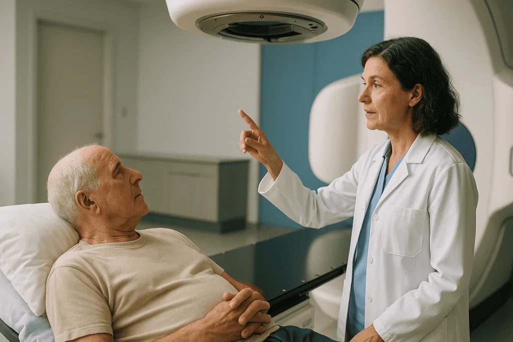
The session itself is painless and silent apart from machine noise. Patients lie still while the gantry rotates around them; the beam cannot be felt and nothing radioactive remains in the body afterward, so there is no risk to family members from contact (NCI). Most people drive themselves home.
Why does the first SBRT follow-up scan wait a few months?
Three practical problems would arise if the first scan were taken a week after SBRT, and understanding them turns an anxious wait into a sensible one.
The first is inflammation. Radiation triggers swelling and an immune response in and around the target. On a CT scan this can enlarge the apparent size of a lung nodule or liver lesion; on a PET scan, which measures glucose uptake, inflammatory cells light up because they are metabolically busy. An early scan can therefore look worse even when every cancer cell in the field has been fatally damaged. The NIH’s National Cancer Institute notes that side effects such as inflammation in the treated area can appear and evolve in the weeks after external beam radiation (NCI).
The second is biology. Because irradiated cells usually die when they attempt to divide, slow-growing cancers simply take longer to shrink. A scan taken before enough cell cycles have passed measures the past, not the effect of treatment.
The third is decision-making. A scan is only useful if it would change what happens next. A few weeks out, almost no result would prompt a new intervention, because the team would want to see the trend on a later scan anyway. Waiting until the picture is interpretable spares people an unnecessary dose of imaging radiation and, often, a frightening but meaningless report.
The interval is not fixed. Teams may scan earlier if new symptoms appear or if a trial protocol requires it, and later for very slow-growing tumors. If a scan has not been scheduled at all, or the planned interval feels unexplained, asking the care team for the reasoning is entirely reasonable.
What doctors measure on the first SBRT follow-up scan
Response assessment sounds subjective, but radiologists and oncologists use shared rules so that two readers looking at the same images reach similar conclusions. The most widely used framework for solid tumors is RECIST 1.1, published after an international review of imaging trials. It compares the longest diameter of measurable lesions on the follow-up scan with the baseline scan taken before treatment (Eisenhauer et al., PubMed).

Under RECIST, a complete response means the target lesions can no longer be seen. A partial response is a decrease of at least 30 percent in the sum of lesion diameters. Progressive disease is an increase of at least 20 percent plus an absolute increase of a few millimeters, or the appearance of a new lesion. Anything in between is called stable disease, which after SBRT is often a perfectly good outcome rather than a disappointment, because a tumor that has stopped growing may be a tumor that is dead but has not yet been cleared.
The team also looks at features that RECIST does not capture. On a PET-CT, a drop in metabolic activity can precede shrinkage. On an MRI of the liver or prostate, changes in how tissue takes up contrast or restricts water movement hint at whether cells are still alive. Radiologists compare the margins of the treated area with the shape of the original radiation field, since a change that exactly matches the planned dose region is far more likely to be treatment effect than new growth.
Finally, the oncologist reads the scan alongside the person in front of them. Weight, energy, pain scores and blood tests all feed into the same conversation. One image rarely settles the question; the trend across two or three scans usually does.
What are good signs that radiation is working?
People naturally search for early clues, and a few are genuinely informative, provided they are read with caution.
Symptom relief is the most noticeable. When SBRT is used for a painful bone metastasis, easing of pain over the following weeks suggests the tumor is no longer pressing on or irritating nearby tissue, though some people experience a temporary flare first. A cough or breathlessness caused by a lung tumor may improve as it shrinks, and a liver lesion pressing on the capsule may stop causing a dull ache.
Falling tumor markers are the second category. For prostate cancer, PSA (prostate-specific antigen, a protein made by prostate tissue) tends to drift downward over many months, and for some liver cancers alpha-fetoprotein behaves similarly. A steady downward trend across several measurements carries more meaning than any single value.
Stability itself is a good sign. A lesion that has stopped enlarging after months of growth before treatment is behaving the way a controlled tumor behaves, even if it never disappears from the scan. Many irradiated tumors leave a permanent scar that looks like a shadow forever, and that shadow is not a failure.
What is not a reliable sign is how severe the side effects were. Fatigue, skin redness or nausea reflect how healthy tissue reacted, not how the cancer responded, and the NHS notes that side effects vary widely between people treated in similar ways (NHS). Feeling fine during treatment does not mean it missed, and feeling rough does not mean it worked harder. The only trustworthy report comes from imaging and markers interpreted by the team over time.
When the scan looks worse but the treatment is working
Few moments are harder than hearing that a post-treatment scan shows something larger or brighter than before. After SBRT this happens often enough that radiologists have specific language for it, and knowing the terms in advance can soften the shock.
Radiation fibrosis is scarring in healthy tissue that received dose. In the lung it appears as a dense, sometimes wedge-shaped patch that can develop over months after treatment and may even grow for a period before stabilizing. Because the patch follows the shape of the treated region rather than the original tumor, an experienced reader can usually tell the two apart. The NIH’s National Cancer Institute lists lung inflammation and scarring among the delayed effects that can follow chest radiation (NCI).
On PET scans, inflammatory cells consume glucose and therefore glow, a phenomenon sometimes called post-treatment uptake. Interpreting a PET too soon after SBRT risks mistaking this for living tumor, which is one more reason the first metabolic scan is often delayed longer than the first CT.
In the liver, the treated zone can show a rim of enhancement where healthy tissue is healing, and in the prostate PSA may rise briefly, a pattern discussed in the next section. In the brain, a related effect after stereotactic radiosurgery is called radionecrosis, and distinguishing it from recurrence sometimes requires advanced imaging or a period of watchful rescanning.
The practical lesson is that an ambiguous first scan usually leads to another scan rather than to a new treatment. Oncologists are trained to tolerate uncertainty for a few months when the alternative is acting on an image that may be lying. Asking, directly, whether a change could be treatment effect is a question every team expects.
PSA after SBRT and other tumor markers: why they wobble
For people treated for prostate cancer, the blood test can feel more important than the scan, because PSA is checked every few months and gives a number to watch. Understanding its behavior after radiation prevents a great deal of unnecessary fear.
PSA is produced by both healthy and cancerous prostate tissue. Radiation damages both, so the level falls gradually as cells stop functioning and die. Unlike surgery, where PSA drops to near zero within weeks because the gland is gone, the decline after radiation is slow, and the lowest point, called the nadir, may not be reached for a long time. The Cleveland Clinic describes this gradual decline as expected after prostate radiation and emphasizes that follow-up relies on the trend rather than a single result (Cleveland Clinic).
Along the way, a temporary rise known as a PSA bounce can occur, often in the first couple of years, before the level resumes its downward path. It is thought to reflect inflammation or delayed release of PSA from damaged cells. A bounce is not, by itself, evidence of recurrence, and oncologists usually respond by repeating the test rather than ordering new treatment.
Other cancers have their own markers. Alpha-fetoprotein in some liver cancers and carcinoembryonic antigen in some bowel cancers can track response, but each has limits: not every tumor produces the marker, and benign conditions can raise them. Markers therefore sit beside imaging, never above it.
If hormone therapy is part of the plan, the medicine itself lowers PSA, which makes the early readings reflect the drug as much as the radiation. The prescribing clinician interprets the numbers in that context, and no change to that medicine should be made on the strength of a single result.
Who SBRT is usually for, and who is usually asked to wait
SBRT earns its place when a tumor is small enough, and well enough defined, to be targeted precisely while sparing what surrounds it. Common settings include early-stage lung cancer in people who are not candidates for surgery, a limited number of metastases in the lung, liver, bone, spine or adrenal gland, selected liver cancers, pancreatic tumors, kidney tumors and localized prostate cancer (Mayo Clinic; NCI). The decision depends on tumor size, location, nearness to sensitive structures such as the bowel or spinal cord, prior radiation to the same area, and the overall treatment plan.
Being asked to wait is not the same as being turned down. Several situations commonly lead a team to delay or reconsider:
- Recent systemic therapy, where the team wants to see how the tumor responds before deciding whether local treatment adds anything.
- Active infection or uncontrolled inflammation in the target area, which could worsen with radiation and blur the planning images.
- A tumor that is currently too large or pressing on a critical structure, where chemotherapy, hormone therapy or surgery may come first to shrink or stabilize it.
- Unclear imaging, where a biopsy or repeat scan is needed to confirm what is being treated.
- Breathing or mobility problems that make it difficult to lie still in the treatment position, which may be addressed with practice sessions or supportive measures.
Alternatives discussed alongside SBRT typically include surgery, conventionally fractionated radiation, ablation techniques that use heat or cold, and systemic therapy alone. Each carries its own balance of benefit and risk, and the National Cancer Institute’s overview of external beam radiation lists factors that guide the choice (NCI). The person’s own priorities belong in that conversation, and the final recommendation rests with the multidisciplinary team.
SBRT side effects timeline: what the first days and weeks usually look like
People who have heard about weeks of daily radiation sometimes expect SBRT to feel proportionally more intense because the dose per session is higher. In practice, because the target is small and the beams are tightly shaped, most short-term effects are confined to the treated area and the surrounding few centimeters.
In the first few days, fatigue is the most common complaint, and it tends to build rather than appear immediately. The NHS describes radiotherapy tiredness as something that can continue for weeks after treatment ends before gradually improving (NHS). Skin over the treated area may redden or feel tender when the target is near the surface, though with deep tumors this is often absent.
Site-specific effects follow in the first one to three weeks. Chest treatment can bring a dry cough or mild chest discomfort; abdominal treatment can cause nausea, loss of appetite or loose stools; pelvic treatment can increase urinary frequency or urgency; spine or bone treatment sometimes produces a temporary pain flare in the first days before relief begins. These generally peak and then fade over the following weeks.
When people ask about the hardest days, the honest answer is that there is no single pattern. For many, the lowest energy arrives one to two weeks after the final session, when the cumulative effect of inflammation meets the emotional let-down of finishing active treatment. For others the hardest day is scan day, months later.
Later effects, which the National Cancer Institute groups as those appearing months after treatment, can include lung scarring, rib pain, changes in liver blood tests or bowel irritation, depending on the site (NCI). The team will have discussed the ones relevant to the specific target, and any new or persistent symptom deserves a report at the next visit, or sooner if it is severe.
A month-by-month view of what happens after SBRT
The sequence below gathers the timelines discussed so far into one place. The intervals are typical ranges reported by the cited sources, not promises, and individual plans differ according to tumor type, location and how the first scan looks.
| Period after final session | What is usually happening inside the body | What the care team is usually doing |
|---|---|---|
| First week | Inflammation begins in the treated area; cancer cells carry fatal DNA damage but mostly still look intact | Phone or clinic check for acute side effects; no routine imaging |
| Weeks 2 to 6 | Fatigue and site-specific effects peak then ease; damaged cells begin to die as they attempt to divide (Mayo Clinic; NHS) | Symptom management; blood tests if markers or organ function are relevant |
| Around months 2 to 3 | Early shrinkage or stability becomes measurable; inflammation has usually settled enough for the picture to be readable | First follow-up imaging, compared with the pre-treatment baseline using RECIST or similar criteria (PubMed) |
| Months 3 to 12 | Continued slow shrinkage or scar formation; PSA or other markers trend down with possible temporary bumps (Cleveland Clinic) | Repeat imaging at intervals set by cancer type; watch for late effects such as fibrosis |
| Beyond the first year | Treated area typically stable; late tissue changes may still evolve | Surveillance continues at longer intervals; any new growth prompts further assessment |
Two features of this table deserve emphasis. First, the busiest clinical period is the first few weeks, when side effects need attention, while the most informative period is the first few months, when the scan happens. The quiet stretch between them is where people often feel abandoned, yet nothing useful would be learned by filling it with tests. Second, the surveillance phase lasts years rather than months, because late recurrence and late tissue effects are both possible. The team’s schedule reflects that long horizon.
What is the success rate of SBRT? Why one number misleads
Search engines are full of percentages attached to SBRT, and almost every one of them describes a different thing. Before any figure is taken to heart, it helps to know which question it answers.
Local control means the treated tumor did not grow back at that spot within a defined period, often a few years. It says nothing about whether cancer appeared elsewhere. Progression-free survival counts time until the cancer grows anywhere or the person dies from any cause. Overall survival counts time until death from any cause. A high local control rate can coexist with a modest survival rate when the disease has already spread microscopically, which is common in metastatic settings.
Numbers also depend heavily on who was studied. Early-stage lung cancer in people fit for surgery, a single liver metastasis from bowel cancer, and painful spinal disease are three entirely different populations, and figures from one cannot be transplanted to another. Study design matters too: single-center reports tend to produce more favorable results than large registries, and short follow-up flatters any treatment whose failures appear late.
This article deliberately quotes no success percentage, because a responsible figure would need to be tied to a specific cancer type, stage and named guideline or systematic review, and even then would describe a group rather than an individual. The National Cancer Institute’s overview of radiation therapy describes benefits and risks in terms of goals, whether aiming to eliminate a tumor, slow its growth or relieve symptoms, rather than universal rates (NCI).
The more useful question to bring to the clinic is specific: for this tumor, in this location, with this history, what is the team hoping SBRT will achieve, and what would make them conclude it has not? That answer can be honest in a way a single percentage never is.
Does SBRT shrink tumors? What people often get wrong
Several beliefs circulate among people recovering from SBRT, and most of them make the waiting harder than it needs to be.
Myth: the tumor should vanish from the scan if the treatment worked. Many irradiated tumors leave a permanent scar or shadow. Radiologists expect this and read stability, not disappearance, as the usual sign of control. A visible remnant months later is often dead tissue (Mayo Clinic).
Myth: a bigger-looking area on the first scan means the cancer is growing. As explained earlier, inflammation and fibrosis can enlarge the treated zone, especially in the lung. The pattern and shape matter more than the size alone, which is why a repeat scan rather than a new treatment is the common response.
Myth: worse side effects mean the radiation hit harder. Side effects reflect how healthy tissue reacted, which varies between people treated identically (NHS). They carry no information about how the tumor responded.
Myth: a rising PSA means the prostate treatment failed. A temporary bounce is a recognized phenomenon, and oncologists interpret the trend across multiple readings over an extended period (Cleveland Clinic).
Myth: scanning sooner would be safer. An early scan would mostly show treatment effect, risk a false alarm, and rarely change management. Timing the scan to when it can be interpreted is the safer choice.
Myth: feeling well afterward means the follow-up schedule can be relaxed. Late effects and late recurrence are both silent at first. Surveillance visits exist precisely because feeling well is not the same as being well.
None of these corrections should be read as reassurance that every scan will be fine. They are a reminder that the first picture is a draft, and that the team is trained to read drafts without panicking.
Questions to ask your care team before and after the first scan
Clinic appointments are short, and the questions that matter most tend to surface in the car afterward. Writing a list in advance, and bringing someone to take notes, changes the quality of the conversation. The following are worth considering; not all will apply to every situation.
- What is the goal of my SBRT: to eliminate this tumor, to control it for as long as possible, or to relieve a symptom?
- When is my first follow-up scan planned, what type of scan is it, and why that interval?
- What would you expect to see on that scan if the treatment is doing its job, and what would you expect if it is not?
- Could the scan show changes from the radiation itself, and how will you tell those apart from the cancer?
- If the first scan is unclear, what happens next: another scan, a different kind of imaging, a biopsy, or waiting?
- Which blood tests, if any, will track my response, and how much should I read into a single result?
- Which side effects should I expect in the first weeks, which are possible months later, and which should prompt a call rather than waiting for the next visit?
- Are there any medicines I take that could affect the scan or the markers, and should any of them be reviewed with the prescriber?
- How long will surveillance continue, and how often will I be seen after the first year?
- If the cancer comes back in the same place or somewhere else, what options would be on the table?
MedlinePlus offers a general guide to questions people can raise about radiation therapy and encourages writing down answers and asking for repetition when something is unclear (MedlinePlus). A good team welcomes the list. If a question is met with impatience rather than an answer, it is reasonable to ask again at the next visit or to request a dedicated appointment.
When to call your doctor after SBRT
Most of the period between the last session and the first scan is uneventful, and most symptoms that arise are mild and expected. A few are not, and the care team would rather hear about them early than at the scheduled visit.
Contact the oncology team the same day, or use emergency services where the symptom is severe, for any of the following red flags:
- Fever or shaking chills, especially if chemotherapy or immunotherapy is also part of the plan, since infection can progress quickly when immunity is lowered.
- Sudden or worsening breathlessness, chest pain, or coughing up blood after treatment to the chest.
- Severe or escalating pain that is not controlled by the measures already discussed, or new pain at a site that was not treated.
- New weakness, numbness, difficulty walking, or loss of bladder or bowel control after treatment to the spine, which can indicate pressure on the spinal cord and is a medical emergency.
- Persistent vomiting, inability to keep fluids down, severe abdominal pain, or black or bloody stools after treatment to the abdomen or pelvis.
- Yellowing of the skin or eyes, or dark urine, after liver treatment.
- A skin reaction that blisters, weeps, or shows spreading redness and warmth.
- Swelling, pain or redness in one calf, or sudden breathlessness, which can signal a blood clot, a recognized risk for people with cancer.
Fatigue that improves with rest, mild nausea, a dry cough without blood, and tenderness over the treated area usually belong to the expected list, and the NHS advises reporting them at the next review unless they worsen (NHS). When in doubt, the nurse line exists for exactly this purpose. No symptom is too small to mention if it is causing worry, and every decision about investigation or treatment rests with the team who knows the plan.
Frequently asked questions
How long does it take for SBRT to work?
Biologically, SBRT starts working during the first session, because the DNA damage that stops cancer cells dividing happens immediately. Visible results take longer: tumors shrink over weeks to months as damaged cells die and are cleared, which is why the first assessment scan is usually planned a few months after the final treatment. Symptoms such as bone pain can improve sooner, while markers like PSA fall gradually over a much longer period.
What are the hardest days after radiation treatment?
There is no universal pattern, but many people report the lowest energy one to two weeks after the final session, when fatigue and local inflammation peak and the structure of daily appointments has ended. Site-specific effects such as cough, nausea or urinary urgency also tend to be most noticeable in that window before easing. For others, the hardest day is emotionally the first follow-up scan, months later.
What are good signs that radiation is working?
The most reliable signs are measured ones: a tumor that is smaller or stable on follow-up imaging compared with the pre-treatment scan, reduced metabolic activity on PET, and a downward trend in any relevant tumor marker across several tests. Relief of symptoms caused by the tumor, such as pain or breathlessness, is encouraging but can be delayed or fluctuate. How intense the side effects feel is not a sign of effectiveness either way.
What is the success rate of SBRT radiation therapy?
No single figure applies, because results depend on the cancer type, stage, location, prior treatment and how success is defined. Local control, progression-free survival and overall survival are different measures, and studies of early lung cancer, liver metastases and spinal disease describe entirely different populations. The most useful question for the treating team is what they hope SBRT will achieve for your specific situation and how they will judge whether it has.
Why is my SBRT follow up scan scheduled so far away?
The delay is intentional. In the first weeks, radiation-induced inflammation can make the treated area look larger or brighter on CT or PET, which risks a false alarm, while slow-dividing cancer cells have not yet had time to die and shrink the tumor. A scan taken too early would rarely change the plan. Waiting until the picture is interpretable gives a clearer answer; the exact interval is set by your team.
Does SBRT shrink tumors completely?
Sometimes the treated tumor disappears from imaging, but very often it leaves a permanent scar or shadow, particularly in the lung and liver. Radiologists expect this and judge success by stability over successive scans and by the pattern of change, not by complete disappearance. A visible remnant months after SBRT frequently represents dead or scarred tissue rather than surviving cancer, and the team will say which interpretation fits your scans.
What does a rising PSA after SBRT mean?
A temporary rise, known as a PSA bounce, is a recognized phenomenon after prostate radiation and does not by itself indicate the treatment failed. PSA declines slowly after radiation because both healthy and cancerous prostate tissue are damaged gradually, and inflammation can cause brief upward blips along the way. Oncologists interpret the trend over multiple readings and may simply repeat the test; any decision belongs with the treating clinician.
What is the SBRT side effects timeline?
Fatigue usually builds over the first days and can last weeks before improving. Site-specific effects such as cough, nausea, loose stools, urinary urgency or a brief pain flare commonly appear within one to three weeks and then fade. Later effects, including lung scarring, rib discomfort or bowel irritation, can develop months after treatment depending on the site. Your team will have explained which of these apply to your target area.
Can the first scan after SBRT look worse even if the treatment worked?
Yes. Inflammation, fluid and healing scar tissue can enlarge the treated area on CT and increase glucose uptake on PET, mimicking growth. Radiologists look at whether the change follows the shape of the radiation field and how it evolves on later scans. An ambiguous first result usually leads to repeat imaging rather than new treatment, because acting on a picture that may reflect healing could do more harm than waiting.
How long does follow-up continue after SBRT?
Surveillance typically continues for years rather than months, with imaging and clinic visits at intervals that lengthen as scans remain stable. The long horizon exists because both late recurrence and late tissue effects can appear quietly, well after a person feels fully recovered. The schedule depends on the cancer type and the findings on early scans, and the treating team sets and adjusts it over time.
References
This article is for general information only and is not a substitute for professional medical advice. Please consult a qualified doctor about your individual situation.

More from the Blog

Sarcoma in Teenagers and Young Adults: How Treatment Planning Considers Growth and School
Sarcoma treatment in teenagers usually combines chemotherapy with surgery, and sometimes radiation, sequenced over many months by a specialist sarcoma team. Because young bodies…

Which Blood and Genetic Tests Guide Lymphocytic Leukemia Treatment Choices?
Treatment choices in chronic lymphocytic leukemia rest on a small set of tests: a complete blood count with flow cytometry to confirm the diagnosis,…

When Is Graft-Versus-Host Disease Suspected? Fever, Rash and Diarrhea After Transplant
Graft-versus-host disease is suspected when someone who has received donor stem cells develops a new rash, persistent watery diarrhea, nausea, or rising liver tests,…

Mohs Surgery Recovery: Pressure Dressing, Stitches and the First Two Weeks of Wound Care
Mohs surgery recovery usually begins with a firm pressure dressing worn for about one to two days, followed by daily gentle cleaning, ointment and…

Why Genetic and Molecular Testing Comes First in Acute Myelogenous Leukemia Treatment
Genetic and molecular testing comes first in acute myelogenous leukemia because the chromosome changes and gene mutations found in the leukemia cells decide which…
Why a Dental Check Comes Before IMRT for Head and Neck Cancer
A dental check comes before IMRT for head and neck cancer because radiation permanently reduces the jawbone's blood supply and its ability to heal.…