

How Is Otoplasty Performed? Cartilage Scoring, Sutures and the Incision Behind the Ear

Key Takeaways
- The standard otoplasty incision sits in the crease behind the ear, where loose skin allows access to the cartilage and a natural shadow hides the scar.
- Ears usually protrude because the antihelical fold never formed, because the conchal bowl is too deep, or both, and each problem calls for a different surgical technique.
- Cartilage scoring works because partially cut cartilage curls away from the scored surface, while permanent internal sutures pin a new fold or draw the bowl toward the head.
- Patient guides from the NHS and Mayo Clinic describe otoplasty as taking roughly one to three hours, usually as a same-day procedure under local or general anesthesia.
- Surgery is generally not offered before around age five, when the cartilage has reached close to adult size and firmness according to NHS and Mayo Clinic guidance.
- The NHS advises avoiding swimming for about four to six weeks and contact sports for about twelve weeks, and a night headband is typically worn for several weeks to protect the healing shape.
Otoplasty is usually performed through a small incision in the crease behind the ear. The surgeon lifts the skin, then reshapes the cartilage by scoring it so it bends more easily, placing internal sutures that create or deepen the ear's natural fold and draw the ear closer to the head, or a combination of both. The procedure typically takes one to three hours under local or general anesthesia, and most people go home the same day.
A father sits in a waiting room turning his son’s school photo over in his hands. The boy is grinning, but his ears stand a little proud of his head, and last term someone gave him a nickname that stuck. The father is not looking for a miracle. He simply wants to know, in plain words, what the surgeon would actually do.
That question, how is otoplasty performed, has an oddly satisfying answer once the jargon is stripped away. The operation is small in scale and mechanical in logic. Ear cartilage behaves like a stiff spring. A surgeon can weaken the spring, tie it into a new shape, or trim a piece of it, and almost everything happens behind the ear where a scar hides in a natural crease.
What follows is the honest version: the anatomy, the incision, the two main techniques, what recovery really involves, and the questions worth asking before anyone agrees to anything.
How is otoplasty performed, step by step?
Strip away the operating room theater and otoplasty follows a sequence that has barely changed in decades. Understanding the order makes the rest of this article easier to follow.
First comes marking. With the patient sitting up, the surgeon draws lines on the ear showing where the new fold should run and how far the ear should move toward the head. Ears are then compared side by side, because symmetry is judged from the front, not from the side.
Second, anesthesia. This is either a local anesthetic injected around the ear, sometimes with a sedative, or a general anesthetic in which the patient is fully asleep. The NHS notes that children are usually treated under general anesthesia while adults often have local, a point the next sections return to.
Third, the incision. A cut is made in the groove where the back of the ear meets the scalp, and the skin is lifted off the cartilage like peeling a label off a jar. This exposes the elastic framework that gives the ear its shape.
Fourth, reshaping. The surgeon may score the cartilage to soften its resistance, place permanent internal stitches that fold or pull it, remove a thin strip of cartilage, or blend these methods. Which combination is used depends on what the ear needs and on the surgeon’s training.
Fifth, closure and dressing. The skin is stitched, a soft padded dressing is molded around both ears, and a bandage or headband holds everything still.
Patient guides from Mayo Clinic describe the whole operation as taking roughly one to three hours, and the NHS gives a similar one-to-two-hour range, with most people going home the same day. The rest of this piece unpacks each of those steps in detail.
What is the surgeon actually reshaping? Ear anatomy in plain language
The outer ear is a single sheet of cartilage, thin as a credit card in places, covered by skin that is almost glued to it on the front and slightly looser at the back. Cartilage is the firm, rubbery tissue that also shapes the tip of the nose. It has no blood vessels of its own and holds its form stubbornly.

Four landmarks matter for otoplasty. The helix is the outer rim, the curled edge you run a finger along. The antihelix is the ridge just inside that rim, roughly Y-shaped, and it is the fold most often missing in ears that stick out. The scapha is the shallow trough between helix and antihelix. The concha is the deep bowl that funnels sound toward the ear canal.
Ears protrude for one or both of two reasons. Sometimes the antihelical fold never formed properly during development, so the top of the ear flares flat instead of curling back. Sometimes the conchal bowl is unusually deep or angled, pushing the whole ear away from the skull like a cupped hand held out from the head. Many people have a bit of each.
This distinction is not academic. A surgeon who diagnoses a missing fold will reach for a technique that creates a fold. A surgeon who sees a deep bowl will reach for a technique that sets the bowl back. Diagnosing the wrong problem produces a result that looks pinned rather than natural.
Cleveland Clinic and Mayo Clinic both describe otoplasty as a procedure for the outer ear only. It does not touch the ear canal, the eardrum, or the tiny bones of the middle ear, which is why hearing is not expected to change. The work is entirely structural, on the visible shell.
The incision behind the ear: where it sits and why
Ask most people where the scar from otoplasty would be and they point vaguely at the front of the ear. In practice the standard incision runs along the back of the ear, in the crease where cartilage meets the scalp, a site known as the postauricular sulcus. Postauricular simply means behind the ear.
The location is chosen for three reasons. Skin at the back of the ear is looser than at the front, so it can be lifted and later closed without tension. The crease is naturally shadowed, so even a slightly raised scar is hard to see unless someone deliberately looks. And the back approach lets the surgeon reach both the conchal bowl and the future antihelical fold without cutting through the visible front surface.
Through this incision the surgeon removes a thin ellipse of skin, then works forward with fine instruments to separate skin from cartilage. Some surgeons keep the incision short, roughly the length of the ear’s upper two-thirds. Others extend it toward the earlobe when the lobe itself also needs to be tucked in.
The NHS lists a small scar behind the ear among the expected effects of the operation, and notes that scars generally fade over time although they do not disappear entirely. Occasionally scar tissue thickens into a raised, firm ridge, which surgeons call a hypertrophic or keloid scar. People with a personal or family history of keloids are usually asked about it beforehand.
One variation deserves mention because it changes where the scar sits. When cartilage is scored from the front, a technique discussed below, some surgeons make a second tiny incision at the front rim of the ear or thread a needle through the skin without a full cut. Your surgeon should be able to draw exactly where any incision will fall.
Cartilage scoring: weakening the spring so the fold bends
Here is a simple kitchen experiment that explains cartilage scoring. Take a strip of firm plastic packaging and try to fold it. It resists and springs back. Now scratch a series of shallow lines along one side and fold it again. It bends toward the scratched side and stays there.
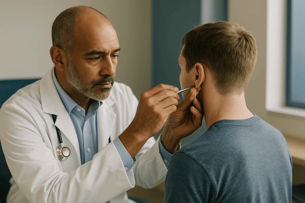
Ear cartilage behaves the same way. Scoring means making deliberate partial-thickness cuts or abrasions on one surface of the cartilage. This releases tension on that side and causes the cartilage to curl away from it. When scoring is applied to the front surface along the line where the antihelix should be, the cartilage naturally rolls backward into a fold.
Two classic descriptions exist in the surgical literature. In one, the surgeon works through the incision behind the ear, tunnels forward under the skin, and rasps the front surface with a fine instrument. In another, the surgeon makes small cuts along the front of the cartilage through a tiny access point. Both aim for the same effect: a fold that forms because the tissue wants to bend, not because it is forced.
Scoring is often paired with a few sutures to hold the fold at the right height while healing locks it in. Used alone it can be unpredictable, because cartilage varies in thickness and stiffness from one person to another. Overscoring can leave sharp, angular edges visible through the thin front skin, the so-called cornered look that experienced surgeons work hard to avoid.
Scoring is described as a cartilage-cutting approach, in contrast to suture-only methods. That word sounds alarming but means something modest here: the cartilage is scratched or partly incised, not removed in bulk. Whether scoring is appropriate depends on how stiff the cartilage feels during the consultation.
Sutures: the stitches that create a fold and set the ear back
If scoring changes what the cartilage wants to do, sutures tell it exactly where to go. Two named suture patterns appear in almost every description of how otoplasty is performed, and they solve the two different problems discussed earlier.
The first is often called a Mustardé suture after the surgeon who popularized it. It is a horizontal mattress stitch, meaning the thread passes through the cartilage in a rectangle rather than a simple loop. Placed across the scapha and the concha, and tightened, the two edges draw together and the cartilage between them buckles backward into a new antihelical fold. Several of these stitches in a row build the entire ridge, like pinching a fold into fabric with a line of pins.
The second is often called a conchomastoid or Furnas suture. This one passes from the conchal bowl to the tough tissue covering the mastoid bone behind the ear. Tightening it pulls the whole bowl inward, rotating the ear toward the head. It is the stitch that fixes protrusion caused by a deep concha rather than a missing fold.
Shaping sutures are typically made of permanent, non-absorbable material, because the cartilage needs to be held while scar tissue slowly forms and takes over the job. That permanence has a downside. Occasionally a knot works its way toward the skin and becomes visible or pokes through, sometimes months later. Mayo Clinic and the NHS both list this among recognized complications.
Suture-only techniques are sometimes described as cartilage-sparing. They tend to be favored for softer cartilage. When cartilage is very stiff, sutures alone may not hold and the ear can drift back, so many surgeons combine them with scoring or with removal of a small strip of cartilage from the bowl.
Cutting, scoring, or sutures alone? A comparison
Patients often arrive having read that one technique is best. The honest position from the surgical literature is more nuanced: each approach suits particular cartilage and particular anatomy, and most surgeons combine methods rather than pledging loyalty to one. The table below summarizes the trade-offs as they are generally described in the literature. None of the entries is a prediction about your ear.
| Approach | What is done | Best suited to | Trade-offs described in the literature |
|---|---|---|---|
| Suture-only (cartilage-sparing) | Permanent internal stitches fold the antihelix or pull the concha back; cartilage is not cut | Thin, pliable cartilage; younger patients | Lower risk of sharp edges; higher chance of gradual loosening or suture problems if cartilage is stiff |
| Scoring (partial-thickness cartilage cutting) | Shallow cuts or abrasions on one surface so cartilage curls into a fold | Stiff cartilage that resists sutures | Durable fold; risk of angular contour if overdone; often needs a few sutures to set height |
| Cartilage excision | A strip of conchal cartilage is removed to reduce a deep bowl | Very deep or wide concha | Powerful setback; more swelling; irregularity possible if edges are uneven |
| Combined | Any two or three of the above tailored to each ear | Most adult and many pediatric ears | Most flexible; result depends heavily on surgeon judgment |
A note on the word cutting. In lay conversation it evokes removal of large pieces. In otoplasty it usually means partial-thickness scoring or excision of a narrow strip, with the ear’s overall size unchanged.
What matters most is not the label but whether the chosen method matches the diagnosis. A missing fold and a deep bowl are different problems and, when both are present, both need addressing or the result will look incomplete.
Local or general anesthesia: what happens on the day
The morning of surgery is usually unhurried. Most otoplasty is day-case, meaning the patient arrives, is operated on, recovers for a few hours, and goes home in the same day, a pattern described by the NHS and Mayo Clinic alike.
The choice of anesthesia turns on age and temperament more than on the surgery itself. Local anesthesia means numbing medication injected around the ear; the patient is awake, sometimes lightly sedated, and feels pressure and movement but not pain. It is commonly used for adults and older teenagers. General anesthesia means being fully unconscious with breathing monitored by an anesthesiologist. It is routine for younger children, who cannot be expected to lie still with instruments working near their face for over an hour.
Under general anesthesia the person will be asked not to eat for a period beforehand. The NHS explains that fasting reduces the risk of stomach contents entering the lungs while reflexes are suppressed. The care team gives specific timings; they vary with age and with whether the intake is solid food, milk, or clear fluids, so it is best to follow the written instructions rather than a general rule.
Because the operation happens beside the ear, surgeons usually infiltrate local anesthetic even when the patient is asleep. This numbs the area for several hours after waking and shrinks small blood vessels, which reduces bleeding during the procedure.
Once finished, a bulky soft dressing is shaped around both ears and held with a bandage that wraps around the head. It looks dramatic in the mirror. Its job is simply to keep the ears still, absorb any oozing, and protect fresh sutures from a stray pillow or hand during sleep.
Someone else should drive home and stay the first night, whichever anesthesia is used.
Who is otoplasty usually for, and who is asked to wait?
Otoplasty is most often performed for ears that protrude noticeably from the head, a trait present from birth and usually shared with at least one relative. It is also used for ears with an unusual shape such as a folded-over top, and occasionally to correct asymmetry after injury or a previous operation.
Age is the first gate. Both the NHS and Mayo Clinic advise that surgery is generally not carried out before around age five, because the ear cartilage needs to reach close to adult size and firmness before it can be shaped reliably. Operating too early risks a result that changes as the ear grows.
Motivation is the second gate, and it matters more than many people expect. For children, surgeons look for a child who has expressed a wish for the change in their own words and can cooperate with dressings afterward. A parent’s distress alone is not usually considered sufficient reason. For adults, the question is whether expectations are realistic. Otoplasty changes the position and contour of the ear. It does not make ears smaller in any meaningful way, and it does not change the face.
Some people are asked to wait or reconsider. Active ear infections or skin conditions over the incision site are postponed until settled. Uncontrolled bleeding disorders, or anticoagulant medicines that cannot be safely paused, are discussed with the prescribing clinician first, and the surgical team decides. A history of keloid scarring prompts a frank conversation about scar risk. Smoking impairs wound healing, and many surgeons ask patients to stop for a period around surgery. Anyone with unstable mental health or body-image distress is usually offered support before elective surgery is scheduled.
None of these is a permanent no. They are reasons to pause and plan.
How is otoplasty performed differently for a child?
Otoplasty for children follows the same anatomical steps as for adults, yet the experience is deliberately different, and parents deserve to know how.
Anesthesia is almost always general, as described earlier. Many children’s units let a parent stay until the child is asleep and be present when they wake. Preparation often includes a walk-through beforehand, with masks and gowns shown in advance, because unfamiliar equipment is more frightening than the idea of surgery itself. Pain relief and any sedation are chosen and given by the anesthesia team; this article does not describe doses, and parents should follow the written plan they are given.
Technique tends to lean toward cartilage-sparing sutures, because young cartilage is softer and bends willingly, though the surgeon adjusts to what is found. The operation is typically performed on both ears in one session even when only one looks prominent, since correcting one side alone often makes the other suddenly appear to protrude.
Comfort at home revolves around the head bandage. Children usually wear it for a few days, then switch to a soft headband over the ears at night for several weeks, a timeline both Mayo Clinic and the NHS describe. Sleeping on the back or with a travel pillow helps stop them rolling onto an ear. Hair washing waits until the surgical team says the wound can get wet.
School return is guided by comfort and by activity. The NHS suggests most children can go back within a week or two but should avoid swimming for about four to six weeks and contact sports for roughly twelve weeks. Teachers can be told to expect a headband, and a quiet word about avoiding playground tussles goes a long way.
Above all, the child should have asked for this. Surgeons routinely speak to the child alone for a moment to be sure.
Otoplasty recovery time: the first days and weeks
Recovery from otoplasty is more about protecting the result than about pain, though the first few days are uncomfortable. Timelines below are typical ranges drawn from NHS and Mayo Clinic guidance; individual experience varies and your team’s instructions override any figure here.
The first two to three days bring throbbing and a feeling of tightness, controlled with pain relief chosen by the surgical team. The head bandage stays on and gets warm and itchy. Sleeping propped up on two pillows helps limit swelling. Ears may feel numb or oddly sensitive, both of which are expected as small nerves recover.
Around day three to seven, the bulky dressing is removed at a follow-up visit, and the ears are seen for the first time. They typically look swollen, bruised, and pinker than expected, and often appear more tightly set than the final result because swelling behind the ear pushes them inward. The NHS notes that bruising and soreness usually settle over the first couple of weeks. A soft headband is then worn over the ears at night, and often during the day at first, for a period Mayo Clinic describes as two to six weeks.
By one to two weeks most adults return to work and most children to school, according to the NHS. Skin stitches are removed or dissolve during this window.
From weeks four to twelve the rules loosen. Swimming waits roughly four to six weeks and contact sports about twelve weeks, per the NHS, because a blow to a healing ear can bleed under the skin or disrupt sutures. Glasses can usually be worn once the team confirms the incision has sealed, sometimes with padding on the arms.
Final contour takes months. Cartilage settles slowly and residual firmness behind the ear softens over the year.
Otoplasty risks and complications, in neutral terms
Otoplasty is widely described as a relatively low-risk operation, but low risk is not no risk, and an honest explainer lists what can go wrong. The NHS, Mayo Clinic, and MedlinePlus each publish complication lists; the items below appear on all of them.
Bleeding and hematoma. A hematoma is a collection of blood under the skin. It shows up as rapidly increasing pain and swelling on one side in the first day or two, and it needs prompt attention because pooled blood can damage cartilage.
Infection. Redness spreading beyond the incision, warmth, pus, fever, or a bad smell from the dressing point to infection. Cartilage infection, called chondritis, is uncommon but can distort the ear if untreated, which is why surgeons take early signs seriously.
Asymmetry. Perfect symmetry does not exist in nature and cannot be manufactured. Small differences are normal; larger ones may prompt revision.
Overcorrection. Ears set too close to the head can look pinned, especially from behind, where the helix should still be visible.
Recurrence. Cartilage has memory. Over months or years an ear can partially drift back toward its old position, particularly with suture-only technique in stiff cartilage. Some people choose a second procedure.
Suture problems. Permanent knots may become palpable, visible, or push through the skin, requiring removal.
Contour irregularities. Overscoring can leave sharp ridges visible through the front skin.
Scarring. A thin line behind the ear is expected. Thick or keloid scars are a recognized but less common outcome.
Numbness or altered sensation. Usually temporary, occasionally persistent.
Anesthesia risks. Nausea, sore throat, and rarely more serious events; the NHS notes that serious anesthetic complications are uncommon and are discussed individually.
Percentages for these outcomes vary widely between published series and depend on technique, so no single figure is offered here.
Ear pinning surgery: what people often get wrong
The phrase ear pinning surgery has stuck in popular use, and it carries several misunderstandings worth correcting.
Myth: the ears are literally pinned. Nothing metal is left behind and nothing is stapled to the skull. Permanent stitches inside the cartilage hold a new shape while scar tissue forms; the phrase is a figure of speech.
Myth: the ears are made smaller. Standard otoplasty changes position and contour, not size. An ear that protrudes often looks large because it catches the light from the front; once set back it appears smaller without losing any real dimension. Ear reduction is a separate, less common operation.
Myth: hearing will change. The outer ear does help gather sound, but the amount of change from repositioning is not clinically meaningful. Cleveland Clinic and Mayo Clinic both describe hearing as unaffected.
Myth: results are visible immediately. What appears when the dressing comes off is a swollen, over-tight version of the final look. Surgeons often set ears slightly closer than the goal, expecting some relaxation. Judging the result before several weeks have passed leads to unnecessary worry.
Myth: it is only for children. Adults have otoplasty in large numbers. The cartilage is stiffer and may need scoring rather than sutures alone, but the operation is otherwise the same.
Myth: the scar is on the front of the ear. The standard incision hides in the crease behind it.
Myth: once done, the shape is fixed forever. Cartilage can drift back partially over time, and a minority of people choose revision. Anyone promising permanence beyond doubt is overstating what the evidence supports.
Myth: it is trivial because it is small. Any operation carries anesthesia risk, bleeding risk, and infection risk. Small does not mean casual, and the consent conversation should treat it accordingly.
Alternatives to surgery: molding, incisionless methods, and doing nothing
Surgery is not the only route, and a good consultation lays out the alternatives in the same neutral tone used for the operation itself.
Newborn ear molding. In the first weeks of life, ear cartilage is unusually soft because of hormones passed from the mother during pregnancy. A pediatric clinician can fit a small splint or molding device that gently holds the ear in an improved shape while the cartilage firms up. Effectiveness falls off quickly as the weeks pass, which is why pediatricians examine ear shape at the first checks. Molding is not a fix for an older child; by the time a school-age child is teased, the window has long closed.
Incisionless suture techniques. Some surgeons place shaping sutures through needle punctures without a formal incision, mainly for ears with soft cartilage that need only a fold created. Reported advantages are quicker recovery and no visible scar. Drawbacks described in the literature include a higher chance of sutures loosening and less ability to address a deep concha. Long-term comparative evidence against traditional otoplasty is limited, so this should be described as an option with less data rather than as equivalent.
Hairstyle and headwear. Not a medical alternative, but for many people, especially adults who have lived comfortably with their ears, it is the honest answer. Prominent ears are a normal variant, not a disease, and no clinician should imply otherwise.
Psychological support. When distress about appearance is severe or out of proportion, talking therapies are recommended before or instead of surgery. Elective procedures rarely resolve body-image concerns rooted elsewhere, and reputable surgeons screen for this.
Waiting. For a child who has not asked for surgery, doing nothing for now keeps every option open. The cartilage will still be there next year.
Questions to ask your care team before otoplasty
A consultation is a two-way examination. The surgeon assesses your ears; you assess whether you understand the plan. These questions, drawn from what patients most often wish they had asked, help.
- Which problem do you see in my ears: a missing antihelical fold, a deep conchal bowl, or both? How does that shape your choice of technique?
- Will you use sutures only, scoring, cartilage removal, or a combination, and why for my cartilage specifically?
- Exactly where will the incision be? Can you draw it? Will there be any incision at the front of the ear?
- Will the shaping sutures be permanent? What happens if a knot becomes visible later?
- Which anesthesia do you recommend for me or my child, and who will administer it?
- What will the ears look like when the dressing comes off, and how long before I should judge the result?
- How long will a headband be needed, and what activities are off-limits, for how long?
- What is your policy if an ear drifts back or the two sides end up noticeably different?
- What signs after surgery would you want a phone call about, and who answers that phone at night?
- How many otoplasties do you perform, and do you have photographs of your own results, including from behind?
- For a child: may you speak with them alone to hear what they want?
- Is there any reason you would advise waiting or not operating at all?
Write the answers down or ask permission to record them. Decisions made in a consultation room feel clear at the time and blurry a week later. A surgeon who welcomes these questions is showing you something about how the rest of the process will go.
When to call your doctor after otoplasty
Most recovery is dull, which is exactly what everyone wants. A few developments, though, need a same-day call to the surgical team or, when severe, emergency care. Do not wait for a scheduled appointment if any of the following appears.
Call urgently for pain that is rapidly worsening on one side, especially with tightening swelling under the dressing. This pattern can signal a hematoma, a collection of blood under the skin that can damage cartilage if not released promptly.
Seek care for bleeding that soaks through the bandage rather than lightly staining it, or for bleeding that does not stop with gentle pressure.
Signs of infection also warrant a call: spreading redness beyond the incision, skin that feels hot, pus or cloudy fluid, a foul smell from the dressing, or a fever. Cartilage infection is uncommon but can permanently distort the ear, so early treatment matters.
Contact the team if a suture pokes through the skin, if the wound edges separate, or if an area of skin over the ear turns dusky, purple, or black, which may indicate compromised blood supply.
After general anesthesia, seek immediate emergency help for difficulty breathing, chest pain, persistent vomiting preventing fluids, or a new severe headache.
Also call, without embarrassment, if the dressing slips and you are unsure how to replace it, if the ears look markedly uneven once uncovered, or if anything simply feels wrong. Surgical teams would far rather answer an unnecessary call than miss a necessary one.
Keep the after-hours number provided at discharge somewhere visible. Every judgment about whether a symptom is expected or concerning belongs to the treating team, who know what was done and what your ears looked like when the bandage went on.
Frequently asked questions
How is otoplasty performed under local anesthesia, and does it hurt?
Under local anesthesia, numbing medication is injected around the ear so the patient stays awake but feels no sharp pain, only pressure, tugging, and the sound of instruments. Some adults also receive a light sedative. The incision, cartilage scoring, and suturing proceed exactly as under general anesthesia. Afterward the area stays numb for several hours, and the surgical team advises on pain relief. Local anesthesia is common for adults and older teens; younger children typically have general anesthesia.
What is the typical otoplasty recovery time before I can go back to work or school?
The NHS suggests most people return to work or school within about one to two weeks, once the bulky dressing has been replaced by a lighter headband and soreness has eased. Swimming usually waits four to six weeks and contact sports around twelve weeks. Final ear shape continues to settle for several months as swelling resolves and cartilage firms in its new position. Your surgeon’s instructions take precedence over these general ranges.
Is ear pinning surgery the same thing as otoplasty?
Yes, ear pinning is the everyday name for otoplasty performed to set protruding ears closer to the head. The term is a figure of speech: nothing is literally pinned, and no metal is left behind. Permanent internal sutures, cartilage scoring, or removal of a small cartilage strip reshape the ear, and scar tissue then holds the new position. Otoplasty also covers reshaping for other ear differences, so the medical term is slightly broader.
At what age is otoplasty for children usually considered?
Both the NHS and Mayo Clinic describe about age five as the earliest usual point, because ear cartilage has reached close to adult size and firmness by then. Beyond age, surgeons look for a child who has expressed the wish themselves and can cope with dressings and a headband. Many families choose the years just before or during early school. For newborns, a molding splint fitted in the first weeks is a different, non-surgical option.
What are the main otoplasty risks and complications I should know about?
Recognized risks include bleeding or a hematoma, infection of skin or cartilage, asymmetry between the two ears, overcorrection giving a pinned look, gradual drift back toward the old position, permanent sutures becoming visible or poking through the skin, sharp contour ridges from overscoring, thickened scarring, altered sensation, and general anesthesia risks. Reported rates vary widely between studies and techniques, so your surgeon should discuss the likelihood in your specific case.
Will the scar behind my ear be visible?
The standard incision is placed in the groove where the back of the ear meets the scalp, so the resulting line is hidden unless someone deliberately looks behind the ear. The NHS notes scars fade over time but do not vanish completely. Occasionally scars thicken into raised keloid or hypertrophic tissue, more often in people with a personal or family history of such scarring, which is why surgeons ask about it beforehand.
Does otoplasty change hearing?
No meaningful change in hearing is expected. Otoplasty reshapes only the outer cartilage shell and does not involve the ear canal, eardrum, or middle ear bones. The outer ear does help gather sound, but repositioning it by a small amount does not alter hearing in a way people notice, according to descriptions from Mayo Clinic and Cleveland Clinic. Temporary muffling from the padded dressing lifts as soon as it is removed.
Can the ears go back to sticking out after surgery?
Partial recurrence is a recognized outcome. Cartilage has a memory and can drift back over months or years, especially when stiff cartilage was treated with sutures alone or a stitch loosens. Most drift is modest, but some people choose a revision procedure. Wearing the protective headband as instructed and avoiding blows to the ears during healing helps protect the result, though no surgeon can promise permanence.
Is otoplasty done on both ears even if only one sticks out?
Frequently, yes. Correcting one ear alone can make the untouched side suddenly appear prominent by comparison, so surgeons often adjust both to achieve balance, even if one needs only a minor change. When the ears are genuinely different in structure, the technique can differ on each side. Your surgeon will explain whether one or both ears are planned and why after examining you from the front and behind.
What does incisionless otoplasty involve, and is it as effective?
Incisionless techniques place shaping sutures through needle punctures rather than a formal cut behind the ear, mainly for soft cartilage that needs a fold created. Reported advantages are no visible scar and quicker recovery. Reported drawbacks include a higher chance of sutures loosening and limited ability to correct a deep conchal bowl. Long-term comparative evidence is limited, so it is best described as an option with less supporting data than traditional otoplasty.
References
- NHS: Ear correction surgery, including ear pinning
- MedlinePlus: Cosmetic ear surgery
- NHS: General anaesthesia
- CDC: About surgical site infections
This article is for general information only and is not a substitute for professional medical advice. Please consult a qualified doctor about your individual situation.

More from the Blog

Is Genioplasty Painful? General Anesthesia, Chin Tightness and What Helps in the First Days
Genioplasty is usually described as uncomfortable rather than severely painful. Because it is performed under general anesthesia, nothing is felt during the operation itself.…

Forehead Lift for Men: Hairline, Brow Shape and Why the Plan Differs for Male Faces
A forehead lift for men lifts sagging brows and softens deep forehead lines by repositioning the tissue beneath the skin, but the plan usually…

Lip Lift vs Corner Lip Lift vs Italian Lip Lift: How the Incision Patterns Differ
The main types of lip lift differ by where the surgeon places the cut. A standard subnasal lip lift removes a strip of skin…

How Sliding Genioplasty Is Performed: The Osteotomy, Repositioning and Plate Fixation
A sliding genioplasty procedure reshapes the chin by cutting the lower front part of the jawbone through an incision inside the mouth, sliding the…

When Can You Wear Contacts and Makeup After Blepharoplasty? Eye Care in the First Weeks
Most surgeons ask patients to keep makeup off the eyelids until incisions have sealed and any stitches are out, which Mayo Clinic places at…

What to Ask a Rhinoplasty Surgeon at Your First Consultation: Twelve Questions That Matter
The most useful questions to ask a rhinoplasty surgeon cover four areas: their training and how often they perform nose surgery, what change is…