

How Lipedema Is Diagnosed: The Exam, Imaging and Staging That Shape a Treatment Plan

Key Takeaways
- Lipedema has no confirmatory blood test or scan; diagnosis rests on history, physical examination and excluding other causes of limb enlargement.
- The ankle or wrist cuff, where enlarged tissue stops abruptly and leaves the foot or hand slim, is one of the most recognizable features on exam.
- A negative Stemmer sign, meaning a skin fold at the base of the toe lifts easily, points away from lymphedema but can change if lipedema progresses.
- Lipedema fat typically does not shrink with dieting even when the trunk does, and that history is itself a diagnostic clue rather than a personal failure.
- Staging from 1 to 3, plus types I to V describing location, guides whether care centers on compression and movement or also involves a surgical discussion.
- New one-sided swelling, hot red skin, or sudden breathlessness alongside leg symptoms are red flags that need prompt medical attention regardless of a lipedema diagnosis.
Lipedema is diagnosed clinically. A clinician takes a detailed history of when the fat pattern began, examines the limbs for symmetrical, tender, easily bruised fat that spares the feet and hands, checks whether the skin pits or holds a fold, and rules out lymphedema, obesity and medical causes of swelling with targeted tests. Ultrasound or lymphoscintigraphy may support the picture, but no single test confirms it.
She had tried every diet her friends swore by, and each one worked the same way: her waist shrank, her face changed, and her legs did not move an inch. What finally sent her to a doctor was not the mirror but a bruise on her thigh she could not remember earning, and the way the sofa arm hurt when she leaned on it.
That story, with small variations, is one clinicians hear again and again from people who turn out to have lipedema. It is also why the question how is lipedema diagnosed matters so much. The condition has no blood marker, no definitive scan and no biopsy that settles it. Diagnosis rests on a patient’s account, a careful set of hands and a clinician who knows what to rule out.
This explainer walks through that process step by step: the conversation, the physical exam, the imaging that sometimes helps, the staging that shapes a plan, and the myths that keep people undiagnosed for years.
How is lipedema diagnosed when there is no single test?
Lipedema is a chronic condition in which fat accumulates abnormally and symmetrically, usually in the legs and sometimes the arms, and the affected tissue tends to be painful and to bruise easily. The NHS describes it as a long-term condition that almost exclusively affects women and that is frequently mistaken for simple weight gain or for lymphedema, a different problem in which fluid builds up because lymph vessels are damaged or blocked.
Because the disease lives in fat and connective tissue rather than in a measurable chemical, diagnosis is clinical. That word has a specific meaning: the clinician reaches a conclusion from history and examination, using tests mainly to exclude other explanations rather than to prove lipedema itself. Cleveland Clinic and the NHS both frame the workup this way, and a widely cited review in Plastic and Reconstructive Surgery Global Open stresses that the pattern of fat, the pain and the sparing of the feet are the anchors of recognition.
In practice, the process has four moving parts. First comes the story: when the shape changed, whether it tracked a hormonal shift, and what has and has not responded to weight loss. Second is the hands-on exam, which looks for symmetry, tenderness, the ankle cuff and how the skin behaves when pressed or pinched. Third is exclusion, where blood tests and occasionally imaging make sure heart, kidney, thyroid or lymphatic disease are not the real driver. Fourth is staging and typing, which describe how far the tissue has changed and where, so a plan can be built around the person rather than the label.
That sequence takes time. It is common for the first visit to end with a working diagnosis and a request for a few tests, and for the picture to firm up over one or two follow-ups. A careful clinician is slow here on purpose.
What actually happens during a lipedema exam
The appointment usually opens with questions rather than a tape measure. Expect to be asked when your legs or arms began to change shape, whether that coincided with puberty, pregnancy or menopause, and whether a mother, sister or grandmother has a similar build. The NHS notes that lipoedema often starts or worsens at these hormonal turning points and that it can run in families, so the clinician is listening for that rhythm.

Pain gets its own set of questions. People with lipedema often describe a deep ache, heaviness or tenderness to light pressure, and bruising after knocks they barely noticed. You may be asked whether your legs feel worse by evening or after standing, and whether the discomfort is different from ordinary muscle fatigue.
Then the physical exam. You will typically be examined standing and lying down, with legs and arms fully visible. The clinician looks for symmetry between the two sides, a disproportion between the lower body and the trunk, and the characteristic shelf or cuff where enlarged tissue stops abruptly at the ankle or wrist while the foot or hand stays slim. Cleveland Clinic describes this cuff sign as one of the more recognizable features.
Hands do the rest. Pressing a thumb into the shin for several seconds checks whether the skin pits, which suggests fluid rather than fat. Gently pinching the skin at the base of the second toe tests the Stemmer sign; in lipedema the fold lifts easily, whereas in established lymphedema it often cannot be pinched. Palpating the tissue itself may reveal small, pea-like nodules under the skin and tenderness out of proportion to the pressure applied.
Height, weight and often waist-to-hip and limb circumference measurements round out the visit, not to judge but to document a baseline the team can compare against later.
What is the pinch test for lipedema, and what does it miss?
The pinch test is the informal name for two related maneuvers. The first is the Stemmer sign described above: the clinician tries to lift a fold of skin at the base of a toe or finger. A fold that lifts freely is called a negative Stemmer sign and points away from lymphedema; a thickened fold that resists pinching is positive and suggests lymphatic fluid has hardened the tissue. The NHS lists this among the checks used to distinguish lipoedema from lymphoedema.
The second maneuver is a tenderness check. A clinician lightly pinches or presses the fat of the thigh, calf or upper arm and asks how it feels. Many people with lipedema report sharp or disproportionate pain from pressure that would barely register on unaffected tissue. This is why some describe the condition as painful fat.
Both tests are quick and useful, and both have limits worth stating plainly. The Stemmer sign is not perfect: early lymphedema can be negative, and long-standing lipedema that has progressed to secondary lymphatic damage can turn positive. A single pinch therefore never rules a condition in or out on its own. Pain thresholds vary enormously between people, and tenderness is also common in fibromyalgia, venous disease and after injury.
What the pinch test does well is add one brushstroke to a larger picture. When the fold at the toe lifts easily, the foot is slim, the thighs are tender and symmetric, the skin does not pit and the story began at puberty, the strokes line up. When they conflict, the clinician looks harder before naming anything.
The takeaway for a reader is simple: the pinch test is a clinical sign interpreted by someone trained to weigh it, not a verdict you can deliver at home.
Can you self test for lipedema at home?
The honest answer is that you can notice, but you cannot confirm. Search traffic for a lipedema self test reflects a real frustration: many people spend years being told to eat less before anyone examines their legs. Paying attention to your own body is reasonable. Treating that attention as a diagnosis is where it goes wrong.
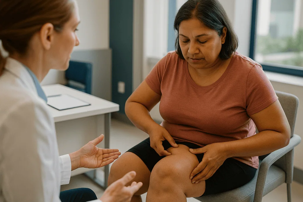
What you can observe at home is pattern and behavior. Does the enlargement affect both legs or both arms about equally? Does it stop at the ankle or wrist, leaving the feet and hands slim? Has the shape stayed stubborn while the rest of you changed with dieting? Do the affected areas hurt when pressed, and do bruises appear without a clear cause? These are the same features the NHS and Cleveland Clinic describe, and noticing them is a good reason to book an appointment.
What you cannot do at home is exclude the alternatives. Fluid from heart, kidney or liver disease, an underactive thyroid, chronic venous insufficiency and lymphedema can all enlarge the legs, some of them dangerously. Blood tests and a trained exam sort those out; a mirror does not. Online quizzes and photo comparisons also tend to over-count, because ordinary fat distribution varies widely and pain is subjective.
A more useful piece of homework is documentation. Write down when the change began and what was happening in your life hormonally. Photograph your legs and arms in consistent lighting from the front and side. Keep a short list of what diets or exercise programs you tried and what did and did not change. Bring that folder to the visit. It shortens the history-taking and, frankly, it is harder for anyone to wave away.
Lipedema vs lymphedema vs obesity: how clinicians tell them apart
The three conditions overlap in appearance, coexist more often than people expect, and call for different plans. Sorting them is the core of the diagnostic work. The distinctions below reflect descriptions from the NHS, Cleveland Clinic and MedlinePlus.
| Feature | Lipedema | Lymphedema | Obesity (generalized) |
|---|---|---|---|
| Distribution | Symmetric; legs, sometimes arms; trunk relatively spared | Often one limb, or asymmetric | Whole body, including trunk and face |
| Feet and hands | Usually spared, with an ankle or wrist cuff | Commonly involved, including toes | Involved in proportion to body |
| Pitting when pressed | Typically absent | Present early; tissue hardens later | Absent |
| Stemmer sign | Negative in most cases | Often positive | Negative |
| Pain and bruising | Tenderness and easy bruising are characteristic | Heaviness; pain variable | Not a defining feature |
| Response to weight loss | Trunk shrinks; affected limbs change little | Fluid not fat; unrelated | Proportional loss |
| Sex distribution | Almost exclusively women | Either sex | Either sex |
Two caveats keep this table honest. First, lipedema and obesity frequently occur together, and a person can have both; the higher body weight does not cancel the lipedema, and the lipedema does not explain all of the weight. Second, long-standing lipedema can strain the lymphatic system until fluid also accumulates, a state often called lipo-lymphedema. At that point the Stemmer sign may turn positive and the feet may swell, which is exactly why the exam is repeated over time rather than done once.
Clinicians also keep venous disease on the list. Varicose veins and chronic venous insufficiency cause aching, evening swelling and skin changes, and they often sit alongside lipedema. A venous ultrasound may be ordered when the story suggests both.
What the lipedema stages and types mean for your plan
Staging describes how much the tissue has changed; typing describes where. Neither is a measure of how much a person weighs or how well they have looked after themselves. Cleveland Clinic and the review by Buck and Herbst describe the commonly used framework, and most clinicians document both at diagnosis.
Stage 1 tissue looks smooth on the surface but feels nodular underneath, like small beads under the skin, and the limb is enlarged but even. In stage 2 the surface becomes uneven, with dimpling and larger, palpable fatty lumps. Stage 3 brings pronounced folds and overhanging lobules, often around the knees and inner thighs, that can interfere with walking and skin care. Some clinicians add a stage 4 to describe lipedema that has progressed to lipo-lymphedema, meaning lymphatic fluid now accumulates alongside the fat.
Types are numbered by region: type I affects the pelvis, buttocks and hips; type II extends from the buttocks to the knees; type III runs from hips to ankles; type IV involves the arms; and type V is confined to the lower legs. Many people have more than one type.
Why does this matter beyond a label? Because stage and type steer the plan. Early stages are usually managed with compression garments, movement, skin care and, where available, manual lymphatic drainage, a gentle massage technique that encourages lymph flow. Later stages call for closer attention to skin folds, mobility and the risk of infection, and may prompt a discussion about whether lymph-sparing liposuction, a surgical technique designed to remove diseased fat while protecting lymph vessels, is worth considering. The NHS lists liposuction among options some people pursue while noting that evidence on long-term outcomes is still developing. Whether it is appropriate for a given person is a decision for the treating team, weighed against risks and alternatives.
Does imaging or a blood test diagnose lipedema?
No test confirms lipedema, but several help exclude what it is not and, in some hands, support the diagnosis. Understanding what each can and cannot show saves disappointment.
Blood tests come first for most people. A clinician will usually check kidney and liver function, thyroid hormones and sometimes markers of heart strain, because fluid from any of these organs can enlarge the legs and mimic the picture. Normal results do not prove lipedema; they clear the field.
Ultrasound is the imaging most commonly used. A venous duplex scan looks at the leg veins for reflux or clots. Soft-tissue ultrasound can show thickened subcutaneous fat with a characteristic texture and, importantly, whether free fluid is present, which tilts toward lymphedema. Ultrasound is safe, repeatable and involves no radiation, but its interpretation is operator-dependent and it cannot draw a hard line between lipedema and ordinary fat.
Lymphoscintigraphy is a nuclear medicine test in which a small amount of tracer is injected into the foot or hand and its path through the lymph vessels is tracked over hours. Cleveland Clinic lists it among tests a clinician may order when lymphedema is suspected. In pure lipedema the lymphatics usually function; in lipo-lymphedema transport slows. The test is not routine for everyone.
MRI and CT can map fat and fluid in detail and are occasionally used when the picture is unclear or surgery is being planned, but they are not standard for diagnosis. DEXA, the bone-density scanner, has been used in research to quantify how fat is distributed between legs and trunk; it remains a research tool in most settings.
The pattern to hold onto: tests answer specific questions the exam raised. If a clinician orders none beyond blood work, it usually means the exam was clear, not that they were cutting corners.
Who is usually diagnosed, and who is asked to wait
Lipedema is overwhelmingly a condition of women, and the NHS notes it typically emerges or accelerates at puberty, during pregnancy or around menopause. Cleveland Clinic cites an estimate that as many as 11% of women may be affected, a figure drawn from older European clinic data and still debated, which is itself a reminder of how under-studied the condition remains. A family history is common. Men are rarely affected, and when they are, clinicians often look for hormonal or liver conditions that shift the balance of estrogen and testosterone.
People most likely to receive a confident diagnosis at a first or second visit share a recognizable profile: symmetric enlargement of the legs or arms that began at a hormonal milestone, feet and hands spared, tenderness and bruising, a trunk that responds to diet while the limbs do not, and normal blood work. When most of those pieces are present, a clinician who knows the condition will usually name it.
Others are asked to wait, and the reasons are sound rather than dismissive. A teenager whose legs have changed shape in the past year may be showing early lipedema or ordinary pubertal development; clinicians often prefer to observe over months before labeling, while still offering support for pain and mobility. Someone with new swelling that pits, involves one leg more than the other or came on over weeks needs venous and lymphatic causes excluded first, and quickly. A person with abnormal thyroid, kidney or heart results is treated for that finding before lipedema is revisited.
Waiting, in this sense, is part of the diagnosis rather than a refusal of one. The right response to a delayed label is to ask what the clinician is watching for and when they want to look again.
Why lipedema is so often missed for years
Ask people with lipedema how long they waited for a name and many will count in decades. Several forces converge to cause that delay, and understanding them helps you push back without becoming adversarial.
The first is training. Lipedema receives little attention in many medical curricula, and Buck and Herbst titled their review around the phrase extremely common misconceptions for that reason. A clinician who has never seen the ankle cuff described will not look for it.
The second is weight bias. Because many people with lipedema also carry extra weight, the enlargement is folded into a general conversation about calories, and the specific features, the pain, the bruising, the disproportion, are never examined. This is not the patient’s failure; it is a pattern the medical community is only now correcting.
The third is the overlap with lymphedema and venous disease. Swelling clinics see plenty of fluid and may reach for compression without asking why fat, not fluid, dominates the picture.
What helps is specificity. Rather than saying my legs are big, describe the features a clinician can verify: both legs equally, stops at the ankle, hurts to press, bruises easily, started at puberty, did not change when I lost weight from my waist. Bring photographs and a timeline. Ask directly whether lipedema has been considered and, if the answer is no, ask what would distinguish it from what they suspect. Requesting a referral to a lymphedema or vascular specialist is reasonable if the primary clinician is unfamiliar with the condition.
None of this guarantees a particular diagnosis. It does raise the odds that the right questions are asked.
What the weeks after a lipedema diagnosis usually look like
A diagnosis does not arrive with a single prescription. What follows is usually a sequence of small, practical steps, each of which the treating team tailors.
In the first days, most people are pointed toward compression. That may mean being measured for medical-grade garments or, for early stages, advice on the class and style of stocking or sleeve that will be tolerated. The NHS lists compression among core measures for managing symptoms and slowing progression, and fitting matters more than most people expect; garments that cut in at the ankle cuff or roll at the thigh do more harm than good.
Over the following weeks, a referral to a lymphedema therapist is common where services exist. Therapists teach skin care to reduce infection risk, gentle self-massage techniques based on manual lymphatic drainage, and movement strategies. Water-based exercise is often suggested because buoyancy reduces joint load while hydrostatic pressure supports the tissue; walking and cycling are frequent alternatives.
Nutrition conversations happen too, framed correctly. The aim is not to shrink the affected limbs, which generally do not respond much to diet, but to manage weight elsewhere, reduce inflammation and protect joints. Any clinician who promises that a particular eating plan will make lipedema fat disappear is speaking beyond the evidence.
If pain is prominent, a discussion of analgesic strategies belongs with the prescribing clinician; this article does not cover specific medicines or how to take them. Where surgery such as lymph-sparing liposuction is raised, most teams spend weeks to months on conservative management and assessment first, and any decision to proceed rests with the surgical team after a full discussion of risks and alternatives.
Expect a follow-up appointment to reassess stage, garment fit and symptoms. Progress in lipedema is measured in comfort and function more than in inches.
Will lipedema go away if you lose weight?
This is the question people most want answered, and the evidence gives a clear if unwelcome reply: lipedema fat generally does not respond to weight loss the way ordinary fat does. The NHS states that lipoedema fat is resistant to diet and exercise, and Cleveland Clinic describes the same pattern. People who lose significant weight typically see their face, waist and upper body change while the affected legs or arms remain disproportionately large.
That does not make weight management pointless. It changes the goal. Reducing overall body weight lightens the load on knees and hips that are already working against extra limb mass, lowers cardiovascular and metabolic risk, and can make compression garments and exercise more comfortable. Some people find that inflammation-related pain eases with dietary changes even when the limb size does not. Those are real benefits, just not the one the mirror was hoping for.
Why the resistance? The mechanism is not fully understood. Research described in the Buck and Herbst review points to abnormal fat cells with increased fibrosis, fragile small blood vessels that leak and bruise, and possible changes in how the tissue handles fluid and hormones. Whatever the precise biology, the practical result is that calorie restriction alone does not remodel the tissue.
Two cautions follow. First, aggressive dieting driven by shame tends to backfire, and a clinician who understands lipedema will not frame weight as the measure of success. Second, the resistance to weight loss is one of the diagnostic clues, so if you have lost weight from your trunk and your legs have not budged, that history belongs in your appointment rather than on a list of personal failures.
What people often get wrong about lipedema diagnosis
Myths about lipedema keep people from care and, occasionally, push them toward the wrong care. Here are the ones clinicians encounter most, with what the evidence actually shows.
It is just being overweight. Obesity affects the whole body and responds to energy balance; lipedema is disproportionate, spares the feet and hands, hurts, bruises and resists dieting. The two can coexist, but they are not the same condition, as the NHS and Cleveland Clinic both make explicit.
A scan or blood test will prove it. No laboratory or imaging test confirms lipedema. Tests exclude other causes and occasionally support the clinical picture. A clinician who diagnoses from history and exam without ordering an MRI is following standard practice, not skipping steps.
If the Stemmer sign is negative, I definitely do not have lymphedema. Early lymphedema can be negative, and lipedema that has progressed can turn positive. Signs are weighed together, over time.
Lipedema always progresses to lymphedema. Some people develop lipo-lymphedema, particularly in later stages, but many do not. Progression is not inevitable, and management aims in part to reduce that risk.
Men cannot have lipedema. It is rare in men but reported, usually alongside hormonal or liver conditions. Rarity is not impossibility.
Surgery is the diagnosis and the answer. Liposuction is one option some people consider after conservative measures and thorough assessment. It is not a diagnostic step, and it is not appropriate for everyone. The NHS notes that long-term evidence is still being gathered. The decision belongs with a treating team who has examined you.
Self-diagnosis is enough. Recognizing the pattern is valuable and often the first step. Confirming it, and excluding the dangerous alternatives, requires a clinician.
Questions to ask your care team about how lipedema is diagnosed
A good appointment is a two-way exchange. Arriving with questions signals that you have done your homework and helps the clinician cover what matters to you. Consider bringing some of the following, adapted to your situation.
- What features in my history and exam point toward lipedema, and which point away from it?
- Have lymphedema, venous disease and hormonal or organ-related causes of swelling been considered, and how were they excluded?
- Which blood tests or imaging, if any, do you recommend, and what specific question will each answer?
- What stage and type would you assign today, and how will we know if that changes?
- Do I show any signs of lipo-lymphedema now, and what would you watch for?
- Which conservative measures do you suggest first, and how will we judge whether they are helping?
- Is a referral to a lymphedema therapist, vascular specialist or dietitian appropriate for me?
- How should I manage pain and bruising, and who is the right person to discuss medicines with?
- If surgery is ever discussed, what assessments would need to happen first, and what are the risks and alternatives?
- How often should I be reviewed, and what should prompt an earlier visit?
Write the answers down or bring someone who can. Diagnostic conversations move quickly, and the details, especially about what was ruled out and why, are easy to lose. If an answer is I am not sure, that is an honest reply worth respecting; ask what the next step to find out would be. Uncertainty handled openly is a better sign of a good clinician than false confidence.
When to call your doctor
Most of lipedema care unfolds at a measured pace, but certain changes should not wait for a scheduled review. The concern is not the lipedema itself so much as the conditions that can sit alongside it or masquerade as it.
Contact your doctor promptly, or seek urgent care, if you notice any of the following, which MedlinePlus, the NHS and Cleveland Clinic flag as warning signs in the context of limb swelling:
- Swelling, pain, warmth or redness in one leg that is new or clearly worse than the other, which can indicate a blood clot in a deep vein.
- Sudden shortness of breath, chest pain or coughing up blood, especially alongside leg symptoms, which requires emergency care.
- An area of skin that becomes hot, red, spreading and tender, with or without fever or chills, which may be cellulitis, a bacterial skin infection to which swollen limbs are prone.
- Rapidly increasing swelling over days, particularly if it now involves the feet, pits when pressed or is accompanied by breathlessness, reduced urine output or weight gain, which can point to heart, kidney or liver causes.
- Open sores, weeping skin or wounds in skin folds that are not healing.
- Numbness, tingling or a limb that becomes pale, cold or difficult to move.
- Pain that has changed character or intensity in a way you cannot explain.
Less urgent but still worth a call: garments that no longer fit or leave marks that do not fade, new lumps that feel different from the familiar nodules, or a stage that seems to be advancing between visits. Your treating team decides how to interpret and act on any of these findings; the purpose of this list is to make sure they hear about them in time.
Frequently asked questions
How do you tell if you have lipedema?
You cannot confirm it yourself, but you can notice the pattern: symmetric enlargement of the legs or arms that spares the feet and hands, tenderness to pressure, easy bruising, onset around puberty, pregnancy or menopause, and limbs that did not shrink when your waist did. Those observations are a reason to see a clinician, who will examine you and rule out lymphedema, venous disease and organ-related swelling.
What is the pinch test for lipedema?
It usually refers to the Stemmer sign, where a clinician tries to lift a fold of skin at the base of a toe or finger. In lipedema the fold typically lifts easily; in established lymphedema it often cannot be pinched. Some also use the term for gently pinching the fat of the thigh to check for disproportionate tenderness. Neither maneuver alone confirms or excludes lipedema.
Is there a reliable lipedema self test?
No. Online quizzes and home checks can highlight features worth raising with a clinician, such as the ankle cuff, symmetry, pain and bruising, but they cannot exclude heart, kidney, thyroid, venous or lymphatic causes of swelling, some of which need prompt treatment. Documenting your timeline and taking consistent photographs is the most useful home preparation.
Will lipedema go away if you lose weight?
Lipedema fat is generally resistant to diet and exercise, so the affected limbs usually change little even when the rest of the body slims down. Weight management still helps by easing joint load, lowering cardiovascular risk and making compression and movement more comfortable, but the goal is health and function rather than reshaping the affected tissue.
What is the difference between lipedema vs lymphedema?
Lipedema is abnormal, painful fat that is symmetric, spares the feet and does not pit when pressed. Lymphedema is fluid accumulation from damaged or blocked lymph vessels, often one-sided, involving the toes, pitting early and hardening later. The two can coexist as lipo-lymphedema, which is why clinicians reassess over time rather than relying on a single exam.
What are the lipedema stages?
Stage 1 shows smooth skin with nodular tissue underneath; stage 2 has an uneven surface with larger fatty lumps; stage 3 involves pronounced folds and overhanging lobules. Some clinicians add stage 4 for lipedema that has progressed to lipo-lymphedema. Types I to V describe which regions are involved. Stage and type together shape the management plan.
Which doctor diagnoses lipedema?
Many people start with a primary care clinician, who takes the history, examines the limbs and orders blood tests. Referral may follow to a vascular specialist, lymphedema clinic, dermatologist or plastic surgeon familiar with the condition, depending on local services and whether lymphedema or venous disease is suspected. Any clinician who examines you and excludes other causes can make the diagnosis.
Does an ultrasound show lipedema?
Ultrasound can show thickened subcutaneous fat and, more importantly, whether free fluid is present, which points toward lymphedema. A venous duplex scan also checks the leg veins for reflux or clots. It supports the clinical picture and excludes alternatives, but it cannot definitively distinguish lipedema fat from ordinary fat on its own.
Can men get lipedema?
Rarely, yes. The condition almost exclusively affects women, but cases in men are reported, usually alongside hormonal imbalances or liver disease that alter estrogen and testosterone levels. When a man presents with the pattern, clinicians typically look for such underlying conditions as part of the workup.
How long does it take to get a lipedema diagnosis?
There is no fixed timeline. Some people receive a working diagnosis at the first visit with a clinician who knows the condition, followed by blood tests and a review to firm it up. Others wait longer because features are early or overlap with other conditions. Historically many people waited years, largely because the condition was unfamiliar to their clinicians.
References
This article is for general information only and is not a substitute for professional medical advice. Please consult a qualified doctor about your individual situation.

More from the Blog

HD Liposculpture Results: Why Definition Takes Months to Appear and How Weight Changes It
HD liposculpture results usually take months to fully appear because swelling, bruising and early healing tissue hide the thin, sculpted fat layer that creates…

Preparing for Vaginoplasty: The Pelvic Exam, Blood Work and Habits to Pause Beforehand
Preparing for vaginoplasty usually involves a pelvic and genital examination, routine blood tests such as a blood count, kidney and liver measures, blood sugar…

Is Tumescent Liposuction Right for You? Small Areas, Stable Weight and Honest Limits
Tumescent liposuction is usually considered for adults at or near a stable, healthy weight who have a few well-defined fat pockets that have not…
Is Classic Liposuction Right for You? Skin Elasticity, Stable Weight and Realistic Areas
A typical classic liposuction candidate is an adult at or near a stable weight for several months, with localized fat pockets that resist diet…

What the Tumescent Solution Does in Tumescent Liposuction: Bleeding, Bruising and Comfort
Tumescent solution is a large volume of very dilute salt water carrying a local anesthetic and epinephrine, infused into the fat before liposuction. It…
How Monsplasty Is Performed: Liposuction, Skin Removal and Where the Incision Sits
Monsplasty reshapes the mons pubis, the soft fatty mound over the pubic bone, using one of two approaches or both together. Liposuction removes excess…