

How Nasal Reconstruction Rebuilds the Nose in Stages: Forehead Flaps and Cartilage Grafts

Key Takeaways
- Forehead skin is fed by the supratrochlear artery, which is why a paramedian forehead flap can stay alive on the nose for weeks before the connecting pedicle is divided.
- A complete nose has three layers, cover, cartilage support and inner lining, and a durable reconstruction replaces every layer that is missing rather than only the visible one.
- Surgeons often replace an entire aesthetic subunit when more than about half of it is lost, because scars hidden along natural borders are far less noticeable than a patch inside a curve.
- Cartilage grafts for nasal reconstruction usually come from the septum, the bowl of the ear or the rib, each with a distinct shape, volume and donor site trade-off.
- Published protocols commonly describe roughly three weeks between stages, with three-stage plans adding an intermediate sculpting operation while the pedicle still guarantees blood supply.
- Nicotine narrows the small vessels a flap depends on, so a nicotine-free period before and after each stage is one of the few modifiable factors that clearly affects flap survival.
Forehead flap nasal reconstruction rebuilds a missing part of the nose using a strip of forehead skin that stays attached to its own blood supply while it heals in place. Surgeons typically work in two or three operations spaced weeks apart, adding cartilage grafts for shape and inner lining for airflow, before dividing the connecting bridge and refining the result.
The bandage comes off in a small room with a mirror, and for the first time since the skin cancer was removed a person sees the gap where the side of their nose used to be. The surgeon is calm about it. Noses, she explains, can be rebuilt. Not patched, rebuilt.
What follows is one of the oldest ideas in surgery, refined by modern anatomy: borrow skin from the forehead, keep it plugged into its own artery, and let it live on the nose until it can survive there alone. Forehead flap nasal reconstruction is slow by design. It unfolds over several operations, and for a few weeks a bridge of living tissue connects brow to nose.
Patients often arrive with two questions: why so many steps, and will it look like me? This explainer answers both with evidence rather than reassurance.
What is forehead flap nasal reconstruction?
A flap is a piece of skin and underlying tissue moved from one part of the body to another while it stays connected to its own blood vessels. That connection is the whole point. A skin graft, by contrast, is shaved free and must pick up new blood vessels from the wound bed, which limits how thick it can be and how well it survives over cartilage or bone.
Forehead flap nasal reconstruction uses a vertical strip of forehead skin fed by a small artery that runs up from the inner corner of the eye. The strip is lifted, swung downward on a stalk called a pedicle, and sewn into the nasal defect. The pedicle stays attached, usually for a few weeks, while the flap grows new connections at its destination. Then the stalk is cut and the forehead is closed.
Why the forehead? Its skin is the closest match to nasal skin in color, thickness and texture, and there is enough of it to cover the whole tip and both nostril rims if needed. Surgeons describe the forehead as the ideal donor site for large nasal defects for exactly these reasons, and the technique has remained the standard approach for larger soft tissue losses of the nose across decades of surgical literature.
Most people who need this operation have lost part of the nose to skin cancer surgery, especially after Mohs micrographic surgery, in which the tumor is removed layer by layer and each layer is checked under a microscope until no cancer cells remain (Mayo Clinic). Trauma, bites, burns and, less commonly, infections or birth differences account for the rest.
Why the nose is rebuilt in three layers, not one
Look at a nostril from the side and you are looking at a sandwich. The outer skin is the cover. Beneath it sits a framework of cartilage that holds the shape and keeps the airway open when you breathe in. Inside is a thin, moist lining that faces the airflow. Remove any one of these and the nose fails in a specific way: missing cover heals into a scar that contracts and pulls; missing support lets the nostril collapse; missing lining scars the airway shut from the inside.

So the surgeon’s first job is not to pick a flap. It is to take inventory. Which layers are gone? A shallow defect after skin cancer removal may involve cover alone. A deep one that punched through the nostril rim involves all three. The reconstruction plan follows that inventory, layer by layer, from the inside out.
The lining is rebuilt first, often with a flap of the moist membrane from inside the septum, the wall dividing the two nostrils, or with a thin skin graft, or with the forehead flap folded over on itself. Cartilage comes next. Cover comes last, and this is where the forehead flap earns its role.
Doing it in one thick piece would defeat the purpose. A flap that had to supply its own lining and support would be bulky, heavy and shapeless, and the airway underneath would be at the mercy of scar contraction. Building in layers lets each part do one job well, and it is the reason the operation takes more than one visit to the operating room.
The nasal subunit idea: why surgeons sometimes remove healthy skin
This surprises almost everyone. A surgeon looks at a defect covering part of the nasal tip and says, in effect, we will take a little more before we start rebuilding.
The reasoning comes from how the eye reads a nose. The surface is not one smooth shape; it is a set of convex and concave zones, called aesthetic subunits, separated by natural shadows and light reflections. Surgeons commonly describe nine: the dorsum (the bridge), the tip, the columella (the central strip between the nostrils), the paired sidewalls, the paired alae (the rounded nostril wings) and the paired soft triangles just above each nostril rim.
Scars that fall along the borders of these zones hide in the shadows. Scars that cross the middle of a zone announce themselves. A widely taught rule of thumb in the reconstructive literature holds that when a defect removes more than about half of a subunit, replacing the entire subunit gives a better match than patching the hole, because the flap then sits as a complete unit with its seams along natural lines.
The trade-off is real. More healthy skin is removed, the flap is larger, and the forehead donor site grows accordingly. Not every surgeon applies the rule rigidly, and small defects are often simply closed as they are. The principle matters most for the tip and alae, where a patch inside a curved surface tends to look like a patch. Ask your surgeon whether the plan involves a full subunit replacement and why, so the size of the operation does not surprise you.
Paramedian forehead flap: what actually happens in the first operation
Paramedian simply means just off the midline. The flap is drawn on one side of the forehead’s center, directly above the inner end of the eyebrow, because that is where the supratrochlear artery emerges and climbs toward the hairline. That artery is the flap’s lifeline. The surgeon often locates it with a handheld Doppler, a small ultrasound probe that makes blood flow audible, before making a single mark.
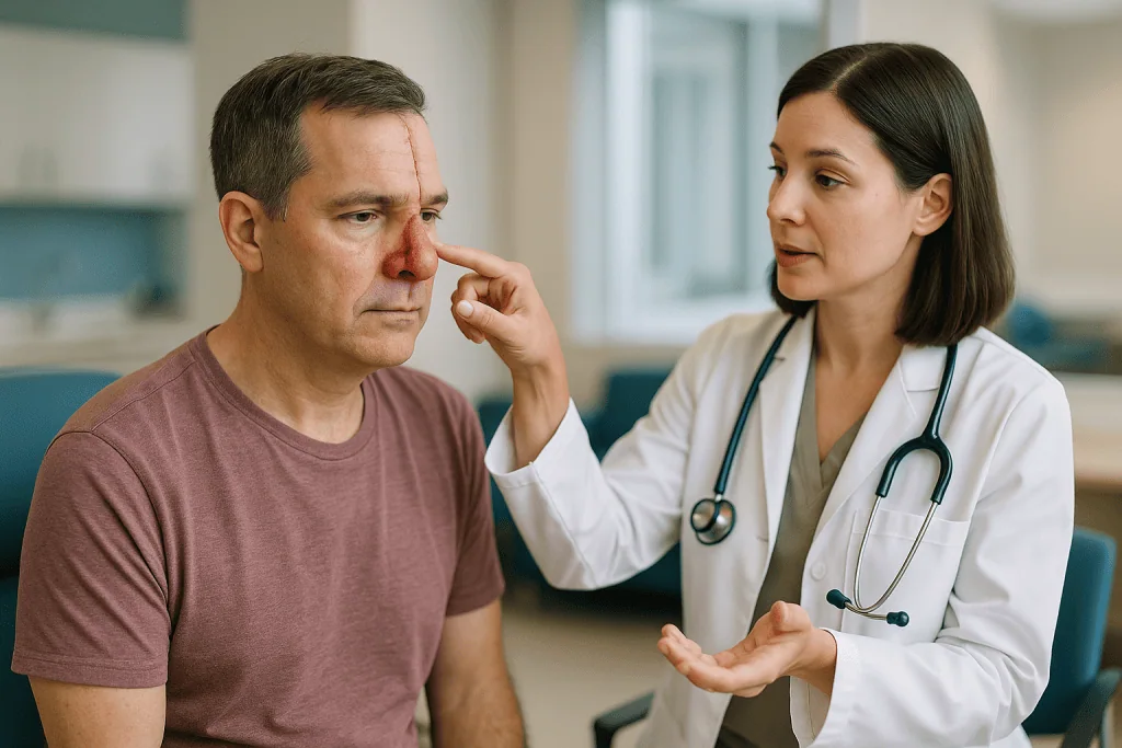
The defect is then measured. A template is cut from foil or dressing material, laid over the nose and shaped to the missing area, then flipped and transferred up to the forehead. The template is placed high enough that, once the flap swings down and rotates, it reaches the tip or rim without tension. On a short forehead, the design may extend into hair-bearing scalp or angle sideways to gain length.
Under general anesthesia or deep sedation, the surgeon lifts the skin paddle with a strip of underlying tissue, carries the pedicle down to the brow and rotates the whole unit roughly 180 degrees onto the nose. Any lining repair and cartilage grafting happens in this same session or has been prepared already. The paddle is thinned where needed and sewn in. The forehead wound is closed directly if it can be; if not, part of it is left to heal on its own over weeks, which usually leaves a surprisingly acceptable scar (MedlinePlus).
The patient leaves with a soft tube of living tissue running from brow to nose. It looks alarming. It is meant to.
Stage two and stage three: dividing the pedicle and sculpting the result
The pedicle is a temporary bridge. Its job is to feed the flap until the flap has grown enough new vessels from the nasal edges to survive on its own. Published protocols commonly describe waiting about three weeks before dividing it, though the interval is a surgical judgment and your team sets the date, not a calendar rule.
In a classic two-stage approach, that division is the second and final operation. The stalk is cut near the brow, the lower end is trimmed and inset into the nose, and the upper end is returned to the forehead to restore the eyebrow’s position. The nose is then left to settle for months before anyone considers refinements.
Many surgeons prefer a three-stage version for full-thickness or large defects. Here the middle stage, roughly three weeks after the first, is a deliberate pause to sculpt. The flap is lifted almost entirely off the nose while still attached at the pedicle, the excess tissue underneath is thinned, cartilage grafts are adjusted or added, and the skin is laid back down. The pedicle is divided at a third operation several weeks later.
Why add an operation? The argument, made in the surgical literature, is that thinning a flap while its blood supply is still guaranteed by the pedicle is safer than thinning it at division, and that the result better mirrors the fine contours of a real nostril. The counterargument is a longer course with more anesthesia. Neither approach is right for every patient; the choice depends on defect depth, the patient’s health and the surgeon’s experience.
Cartilage graft nose reconstruction: where the framework comes from
Skin alone cannot hold a nostril open. Every full-thickness nasal reconstruction, and many partial ones, needs a scaffold, and that scaffold is almost always the patient’s own cartilage. Cartilage is a firm, flexible tissue that has no blood vessels of its own and survives as a graft by soaking up nutrients from surrounding tissue.
Three donor sites cover most needs:
- Septum. The cartilage wall inside the nose is straight, thin and close at hand. It is often harvested through the same operative field and suits struts and small plates.
- Ear. The bowl of the ear, called the concha, is naturally curved, which makes it well suited to rebuilding the rounded alar rim. The ear’s outer shape is unchanged after harvest, and the scar sits behind it.
- Rib. When large amounts are needed, or the septum has already been used, a segment of rib cartilage offers volume. It carries a chest incision and a small risk of lung injury, and rib cartilage can warp over time, so surgeons carve it carefully.
Grafts are shaped into specific parts: a strut along the nostril margin to stop it curling inward on inspiration, a plate along the sidewall, a shield over the tip. Cartilage placed under a forehead flap at the first stage, or during an intermediate stage, is what gives the rebuilt nose a nostril that stays open when the patient breathes in hard.
Cartilage grafts carry their own risks: infection, absorption over years, visible edges under thin skin, and donor site pain. They are, nonetheless, the reason a reconstructed nose can be both a shape and an airway.
Who forehead flap nasal reconstruction is usually for, and who is asked to wait
The operation is generally reserved for defects that are too large, too deep or too awkwardly placed for simpler options. Typical candidates include people who have lost most of the tip, a whole nostril wing or a through-and-through segment after skin cancer surgery or injury. Smaller, shallow losses are usually handled with local flaps from the adjacent nose or cheek, or with a skin graft, none of which require the multi-stage commitment.
Some people are asked to wait, or to prepare first:
- Uncertain margins. If pathology has not confirmed that all cancer has been removed, reconstruction is delayed so that a recurrence is not hidden under a flap. This is especially true for melanoma, where final margin decisions can take longer (NHS).
- Active smoking or nicotine use. Nicotine narrows small blood vessels, and flap survival depends on those vessels. Surgeons routinely ask for a nicotine-free period before and after each stage.
- Poorly controlled diabetes or blood-thinning medication. Both raise the risk of wound problems and bleeding. Managing them is a decision for the prescribing clinician, never something to adjust on your own.
- Planned radiation. If the treated area will receive radiotherapy, the sequence of surgery and radiation is planned jointly by the cancer and reconstructive teams.
Age by itself is not a barrier. Many patients are in their seventies or eighties, because that is when skin cancers of the nose are most common. What matters is fitness for repeated anesthesia and the capacity to manage several weeks with a pedicle in place. A frank conversation about daily life, vision, hearing and support at home is part of the assessment.
Nasal reconstruction after Mohs surgery: how the timing works
Mohs micrographic surgery removes skin cancer one thin layer at a time, with each layer examined under a microscope while the patient waits, until the edges are clear. It is favored on the nose because it spares as much healthy tissue as possible while checking every margin (Mayo Clinic; Cleveland Clinic). The trade-off is that nobody knows the final size of the wound until the last layer is clear.
That uncertainty shapes the reconstruction. In many centers, the Mohs surgeon completes the removal and either repairs the wound the same day or dresses it and refers the patient to a reconstructive surgeon within days. A wound can safely be left open for a short period while plans are made; keeping it clean and moist is the priority (MedlinePlus). For larger defects, a brief delay also allows any swelling to settle so the true shape can be measured.
Basal cell and squamous cell carcinomas, the most common skin cancers, are usually reconstructed once Mohs confirms clear margins (CDC; NCI). Melanoma is different: margins are typically assessed on permanent, fully processed pathology, which takes longer, so reconstruction is often deliberately staged after that result.
People sometimes ask whether covering the area with a flap could hide a recurrence. This is precisely why clear margins are confirmed first, and why follow-up skin checks continue for years after reconstruction. A rebuilt nose is still skin that has proven it can make cancer, and the rest of the face carries the same history of sun exposure. The reconstruction changes appearance; it does not change the need for surveillance.
How the options compare: skin graft, local flap or forehead flap
Surgeons match the repair to the hole. The forehead flap is the most capable option, and also the most demanding, so it is reserved for defects where lesser methods would fall short. The table below summarizes the usual reasoning in plain terms; the fit for any individual is a clinical decision.
| Option | Usually considered for | Operations | Main strengths | Main limitations |
|---|---|---|---|---|
| Healing on its own | Small, shallow, concave areas | None | No donor site, simple care | Slow; scar may contract on convex zones |
| Full-thickness skin graft | Shallow defects with a healthy bed | One | Quick, single stage | Color and thickness mismatch; cannot cover bare cartilage reliably |
| Local nasal or cheek flap | Small to medium defects | One | Excellent color match, single stage | Limited size; may distort nostril or tip |
| Paramedian forehead flap | Large, deep or full-thickness defects | Two or three | Best match for large areas; supports cartilage and lining | Weeks with a pedicle; forehead scar; multiple anesthetics |
Two points deserve emphasis. First, the categories overlap; a medium defect on the tip might be handled either way depending on skin quality and the patient’s priorities. Second, the forehead flap is not chosen because it is fancier. It is chosen because it can carry cartilage, restore lining and resurface an entire subunit in matching skin, tasks the simpler options cannot manage together. When a surgeon proposes it for a modest-looking wound, the usual reason is depth rather than width.
Forehead flap surgery recovery: what the days and weeks look like
The first week is about protecting the pedicle. The stalk is dressed but left visible so the team can check its color and warmth; a pale, dusky or cold flap is a sign of blood flow trouble and needs prompt review. Sleeping on the back with the head raised, avoiding anything that tugs on the face, and keeping glasses off the bridge are standard instructions. Many people go home the same day or after one night, depending on the extent of cartilage and lining work.
Eating and drinking are usually unrestricted, though a wide yawn or a sneeze feels strange with a tube of tissue on the face. Swelling and bruising around the eye on the flap side are common and settle over the following weeks (MedlinePlus). The forehead wound, if closed directly, is often the sorest part. If a portion was left to heal by itself, it will look raw for several weeks before quietly filling in.
Between stages, the flap is bulky, red and shiny. This is normal, and it is not the final result. The interval before pedicle division is often described as about three weeks in published protocols, with the exact date set by the surgeon. People return to desk work and light activity in the interval; heavy exertion, contact sports and swimming wait until the surgeon clears them.
Each later stage brings a shorter recovery, typically a few days of swelling. Full settling is measured in months: scars soften and fade over roughly a year, cartilage grafts firm into place, and the flap’s color slowly matches its neighbors. Photographs taken at each stage help patients see progress that a mirror hides.
What are the risks of a forehead flap?
Every operation carries risk, and this one carries a specific set that is worth understanding before consent.
Flap compromise. The most feared complication is loss of blood supply to part or all of the flap. Partial loss at the tip of the flap is more common than total loss and may heal with dressings or need a small revision. Nicotine, pressure on the pedicle, hematoma (a collection of blood under the skin) and infection are the usual culprits.
Bleeding and infection. Both are possible at the nose and at the forehead, and both are more likely in people with diabetes, on blood thinners or with poor nutrition. Antibiotics may be used around surgery at the team’s discretion; the decision belongs to the treating clinician.
Forehead changes. A vertical scar above the brow is unavoidable. The eyebrow can sit slightly higher or lower on the flap side, and some people notice numbness or altered sensation of the forehead and scalp that improves slowly or partly persists.
Nasal changes. Nostril narrowing, a pulled-up rim, a notch at the flap edge, thickening of the flap (sometimes called pincushioning) and asymmetry are all possible. Cartilage grafts can shift, warp or become visible. Breathing through the reconstructed side may feel different.
Need for revision. Refinement procedures are common enough that surgeons often describe them as part of the process rather than a failure of it. Frequency varies widely with defect size and technique, and any figure should come from your own surgeon’s audited results.
Anesthetic risks and donor site risks from ear or rib harvest add to the list. Weighing these against living with the defect is a personal calculation, and a legitimate answer is to choose a simpler repair or none at all.
How successful is a forehead flap? What the evidence actually shows
This is the question everyone asks, and honesty requires an unsatisfying answer: it depends on how success is defined, and the numbers you find online come from individual surgical series, not from randomized trials.
If success means the flap survives and the defect is covered, the surgical literature consistently reports high survival rates for the paramedian forehead flap, which is why textbooks describe it as the workhorse for large nasal defects. Total flap loss is uncommon in experienced hands, and the forehead’s blood supply is regarded as unusually reliable among facial flaps.
If success means a nose that looks and breathes like the original, the picture is more nuanced. Outcomes are reported in surgeon-rated scales, patient questionnaires and photographs, none of which are standardized across studies. Revisions are frequent. Results in reviews tend to be better for cover-only defects than for full-thickness ones, better in non-smokers, and better when lining and support were addressed rather than skipped.
No national guideline publishes a success percentage for this operation, and this article will not invent one. What can be said with confidence is that the technique has persisted for centuries because it solves a problem nothing else solves as well, and that the greatest variable in outcomes appears to be planning: correct inventory of missing layers, adequate cartilage support and patient willingness to complete every stage.
Ask your surgeon three things: how many of these they perform, how they measure their own results, and what a typical case that resembles yours looked like at one year. Those answers are more informative than any figure pulled from a website.
What people often get wrong about forehead flap nasal reconstruction
Myth: the nose is finished after the first operation. The first stage attaches the flap; it does not finish the nose. Expect two or three procedures spaced weeks apart, then months of settling. Judging the result in the first month is like judging a house by its scaffolding.
Myth: the forehead will be disfigured. The donor scar is vertical, sits in a region where skin is thick and forgiving, and tends to fade substantially over a year. A brow shift can occur, but a disfigured forehead is not the expected outcome (MedlinePlus).
Myth: a skin graft would be quicker and just as good. For shallow wounds, a graft can be an excellent choice. For deep ones, grafts cannot carry cartilage, cannot resist contraction over the nostril rim and often heal paler and shinier than the surrounding skin.
Myth: cartilage can be replaced with an implant. Synthetic implants exist, but they carry higher infection and extrusion risks in a nose that has already been operated on, and most reconstructive surgeons prefer the patient’s own cartilage for this reason.
Myth: once reconstructed, the skin cancer risk is over. The forehead skin has the same sun history as the nose it replaces, and the surrounding face is unchanged. Regular skin checks remain essential (CDC).
Myth: the pedicle can be hidden. It cannot. For a few weeks it is visible, and many people prepare a simple explanation for colleagues and neighbors. Planning for that period, including time off or remote work, is part of realistic preparation, not vanity.
Questions to ask your care team before agreeing to surgery
A good consultation leaves you knowing not just what will happen but why this plan rather than another. These questions help.
- Which layers of my nose are missing, and how will each one be rebuilt?
- Are you planning to replace a full subunit, and how much healthy skin will that remove?
- Two stages or three, and what makes one better for my defect?
- Where will the cartilage come from, and what does that donor site involve?
- Roughly how long will the pedicle stay in place, and what should I look for in its color and warmth?
- Which of my current medicines or supplements might affect bleeding or healing, and who will advise me about them?
- What nicotine-free period do you require, and does that include vaping and patches?
- What is your plan if part of the flap does not survive?
- How often do your patients need revision, and what does a typical revision involve?
- Can I see photographs of a case similar to mine at each stage, including at one year?
- How will breathing on the reconstructed side be checked, and what happens if it narrows?
- What follow-up for skin cancer will I need afterward, and who coordinates it?
Bring someone with you if you can. Multi-stage plans are easy to muddle when heard once, and a second set of ears catches what anxiety misses. Ask for the plan in writing, with the anticipated sequence of stages, so that you can check the team’s expectations against your own life: work, travel, caring responsibilities and events you would rather not attend with a pedicle. Most surgeons welcome these questions; the ones who answer them patiently are usually the ones who have thought hardest about the answers.
When to call your doctor
Most recovery days are uneventful. A few signs should prompt a same-day call to the surgical team, or emergency care if the team cannot be reached.
Flap color change. A flap that turns pale white, dark purple, blue or black, or becomes cold to the touch compared with the surrounding skin, may be losing its blood supply. This is time-sensitive; call immediately.
Bleeding that does not stop. Oozing that soaks through a dressing after ten to fifteen minutes of gentle pressure, or a rapidly enlarging, tense swelling under the flap or forehead wound, needs urgent assessment.
Signs of infection. Spreading redness, increasing pain after the first few days rather than easing, pus or foul-smelling drainage, or a fever should be reported (MedlinePlus).
Breathing problems. Sudden inability to breathe through the reconstructed side, especially with swelling, warrants a call. Any difficulty breathing through the mouth as well is an emergency.
Vision changes or severe eye pain on the operated side, given how close the flap’s origin lies to the eye.
Wound separation. Edges of the flap or forehead wound pulling apart, or a cartilage graft becoming visible through the skin.
Chest pain, shortness of breath or calf pain after any anesthetic, which can indicate a clot and need emergency care regardless of the surgery.
Between these red flags and routine follow-up lies a gray zone of worries that feel small: a stitch that looks loose, a color that seems slightly off. Call anyway. Teams that perform staged reconstruction expect these calls and would rather see a healthy flap than miss a struggling one. Keep the clinic’s after-hours number where you can find it before you need it.
Frequently asked questions
How long does it take to recover from forehead flap surgery?
Recovery runs in phases rather than a single stretch. The first days involve swelling, bruising and protecting the pedicle; light activity typically resumes within a week or two. Stages are commonly spaced around three weeks apart in published protocols, and final settling of scars and contour is measured in months, often close to a year. Your surgeon sets the exact schedule.
How successful is a forehead flap?
Flap survival is high in the surgical literature, which is why the paramedian forehead flap is regarded as the standard for large nasal defects. Success in appearance and breathing is harder to measure, depends on defect depth, smoking status and adequate cartilage support, and often involves revision. No national guideline publishes a success percentage, so ask your surgeon about their own audited results.
What are the risks of a forehead flap?
The main risks are partial or, rarely, total loss of the flap from poor blood flow, bleeding, infection, a visible forehead scar, eyebrow asymmetry, forehead numbness, nostril narrowing or notching, thickening of the flap and the need for revision surgery. Cartilage harvest adds donor site risks. Nicotine, diabetes and blood thinners raise the likelihood of wound problems.
How long does it take for a nose flap to heal?
The flap begins forming new blood vessels at the nose within days, and surgeons typically consider it safe to divide the pedicle after about three weeks, though the timing is a clinical judgment. Skin edges usually seal within two weeks, while the flap remains swollen and shiny for months. Full color match and softening of scars can take up to a year.
What is a paramedian forehead flap?
A paramedian forehead flap is a vertical strip of forehead skin designed just off the midline, over the supratrochlear artery, and rotated down to cover a nasal defect while remaining attached at its base. The word paramedian refers to its position beside the center line. It is the most common form of forehead flap used in nasal reconstruction today.
How long after Mohs surgery can nasal reconstruction be done?
Reconstruction is planned once pathology confirms the cancer margins are clear, which with Mohs surgery is often the same day for basal and squamous cell carcinomas. The wound can be safely dressed for days while a reconstructive surgeon is consulted. Melanoma margins usually require fully processed pathology, so reconstruction is often deliberately delayed until that result is available.
Where does the cartilage come from in nose reconstruction?
Cartilage grafts are usually taken from the patient’s own body: the septum inside the nose for straight, thin pieces; the bowl of the ear for naturally curved pieces suited to the nostril rim; or the rib when larger volumes are needed. Each site has its own scar and risks, and the choice depends on how much support the defect requires.
Can you breathe normally after forehead flap nasal reconstruction?
Many people breathe well after reconstruction, but the airway on the rebuilt side can feel different, and narrowing is a recognized risk. Adequate inner lining and cartilage struts along the nostril margin are the main safeguards against collapse when breathing in. Persistent obstruction can sometimes be improved with revision. Report any sudden change in breathing to your surgical team promptly.
What does the forehead look like after a forehead flap?
The forehead is left with a vertical scar above the inner eyebrow on the flap side. If the wound could not be closed directly, part of it heals on its own over several weeks and usually blends acceptably. The eyebrow may sit slightly differently, and some numbness of the forehead or scalp is common, improving gradually and sometimes persisting in part.
Do you have to stop smoking before forehead flap surgery?
Surgeons routinely require a nicotine-free period before and after each stage because nicotine narrows the small blood vessels a flap depends on, raising the risk of partial flap loss and wound breakdown. This typically includes cigarettes, vaping and nicotine replacement products. The length of the required period and any support for stopping should be discussed with your surgical team and primary clinician.
References
- MedlinePlus: Skin flaps and grafts – self-care
- NHS: Non-melanoma skin cancer – Treatment
- CDC: About Skin Cancer
This article is for general information only and is not a substitute for professional medical advice. Please consult a qualified doctor about your individual situation.

More from the Blog

When Is It Safe to Fly Home After Facial Aesthetic Surgery? Stay Length and Follow-Up Abroad
Most people are asked to wait at least a week before flying home after facial aesthetic surgery, and often longer for a facelift or…

Does Revision Rhinoplasty Take Longer to Heal? Swelling, Splints and a Realistic Timeline
Yes, revision rhinoplasty usually heals more slowly than a first nose operation. The early steps look similar: the external splint typically comes off after…

Can Filler Fix a Dorsal Hump? What Liquid Rhinoplasty Changes and What Only Surgery Can
Filler cannot remove a dorsal hump; it can only camouflage one. In liquid rhinoplasty, a clinician injects a hyaluronic acid gel above and below…

Zygoma Reduction Risks: Cheek Sagging, Nerve Numbness and Bone Healing Concerns Explained
Zygoma reduction, or reduction malarplasty, carries risks that include cheek soft-tissue sagging, numbness in the cheek and upper lip from irritation of the infraorbital…

When Do Forehead Lines Need a Forehead Lift Rather Than Injectables?
Forehead lines usually respond to botulinum toxin injections when they come from muscle movement and the brows still sit at a normal height. A…

Ear Molding for Newborns vs Otoplasty Later: How the Two Approaches Differ
Ear molding and otoplasty treat ear shape at very different ages. Ear molding uses soft splints applied in the first weeks of life, while…