

How Shock Wave Therapy (ESWT) Works on Stubborn Tendons: What Happens in a Session
Key Takeaways
- Shockwave therapy delivers acoustic pressure pulses, not electricity; the energy is released mainly where tissue density changes, such as where a tendon meets bone.
- Long-standing tendon pain is usually degeneration with poor blood supply rather than inflammation, which is why a treatment that deliberately stresses the tissue can make biological sense.
- Focused devices converge energy at depth and suit calcified shoulder tendons; radial devices are strongest at the skin surface and suit superficial sites like the heel and outer elbow.
- Mayo Clinic notes that most plantar fasciitis settles within several months of conservative care, so shockwave is a second-line option for chronic cases, not a first move.
- Evidence is most consistent for calcific shoulder tendinopathy, moderate but mixed for plantar fasciitis and Achilles tendinopathy, and genuinely conflicting for tennis elbow.
- Pain relief felt immediately after a session comes from overstimulated nerve fibers, not a repaired tendon; trials generally judged real outcomes around three months later.
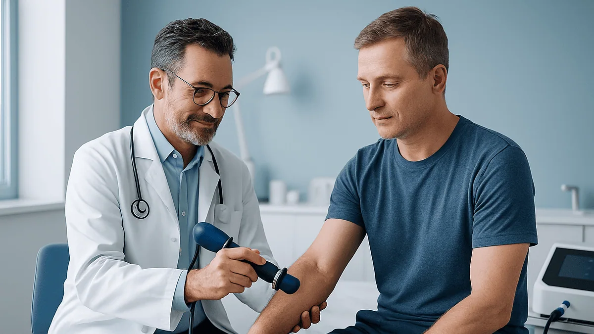
Shockwave therapy (ESWT) sends brief, high-energy pressure pulses through the skin into a painful tendon. The pulses create controlled micro-stress that appears to stimulate new blood vessels, growth-factor release and tissue remodeling, while also quieting pain-signaling nerve fibers. Treatment happens without anesthesia, the pulses themselves take only minutes, and courses are usually spread over several weeks. Evidence is moderate and varies by tendon.
The physiotherapist has already watched you limp across the room twice. Your heel pain started before the summer, you have stretched, iced, bought the sensible shoes, and this morning the first step out of bed still felt like standing on a marble. Then comes the sentence: “We could try shockwave.” You nod, and drive home wondering exactly how does shockwave therapy work, whether it hurts, and why anyone would fire pressure waves at an already sore tendon.
The idea sounds counterintuitive, and the marketing that surrounds it does not help. Some clinics describe it as almost magical; some skeptics dismiss it as an expensive rubber mallet. The truth sits in between and is far more interesting.
This explainer walks through what actually happens on the treatment couch, what the pulses are thought to do inside a tendon that has stopped healing on its own, where the evidence is solid and where it is thin, and the questions worth asking before you say yes.
How does shockwave therapy work on a stubborn tendon?
Extracorporeal shock wave therapy, or ESWT, means a machine outside the body (extracorporeal) generates a very short, very sharp pressure pulse (a shock wave) and aims it at a painful spot. Nothing electrical enters you. The pulse is acoustic, a burst of pressure that travels through skin, fat and muscle and releases most of its energy where tissue density changes, such as the boundary between a tendon and the bone it attaches to.
Why aim that at a tendon that already hurts? Because the problem in a long-standing tendon injury is usually not inflammation. Tendinopathy is the term for a tendon whose collagen fibers have become disorganized, with sparse blood supply and cells that have largely stopped repairing. It is a stalled healing project, not a fire. The pulses are meant to restart the project.
Three mechanisms are proposed, and it is honest to say most of the detailed evidence comes from laboratory and animal studies rather than human tissue samples:
- Mechanotransductionthe process by which cells sense physical stress and change their chemistry. Stressed tendon cells release growth factors and signaling molecules that encourage new small blood vessels (neovascularization) and fresh collagen.
- Hyperstimulation analgesiaoverstimulating local pain nerve endings appears to reduce their output, including the pain messenger substance P, which is one reason discomfort often eases during and after a session.
- Mechanical disruptionin tendons that have developed chalky calcium deposits, the pulses can fragment those deposits so the body can reabsorb them.
Which of these matters most probably depends on the tendon. That uncertainty is not a flaw in the story; it is where the science actually is, and it explains why results differ so much from heel to shoulder.
What actually happens in a shockwave therapy session, step by step
Most people are surprised by how ordinary the appointment feels. There is no changing gown, no drip, no sedation, and you walk in and walk out under your own power.
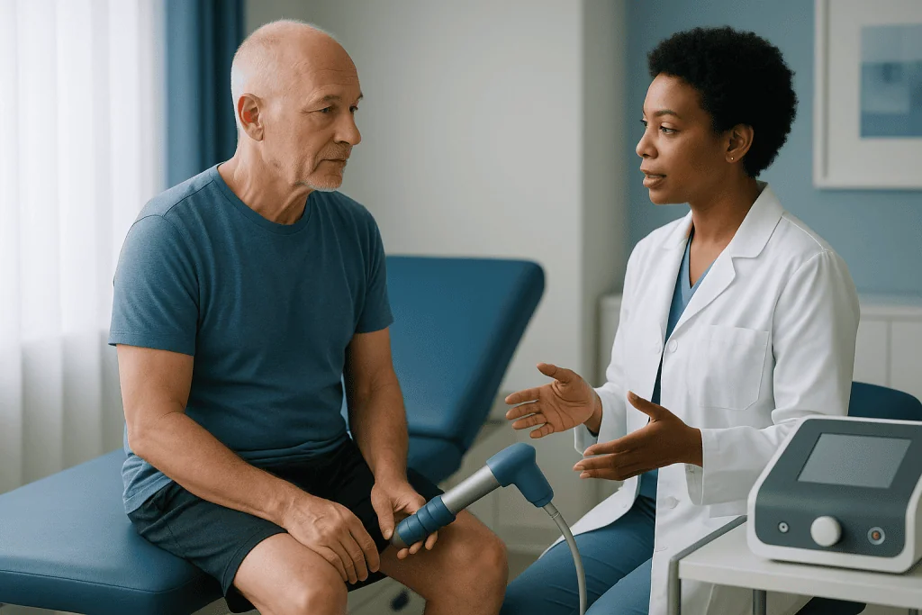
The clinician begins by finding the exact spot that hurts. This is done by pressing along the tendon until you flinch, sometimes with a handheld ultrasound scanner to confirm the target, particularly for deeper structures such as the shoulder. Precision matters, because the energy is delivered to a small area and a pulse aimed two centimeters off target treats healthy tissue.
A layer of water-based gel goes on the skin, the same kind used for pregnancy scans. Gel matters more here than most people realize: shock waves lose energy crossing air, so the gel ensures the pulse passes cleanly from the applicator head into you.
Then the applicator is pressed firmly against the spot and the machine starts delivering pulses, typically several every second, with a rhythmic tapping sound. The clinician usually begins at a low intensity and increases it gradually, checking with you throughout. You stay awake and able to speak, which is deliberate: your feedback guides where and how hard the pulses land.
The pulses themselves take only minutes per area. The clinician may shift the applicator slightly to cover the whole painful zone, then wipe off the gel. Some check the tender spot again straight afterward; many people notice it is already less sharp to press, a short-lived effect of the nerve-quieting described earlier rather than proof the tendon has healed.
You can usually put your shoe back on and go about the day, with advice to ease off strenuous activity for a short while. There is no bandage, no wound and nothing to keep dry.
Is shockwave therapy painful?
Yes, for most people it is uncomfortable, and pretending otherwise does nobody a favor. The sensation is usually described as a deep, rapid tapping or a firm ache that intensifies when the applicator sits directly over the sore point. Bony spots such as the heel or the outside of the elbow tend to feel sharper than fleshier areas because there is less tissue to cushion the pulse.
Two things make it far more tolerable than the description suggests. First, the intensity is ramped. A skilled clinician starts gently, lets the tissue and the nerves adjust, and increases only as you tolerate it. You are in control, and saying “that’s my limit” is a normal part of the appointment, not a failure. Second, the hyperstimulation effect kicks in during the session itself: after the first minute or so, many people report the spot going oddly dull or numb, and the same intensity that stung at the start becomes an ache they can breathe through.
You may reasonably ask why nobody offers a numbing injection first. Local anesthetic is generally avoided, and not only for convenience. In some trials, numbing the area before treatment appeared to reduce the benefit, possibly because the nerve response is part of how the therapy works, and because a numbed patient cannot help the clinician find the precise target. Your own team will explain their approach.
Afterward the area commonly feels bruised or tender for a day or two, occasionally longer, and the skin may look flushed. That soreness is expected. What is not expected is pain that escalates over several days rather than settling, and that belongs in the “when to call” section below.
If pain is a real worry for you, say so beforehand. Slower ramping and shorter bursts are routine adjustments.
Focused vs radial shockwave: what's the difference and does it matter?
Two quite different machines share the name shockwave, and the distinction genuinely affects what can be treated.
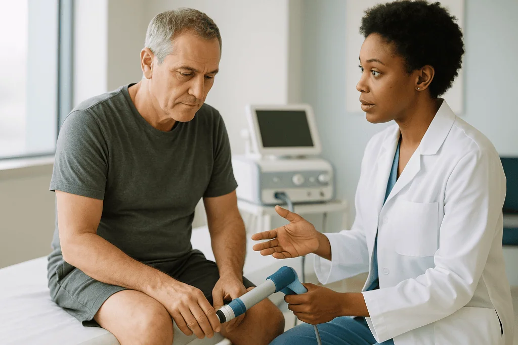
Focused shockwave is the original technology, adapted from the devices used to break kidney stones. A generator, whether electrohydraulic (a spark in water), electromagnetic (a vibrating membrane) or piezoelectric (crystals that snap when charged), creates a true shock wave with an extremely steep rise in pressure. The wave is shaped so that its energy converges at a chosen depth beneath the skin, like sunlight through a magnifying glass. The clinician can treat a target several centimeters deep while the skin above it receives relatively little energy.
Radial shockwave, often called radial pressure wave, works differently. Compressed air fires a small projectile against the applicator head, which then vibrates against the skin. The resulting wave is slower-rising, spreads outward in all directions, and is strongest right at the surface, fading with depth. Strictly speaking it is not a shock wave at all, though the term has stuck.
In practice this means radial devices suit superficial, broad targets: the plantar fascia under the heel, the mid-portion of the Achilles, the tendon attachment at the outer elbow. Focused devices are chosen for deeper or more precise work, such as calcium deposits in the rotator cuff of the shoulder, where fragmenting a specific lump is the aim.
Which is better? The trial literature does not crown a winner overall. Head-to-head comparisons are few and small, and outcomes seem to depend more on choosing the right tool for the depth of the problem, and on accurate targeting, than on the label on the machine. When a clinic offers you “shockwave”, it is fair to ask which type, and why that one for your tendon.
Who is shockwave therapy usually for, and who is usually asked to wait?
ESWT is not a first-line treatment, and guideline-minded clinicians do not present it as one. Its usual place is with tendon and fascia problems that have persisted for months despite the basics: relative rest, activity modification, a structured strengthening program and footwear or equipment changes. Mayo Clinic describes it as an option for chronic plantar fasciitis that has not responded to more conservative treatments, and that framing applies broadly across tendons.
The conditions most often treated include plantar fasciitis (heel), Achilles tendinopathy (back of the ankle), lateral epicondylitis or tennis elbow (outer elbow), calcific tendinopathy of the shoulder, greater trochanteric pain syndrome (outer hip) and patellar tendinopathy (front of the knee). A good candidate has a clear, localizable sore point, a diagnosis confirmed by examination and often imaging, and a willingness to keep doing the rehabilitation exercises alongside.
Some people are asked to wait, and some are steered elsewhere entirely. Typical reasons include:
- Pregnancy, when the treatment area is anywhere near the abdomen or pelvis.
- Bleeding disorders, or taking anticoagulant (“blood-thinning”) medicines, because the pulses can cause bruising and bleeding into tissue; the prescribing clinician decides how to proceed, never the shockwave provider alone.
- A recent corticosteroid injection into the same area; many teams leave a gap of several weeks, since steroids can temporarily weaken tendon.
- Active infection, a skin wound, or a known tumor at or near the site.
- A suspected tendon tear rather than tendinopathy; pulses aimed at a partially torn tendon are a different risk proposition.
- Children and adolescents whose growth plates near the target have not closed.
- Sites overlying the lungs, bowel, spinal cord or major nerves and blood vessels.
- Certain implanted electronic devices, depending on the machine; the clinician checks.
None of these is a judgment about you. They are the reasons a careful team says “not yet” or “not this”, and the final call sits with them.
Shockwave therapy for plantar fasciitis: what the evidence actually shows
Heel pain is where most people first encounter ESWT, so it deserves an honest look. Plantar fasciitis is irritation and degeneration of the thick band of tissue running from the heel bone to the toes. It is the classic “first step in the morning” pain, and it is very common.
The first and most important fact is one that ESWT clinics rarely lead with: most people get better without it. Mayo Clinic notes that the majority of people with plantar fasciitis recover within several months of conservative treatment such as stretching, footwear changes, rest from aggravating activity and ice. That timeline is slower than anyone wants, but it means a good clinician’s first move is patience and a plan, not a machine.
For the minority whose pain drags on past that window, the picture on shockwave is genuinely mixed. Mayo’s summary is measured: some studies show promising results, but the treatment has not been shown to be consistently effective. Cleveland Clinic lists it among options considered when standard measures have not worked. Systematic reviews tend to find that ESWT reduces pain more than placebo treatment in chronic cases, but the size of that benefit varies widely between trials, partly because trials used different machines, energy levels, session counts and definitions of success.
What does that mean at the treatment couch? It means ESWT for a heel that has hurt for many months is a reasonable, evidence-supported thing to try, particularly before anyone mentions surgery, and it means nobody can promise you it will work. Roughly, the fairer statement is that a meaningful proportion of people improve, some do not, and the exercise and load-management work you do around the sessions probably matters as much as the pulses.
If your heel pain is new, weeks rather than months old, the evidence points to giving the simpler measures their fair chance first.
Which tendons respond best? Achilles, tennis elbow and calcific shoulder compared
Tendons are not interchangeable, and the research reflects that. The table below summarizes where mainstream evidence currently sits for the most commonly treated sites. “Consistent” means most good-quality trials point the same way; “mixed” means results genuinely conflict; “limited” means few or small studies.
| Site | Common diagnosis | Where the evidence sits | What is worth knowing |
|---|---|---|---|
| Heel | Plantar fasciitis | Moderate, mixed | Best studied; benefit over placebo in chronic cases, size varies |
| Shoulder | Calcific rotator cuff tendinopathy | Most consistent | Focused devices; aim is fragmenting the deposit; less clear for non-calcific shoulder pain |
| Outer elbow | Lateral epicondylitis | Mixed | Several trials show no advantage over sham; others positive |
| Back of ankle | Achilles tendinopathy | Moderate, mixed | Mid-portion and insertional (at the heel bone) behave differently; usually paired with loading exercises |
| Outer hip | Greater trochanteric pain syndrome | Limited to moderate | Smaller trials, generally favorable |
| Front of knee | Patellar tendinopathy | Limited | Often used in athletes; evidence still thin |
Two patterns stand out. The clearest success story is the calcified shoulder tendon, which makes mechanical sense: there is a physical deposit to break up, and imaging can show whether it has shrunk. The murkiest is tennis elbow, where well-run trials have repeatedly failed to separate real treatment from sham, even though plenty of people report feeling better after either.
The Achilles deserves a note of caution. Insertional tendinopathy, where the tendon meets the heel bone, tends to respond less predictably than pain in the mid-portion a few centimeters higher, and pulses aimed too close to an already thinned insertion are treated with extra care.
The practical takeaway is to ask your clinician not “does shockwave work” but “what does the evidence say for my tendon, at my site”.
How many shockwave therapy sessions do people usually have, and how far apart?
There is no universal recipe, and anyone who quotes you one with total confidence is simplifying. That said, the protocols reviewed in the interventional procedures guidance published by NICE, the UK’s guideline body, most often used a short course of around three sessions spaced roughly a week apart, and that pattern remains the most common starting point in practice. Radial devices are sometimes used in slightly longer courses because each session delivers less energy at depth.
Why spread the sessions out rather than deliver everything at once? The answer comes back to mechanism. If the therapy works by provoking a biological response, that response needs time to unfold. Cells release signaling molecules over hours, new capillaries take days to sprout, and collagen remodeling continues for weeks. Stacking pulses on consecutive days would add tissue stress without giving the repair processes room to operate, and would compound bruising. The weekly rhythm is a compromise between momentum and recovery.
Within each session the clinician sets the number of pulses and the intensity according to the site, the device and your tolerance. These are clinical settings, and they differ between focused and radial machines, so the numbers are not something to compare across clinics or to negotiate.
What happens if a first course does not help? Most teams judge the outcome not on the day of the last session but weeks later, since trials typically measured results around three months after treatment. Some offer a second course; others conclude the tendon is unlikely to respond and turn to other options. A second course is a decision for the treating team, based on whether you improved partially, not at all, or improved then relapsed.
If a clinic proposes an open-ended series with no review point, that is a fair moment to ask why.
What the days and weeks after shockwave therapy usually look like
The first evening is usually the most noticeable. The treated area often feels bruised, warm and tender to touch, and the skin may be reddened or show tiny pinpoint marks. Many people find the tendon itself is briefly less painful to use, thanks to the nerve-quieting effect, which can be misleading; the tissue has just been deliberately stressed, and sprinting up the stairs to celebrate is exactly the wrong response.
Over the next two to three days the soreness typically settles. Clinicians commonly advise relative rest for a short spell, meaning normal daily movement is fine but the activity that originally aggravated the tendon is scaled back. Ice is often discouraged, and some teams ask you to avoid anti-inflammatory painkillers for a period, on the reasoning that the treatment is meant to provoke a healing response and inflammation-suppressing measures might blunt it. The evidence for that caution is not settled. Follow your own team’s instructions, and never stop or change a prescribed medicine on a shockwave provider’s say-so without checking with whoever prescribed it.
Between sessions, the rehabilitation program continues. This is not an optional extra. Progressive loading exercises, the slow, heavy movements that make a tendon work under control, are the part of treatment with the longest track record, and ESWT is best thought of as something that may help a tendon respond to them.
Weeks four to twelve are when the real verdict emerges. Because trials generally measured outcomes around three months after treatment, that is a sensible horizon for judging whether the course helped. Improvement is usually gradual: the morning pain shortens, the tender spot shrinks, walking distance stretches. Sudden dramatic change is not the typical pattern. Some people plateau then improve later; some notice little at all.
Keeping a simple weekly note of pain during a fixed task, such as the first ten steps of the day, gives you and your team something firmer than memory to review.
Shockwave therapy side effects: what is common, what is rare
For a procedure that involves deliberately hammering a sore tendon, ESWT has a reassuring safety record when used within its intended limits. Guideline reviews have found serious complications uncommon. That does not mean nothing happens.
The common effects are local and short-lived:
- Pain during the session, easing as the nerves adapt.
- Tenderness and a bruised feeling for one to three days.
- Redness, mild swelling, and small pinpoint red spots (petechiae) where tiny skin vessels have leaked.
- Visible bruising, more likely in people who bruise easily or take blood-thinning medicines.
- Temporary numbness or tingling over the treated area.
Less common problems include a larger collection of blood under the skin (hematoma), skin damage where the applicator sat, and irritation of a nearby nerve producing longer-lasting tingling or weakness. These are more likely when high energies are used or when the target lies close to a nerve, which is one reason careful anatomical targeting and sensible energy settings matter more than enthusiasm.
The rare event people worry about most is tendon rupture. Cases have been reported, mostly in tendons already weakened, sometimes by previous corticosteroid injections, and mostly with high-energy focused treatment. It is precisely why partially torn tendons and recently injected tendons are usually excluded, and why the Achilles insertion is treated with caution.
Two further points deserve a place here. First, ESWT does not involve radiation and leaves nothing in the body, so there is no cumulative dose in the way there is with imaging. Second, a temporary flare of the original pain in the days after treatment is well recognized and usually settles; a flare that keeps worsening is not typical and warrants a call.
Every risk here is a reason for a conversation, not a reason for alarm. Ask how the clinic minimizes them; a confident answer usually involves ultrasound guidance, graded intensity and clear exclusion criteria.
How does shockwave therapy work alongside injections, exercise and surgery?
Nobody should choose ESWT in a vacuum. Its value depends on where it sits in a sequence of options, and that sequence is fairly well agreed across guideline sources.
Load management and exercise come first and stay throughout. Tendons adapt to controlled stress; a structured, progressive strengthening program is the intervention with the deepest evidence base for most tendinopathies, and NHS and Mayo Clinic guidance both place activity modification and exercise at the foundation. ESWT is an adjunct to that, never a replacement.
Corticosteroid injections deliver an anti-inflammatory steroid to the painful area. They often reduce pain quickly, but the relief is frequently short-lived and, for tendons, comes with a specific caveat: Mayo Clinic notes that multiple injections into the plantar fascia can weaken it and risk rupture, and similar concern applies to other tendons. This is why many clinicians now reserve steroid for particular situations, and why a recent injection usually delays shockwave.
Platelet-rich plasma involves injecting a concentrate of the patient’s own blood platelets. Its evidence in tendinopathy is mixed and varies by site; it is not established as superior to ESWT or to exercise.
Surgery, whether releasing part of the plantar fascia, removing degenerated tendon tissue or clearing a calcium deposit, is generally reserved for persistent, disabling symptoms after other measures have been fully tried, because it carries recovery time and its own complications.
Seen this way, shockwave occupies a specific niche: a non-invasive option for a tendon that has stalled despite months of good rehabilitation, tried before anyone reaches for a scalpel. It does not weaken tissue the way repeated steroid can, and it requires no recovery period beyond a few sore days.
Which of these, in what order, suits your tendon, your health history and your goals is a decision that belongs with your treating team.
What people often get wrong about shockwave therapy
Misunderstandings cluster around this treatment, partly because the name is dramatic and partly because it is sold in very different ways. A few corrections:
“It’s an electric shock.” It is not. No current passes into the body. The “shock” is a pressure wave, the same physical phenomenon as a sonic boom, scaled down and aimed.
“It’s the same as therapeutic ultrasound.” Therapeutic ultrasound uses continuous, low-pressure sound to gently warm tissue. Shockwave delivers pulses thousands of times higher in peak pressure, for microseconds. The two machines may sit side by side in a clinic and share the gel, but they are not related in effect.
“More pain means it’s working better.” Intensity is set to the target and your tolerance, not maximized. Gritting through agony does not improve outcomes and increases bruising and nerve irritation.
“One session and you’re fixed.” Courses are spread over weeks, and results are judged months later. Feeling better walking out of the room is the nerve effect, not a repaired tendon.
“It replaces the exercises.” It does not. Every serious protocol pairs it with loading rehabilitation. Skipping the exercises and expecting the machine to carry the load misreads how tendons heal.
“It works for everything.” Shockwave is marketed for an ever-widening list of conditions, some with almost no supporting trials. The evidence is strongest for calcific shoulder tendinopathy and chronic plantar fasciitis, genuinely mixed for tennis elbow, and thin for many newer uses. A treatment can be well studied for one problem and unproven for another.
“Chronic means it will never heal.” Chronic simply means long-standing. Mayo Clinic’s observation that most plantar fasciitis settles within several months of conservative care is a reminder that persistence, not passivity, is the usual path.
A good clinician will make these points unprompted. If the pitch you hear contradicts them, ask more questions.
Questions to ask your care team before agreeing to ESWT
The most useful thing you can bring to a shockwave consultation is a short list of direct questions. The answers tell you as much about the quality of the clinic as about the treatment. A thoughtful team will welcome them; evasive or breezy responses are informative in their own way.
- What exactly is my diagnosis, and how was it confirmed? Has anyone checked for a partial tear rather than tendinopathy?
- What does the evidence say for shockwave at this site specifically, not for tendons in general?
- Which type of device will you use, focused or radial, and why that one for the depth of my problem?
- Will you use ultrasound to locate the target, or palpation alone?
- How many sessions are you proposing, how far apart, and at what point will we sit down and judge whether it has helped?
- What should I do, and not do, in the days after each session?
- Are there reasons in my medical history, medicines or recent injections that make this a poorer option for me?
- What is the plan if it does not help, and what would you suggest instead?
- How will the exercise program change during and after the course?
- Which symptoms afterward would you want to hear about straight away?
Bring your medication list, including anything bought without a prescription and any supplements, and mention any injection you have had into the area in the past few months, even one you think is unrelated. Say plainly if you have a bleeding tendency or bruise easily. Mention pregnancy or the possibility of it.
It also helps to be honest about your goal. “I want to walk the dog without limping” and “I want to run a marathon in the spring” lead to different conversations about what a realistic outcome looks like and what the exercise plan should build toward.
Finally, ask who is delivering the treatment and who is clinically responsible for your care overall. Shockwave is one tool; the plan around it is what matters.
When to call your doctor after shockwave therapy
Most people need nothing more than a couple of quiet days after a session. A short list of signs, though, should prompt a call to your treating team or, in the more urgent cases, same-day medical attention rather than waiting for the next appointment.
Contact your clinician promptly if you notice:
- A sudden snap or pop in the tendon, a feeling of being kicked in the back of the ankle, or a new inability to push off the foot, stand on tiptoe or lift the arm: these can signal a tendon tear and need assessment quickly.
- Pain that keeps escalating beyond the second or third day instead of easing, or pain that wakes you from sleep and is out of proportion to what you were told to expect.
- Spreading redness, heat or swelling, especially with fever, chills or feeling generally unwell, which can indicate infection.
- A large or rapidly enlarging bruise or swelling under the skin, particularly if you take blood-thinning medicines or have a bleeding condition.
- Numbness, tingling or weakness that persists beyond a day or two, or that spreads beyond the treated area.
- New calf swelling, tenderness or warmth, or breathlessness or chest pain, which are unrelated to the treatment itself but are never signs to sit on.
- Skin breakdown, blistering or an open area where the applicator was placed.
None of these is common. Listing them is not fear-mongering; it is the same courtesy a good clinician extends before any procedure, so that the rare problem is caught early rather than explained away as “probably normal”.
Between sessions, use your judgment generously. If something feels wrong, or if you are simply unsure whether a symptom is expected, a phone call costs nothing and is what the team would prefer. And if the treatment course finishes and your tendon has not improved, that too is a reason to go back rather than to give up: the next step in the sequence is a decision for you and your treating team together.
Frequently asked questions
Is shockwave therapy painful?
It is uncomfortable for most people, usually described as a deep, rapid tapping or firm ache that is sharpest over the tender point. Clinicians start at low intensity and increase only as you tolerate it, and many people find the area goes dull or numb within the first minute. Afterward the spot commonly feels bruised for a day or two. Local anesthetic is generally not used because numbing may reduce the benefit.
Does shockwave therapy for plantar fasciitis actually work?
It can help some people with chronic heel pain, but the evidence is mixed. Mayo Clinic states that some studies show promising results while the treatment has not proven consistently effective, and reserves it for cases that have not responded to conservative care. Since most plantar fasciitis settles within several months with stretching, footwear changes and rest, shockwave is usually considered only after those measures have had a fair trial.
What are the most common shockwave therapy side effects?
Temporary pain during treatment, tenderness for one to three days, redness, mild swelling, pinpoint red skin spots and bruising are the usual effects, and they settle on their own. Less common problems include a blood collection under the skin, skin damage or nerve irritation. Tendon rupture has been reported rarely, mostly in tendons already weakened by tears or previous steroid injections, which is why those situations are normally excluded.
Focused vs radial shockwave: which one is better?
Neither is better across the board; they suit different depths. Focused shockwave concentrates energy at a chosen depth and is preferred for deeper or calcified targets such as the rotator cuff. Radial pressure waves are strongest at the surface and spread outward, suiting superficial sites like the plantar fascia, mid-Achilles and outer elbow. Head-to-head trials are few, and accurate targeting appears to matter more than the machine type.
How many shockwave sessions will I need?
Protocols reviewed in NICE interventional procedures guidance most often used around three sessions spaced roughly a week apart, and that remains a common starting point, though radial protocols are sometimes longer. Sessions are spaced so the tissue can mount a repair response between them. Your clinician sets the plan and should agree a review point, usually weeks after the last session, to judge whether a further course is worthwhile.
How long after shockwave therapy do you see results?
Improvement is usually gradual rather than immediate. The pain relief felt straight after a session is a short-lived nerve effect. Because trials generally measured outcomes around three months after treatment, that is a realistic horizon for deciding whether it has helped. Typical signs of progress include shorter morning pain, a shrinking tender spot and longer comfortable walking. Some people plateau and improve later; others notice little change.
Can I exercise after a shockwave session?
Normal daily movement is generally fine, but clinicians usually advise scaling back the specific activity that aggravated the tendon for a short spell after each session while soreness settles. The structured strengthening program prescribed by your physiotherapist continues throughout the course, because progressive loading is the part of tendon treatment with the strongest evidence. Follow your own team’s instructions, since advice varies by site and device.
Can I have shockwave therapy if I take blood thinners?
Possibly, but it needs a conversation first. The pulses can cause bruising and bleeding into tissue, so anticoagulant medicines and bleeding disorders are common reasons to pause or decline treatment. The decision belongs with the clinician who prescribes your medicine together with the treating team; never stop or alter a blood thinner on a shockwave provider’s advice alone. Tell the clinic about every medicine and supplement you take.
Is shockwave the same as therapeutic ultrasound or TENS?
No. Therapeutic ultrasound uses continuous low-pressure sound to gently warm tissue, and TENS passes a mild electrical current through skin electrodes to modulate pain. Shockwave delivers brief acoustic pressure pulses with far higher peak pressure and no electricity entering the body. They may share a clinic room and the same coupling gel, but their mechanisms and the evidence behind them are entirely different.
Does shockwave therapy work for calcific shoulder tendinopathy?
This is the site where evidence is most consistent. Focused shockwave can fragment calcium deposits within the rotator cuff tendons so the body can reabsorb them, and imaging can show whether a deposit has shrunk. Results are less clear for shoulder pain without calcification. As with other sites, it is usually offered after conservative measures have been tried and alongside a rehabilitation program, with the treating team deciding suitability.
References
This article is for general information only and is not a substitute for professional medical advice. Please consult a qualified doctor about your individual situation.

More from the Blog
Diagnostic Nerve Blocks: How a Temporary Block Helps Find the Source of Your Pain
A diagnostic nerve block is a short-acting local anesthetic injected near a specific nerve to test whether that nerve carries your pain. If the…

The Day After an Epidural Injection: Soreness, a Brief Pain Flare and Getting Back to Activity
Mild soreness at the injection site and a short-lived increase in pain are common in the first one to three days after an epidural…

Spinal Cord Stimulator Recovery: Bending and Lifting Limits, Wound Checks and Programming
Spinal cord stimulator recovery usually means walking on the day of surgery, keeping incisions dry and inspected daily, and avoiding bending, twisting and lifting…

Living With a Spinal Cord Stimulator: Charging, Travel Scanners, MRI Rules and Daily Habits
Living with a spinal cord stimulator mostly means building a few routines: charging a rechargeable unit on a regular schedule, carrying your device ID…
When a Spinal Cord Stimulator Stops Helping: Reprogramming, Lead Checks and Revision
A spinal cord stimulator that has stopped helping is usually assessed in stages. The care team first reviews the device log and tries reprogramming,…

Protecting a Numb Limb After a Nerve Block: Heat, Pressure, Driving and Signs to Report
After a nerve block, a limb can stay numb, weak and unable to sense heat or pressure for several hours to about a day.…