Shock Wave Therapy (ESWT)
Shock wave therapy, or extracorporeal shock wave therapy (ESWT), uses pressure waves applied through the skin to treat long-standing tendon and soft tissue pain, including plantar fasciitis, Achilles and patellar tendinopathy, tennis…
Quick answer
Shock wave therapy (ESWT) is a non-surgical outpatient treatment that delivers pressure waves through the skin to stimulate healing in chronic tendon and soft tissue problems such as plantar fasciitis, tennis elbow, and calcific shoulder tendinitis. Sessions take about 15 to 30 minutes, usually in a course of three to five, with minimal downtime and mostly mild, temporary side effects.
What is shock wave therapy (ESWT)?
Shock wave therapy, also called extracorporeal shock wave therapy or ESWT, is a non-surgical treatment that uses pressure waves delivered through the skin to treat painful, long-lasting problems in tendons, ligaments, and other soft tissues. The word extracorporeal simply means “outside the body”: the waves are generated by a hand-held device pressed against the skin, so nothing is cut or injected. The waves travel into the tissue and are thought to stimulate the body’s own healing response, increase local blood flow, and reduce pain signals in the treated area.
Shock wave therapy is most often used for chronic tendinopathies, which are tendon problems that have lasted for several months and have not improved with rest, stretching, and standard physical therapy. Common examples include:
- Plantar fasciitis (pain in the thick band of tissue under the heel and arch of the foot)
- Achilles tendinopathy (pain in the large tendon at the back of the ankle)
- Lateral epicondylitis, commonly called tennis elbow
- Calcific tendinitis of the shoulder (calcium deposits inside the rotator cuff tendons)
- Patellar tendinopathy, sometimes called jumper’s knee
- Greater trochanteric pain syndrome (pain on the outside of the hip)
Higher-energy forms of shock wave therapy are also used in some centers for slow-healing bone fractures, known as non-unions. A related but separate technology, lithotripsy, uses shock waves to break up kidney stones; this page is about the musculoskeletal use only. In many hospitals, including Acibadem, shock wave therapy is provided through the Physical Medicine & Rehabilitation department or an orthopedic or sports medicine clinic.
Who needs shock wave therapy: candidates and limits
Shock wave therapy is generally considered when a tendon or soft tissue problem has become chronic, meaning it has typically lasted three to six months or longer, and first-line measures have not been enough. Those first-line measures usually include relative rest, activity changes, stretching and strengthening exercises, supportive footwear or braces, and over-the-counter pain relief. Your doctor may suggest shock wave therapy as a step before considering injections or surgery.
You may be a candidate if:
- Your pain is localized to a specific tendon or attachment point and a clinician has confirmed the diagnosis, often with an examination and sometimes with ultrasound or other imaging.
- You have completed an adequate period of conservative treatment without satisfactory improvement.
- You are able to continue an exercise program alongside treatment, because shock wave therapy usually works best when combined with rehabilitation.
Shock wave therapy is not usually suitable in the following situations, and your doctor will check for them beforehand:
- Pregnancy, particularly for treatment near the abdomen or pelvis
- A bleeding disorder or use of blood-thinning medication, because of the risk of bruising and bleeding in the tissue
- Infection, an open wound, or a skin condition over the treatment area
- A tumor in or near the treatment area
- Treatment directly over the lungs, major nerves, large blood vessels, or the growth plates of children whose bones are still developing
- A recent steroid injection into the same area, since many clinicians recommend waiting several weeks
- A pacemaker or similar implanted electronic device, depending on the type of device and treatment location
Shock wave therapy is not intended for acute injuries such as a sudden tendon tear, and it does not replace surgery when a tendon is fully ruptured.
How the shock wave therapy procedure works
The shock wave therapy procedure is an outpatient treatment carried out in a clinic room. It does not require a hospital stay, and most people walk in and walk out on the same day.
Before the session. A physician or trained therapist confirms the exact spot to be treated by pressing on the area and asking where the pain is greatest. Some clinicians use ultrasound imaging to locate the tendon precisely. A water-based gel is applied to the skin so the waves pass efficiently from the device into the tissue.
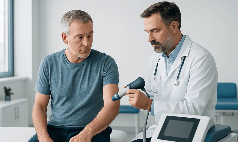
During the session. The applicator, which looks like a small handheld probe, is pressed firmly against the skin over the painful area. The device then delivers a series of rapid pulses. You will feel and hear a tapping or thumping sensation. Many patients describe it as uncomfortable but tolerable; the energy level can usually be adjusted so that it stays within your comfort range. A single session commonly delivers a few thousand pulses and takes around 5 to 15 minutes of actual treatment, with the whole appointment typically lasting 15 to 30 minutes.
There are two broad types of devices. Focused shock waves concentrate energy at a set depth and are often used for deeper tissues or calcium deposits. Radial pressure waves spread out from the skin surface and are commonly used for more superficial tendons. Your clinician chooses the type based on the condition being treated.
After the session. The gel is wiped off and you can usually leave immediately. Local anesthetic is generally not used, because numbing the area can make it harder to target the correct spot and may reduce the treatment effect. A typical course consists of about three to five sessions spaced roughly one week apart, although your treatment plan may differ.
Preparation for shock wave therapy
Preparation for shock wave therapy is simple compared with surgical procedures, but a few steps help the treatment go smoothly and safely.
- Share your full medical history. Tell your doctor about any blood-thinning medication, bleeding disorders, pregnancy or possible pregnancy, implanted devices, previous steroid injections, and any cancer history.
- Ask about anti-inflammatory medicines. Many clinicians advise avoiding non-steroidal anti-inflammatory drugs (NSAIDs such as ibuprofen) for a period before and after each session, because the treatment is thought to work partly by triggering a controlled inflammatory healing response. Follow your own doctor’s advice rather than stopping any medication on your own.
- Bring or wear suitable clothing. The treatment area needs to be exposed, so loose clothing or shorts are helpful for knee, hip, or ankle treatment.
- Bring imaging reports. If you have had X-rays, ultrasound, or MRI scans of the area, bring the reports or images.
- Plan your day normally. No fasting is needed and you can typically drive yourself home afterward.
Recovery and aftercare: shock wave therapy recovery time
Shock wave therapy recovery time is short in terms of daily activity, but the healing response it aims to trigger takes longer to unfold. Understanding both parts helps set realistic expectations.
The first 24 to 72 hours. Many patients notice mild soreness, redness, or slight swelling in the treated area for a day or two. Some experience a brief increase in pain before improvement begins. Applying a cool compress can be soothing, although your clinician may advise against icing if they want to preserve the inflammatory response. Simple pain relief with acetaminophen is often acceptable; check with your clinician before using NSAIDs.
Activity. Most people return to normal daily activities, including work, the same day or the next day. Your clinician will typically ask you to avoid strenuous or high-impact exercise involving the treated area for a short period after each session, often around 48 hours, and to avoid sudden increases in training load during the course of treatment.
Continuing rehabilitation. Shock wave therapy is usually one part of a broader plan. Stretching and progressive strengthening exercises, footwear or orthotic changes, and load management remain important. Many patients continue a home exercise program throughout and after the course of sessions.
Timeline for improvement. Pain relief is not always immediate. Some patients feel better after the first or second session, but for many the most noticeable improvement develops gradually over several weeks to a few months after the course is complete, as the tissue remodels. Clinicians often reassess response around 6 to 12 weeks after the final session before deciding on next steps.
Shock wave therapy risks and side effects
Weighing shock wave therapy risks and benefits is an important part of the decision. The treatment is widely regarded as low risk when performed by trained staff on appropriately selected patients, but it is not risk-free.
Common, usually short-lived side effects include:
- Pain or discomfort during the session
- Temporary soreness, tenderness, or aching afterward
- Redness of the skin over the treated area
- Mild swelling
- Bruising or small pinpoint bleeding spots under the skin (petechiae)
- Numbness or tingling that typically resolves within hours to days
Less common risks include a temporary flare of symptoms lasting longer than expected, skin irritation or small skin breaks, and, rarely, tendon injury when high energy levels are used over already weakened tissue. Nerve irritation can occur if treatment is applied too close to a superficial nerve. These risks are one reason the treatment is not used in people with bleeding disorders, over infected or damaged skin, or in children’s growth plates.
Benefits include the non-invasive nature of the treatment, the absence of incisions, medication, or anesthesia, minimal downtime, and the possibility of avoiding or postponing injections or surgery. For many chronic tendon problems, shock wave therapy offers a middle step between exercise-based care and more invasive options.
It is also worth understanding the limitations: some people do not respond, results can take time to appear, and the treatment does not correct underlying causes such as training errors, poor footwear, or biomechanical problems, which still need to be addressed.
Results and outlook
The research on shock wave therapy varies by condition. The evidence is generally considered strongest for chronic plantar fasciitis and calcific tendinitis of the shoulder, where clinical studies and reviews have reported meaningful pain reduction and improved function for many patients compared with placebo or no treatment. Evidence for tennis elbow, Achilles tendinopathy, patellar tendinopathy, and hip tendon pain is more mixed, with some studies showing benefit and others showing little difference from exercise therapy alone. Overall, professional guidelines in several countries describe shock wave therapy as a reasonable option for chronic tendinopathy when conservative care has failed, while noting that response is not guaranteed.
Factors that may influence the outcome include how long the problem has existed, whether the diagnosis is accurate, the energy level and number of sessions used, and whether rehabilitation exercises are followed. Your doctor may recommend a second course if you had a partial response, or may discuss alternative treatments if there is no improvement after an adequate trial.
Cost considerations
Because shock wave therapy is an outpatient treatment without anesthesia, implants, or a hospital stay, its overall cost structure is different from that of surgery. The main factors that influence cost include:
- Number of sessions. Treatment is usually priced per session or per course, so the number of sessions your clinician recommends is the largest driver.
- Type of device. Focused shock wave equipment is generally more complex than radial devices, which can affect pricing.
- Imaging guidance. Ultrasound localization or diagnostic imaging before treatment may be billed separately.
- Specialist consultation and follow-up visits. Initial assessment and reassessment after the course are often separate items.
- Accompanying physical therapy. A structured rehabilitation program may add to the total.
Insurance coverage varies widely: some plans cover shock wave therapy for specific conditions after documented failure of conservative care, while others consider it elective. Checking coverage in advance and asking the clinic for a written treatment plan can help avoid surprises.
Frequently asked questions
Does shock wave therapy hurt?
The shock wave therapy procedure is usually uncomfortable rather than severely painful. Most people describe a firm tapping or snapping sensation over the sore spot. Clinicians typically start at a low energy level and increase gradually, and they can lower it if the discomfort becomes too much. Mild soreness for a day or two afterward is common.
How many sessions of shock wave therapy are needed?
A typical course is three to five sessions, usually spaced about one week apart, although this depends on the condition, the type of device, and your response. Some patients need fewer sessions and others may be offered a second course if they improve partially. Your clinician will usually reassess before recommending more treatment.
What is the shock wave therapy recovery time?
Return to everyday activities is typically immediate or within a day. Strenuous or high-impact exercise on the treated area is often avoided for about 48 hours after each session. The healing response itself develops more slowly, and many patients notice the greatest improvement several weeks to a few months after finishing the course.
Who needs shock wave therapy rather than other treatments?
Shock wave therapy is generally reserved for people with chronic tendon or soft tissue pain that has not improved after several months of rest, exercise therapy, and other conservative measures. It is not usually a first-line treatment and is not appropriate for acute tears, infections, or people with certain medical conditions. A specialist can help decide whether it fits your situation.
What are the main shock wave therapy risks and benefits?
The main benefits are that it is non-invasive, requires no anesthesia or downtime, and may help some people avoid injections or surgery. The main risks are temporary pain, redness, swelling, bruising, and, rarely, nerve irritation or tendon injury. Serious complications are uncommon when patients are screened properly and the treatment is applied by trained staff.
Can I take painkillers after shock wave therapy?
Acetaminophen is often considered acceptable for post-session soreness. Many clinicians advise avoiding anti-inflammatory medicines such as ibuprofen for a period around treatment because the therapy is thought to rely on a controlled inflammatory healing response. Always follow the specific advice of your own doctor, especially if you take medication for other conditions.
Is shock wave therapy the same as ultrasound therapy?
No. Therapeutic ultrasound uses continuous high-frequency sound waves at low energy, mainly to warm tissue. Shock wave therapy delivers brief, high-pressure pulses that produce a distinct mechanical effect on tissue. The two treatments feel different and are used for different purposes, although ultrasound imaging may be used to guide where shock waves are applied.
When to see a doctor
You should consider being assessed by a specialist in physical medicine, orthopedics, or sports medicine if you have tendon or heel pain that has lasted more than a few months despite rest and basic care, if pain limits walking, work, or sleep, or if you are unsure of the cause of your pain. A proper diagnosis matters because shock wave therapy is only appropriate for specific conditions, and other problems, such as stress fractures, nerve entrapment, or inflammatory arthritis, need different treatment.
After a shock wave therapy session, mild soreness and redness are expected. However, seek prompt medical attention if you notice any of the following:
- Severe or rapidly worsening pain that does not settle within a few days
- Marked swelling, spreading bruising, or a large lump under the skin
- A sudden pop or snap in the tendon, followed by weakness or inability to bear weight
- Numbness, tingling, or weakness that persists beyond a day or two
- Signs of infection over the area, such as increasing redness, warmth, discharge, or fever
- Any new symptom that concerns you or feels out of proportion to what you were told to expect
These situations are uncommon, but they should be evaluated by a clinician rather than waiting for the next scheduled session.
Preparation
- Tell your doctor about blood thinners, bleeding disorders, pregnancy, implanted devices, recent steroid injections, or cancer history. Ask whether to pause anti-inflammatory medicines before sessions and follow that advice rather than stopping medication on your own. Wear loose clothing that allows access to the treatment area and bring any imaging reports. No fasting is required.
Aftercare
- Expect mild soreness, redness, or slight swelling for a day or two after each session. Avoid strenuous or high-impact activity involving the treated area for about 48 hours, and check with your clinician before using anti-inflammatory painkillers. Continue your prescribed stretching and strengthening program, and report severe pain, marked swelling, persistent numbness, or signs of infection promptly.
Medically reviewed by the Acıbadem International Medical Board September 13, 2026
See our medical review board →
Update history
- PublishedSeptember 13, 2026
- Medical review approvedSeptember 13, 2026
- Last content updateSeptember 13, 2026
References2
From the Acibadem Blog

Who Is a Candidate for Shock Wave Therapy? Plantar Fasciitis, Tennis Elbow and More
Shockwave therapy is usually considered for adults with a chronic tendon or fascia problem, most often plantar fasciitis, tennis elbow or calcific shoulder tendinopathy,…
Shock Wave Therapy Side Effects: Redness, Soreness and What the First Two Days Feel Like
Shockwave therapy side effects are usually mild and short-lived: temporary redness, warmth, slight swelling, small pinpoint bruises and a deep, bruised-feeling soreness at the…