

How Soon Does Clubfoot Need to Be Corrected: What It Means, What to Expect and When to See a Specialist
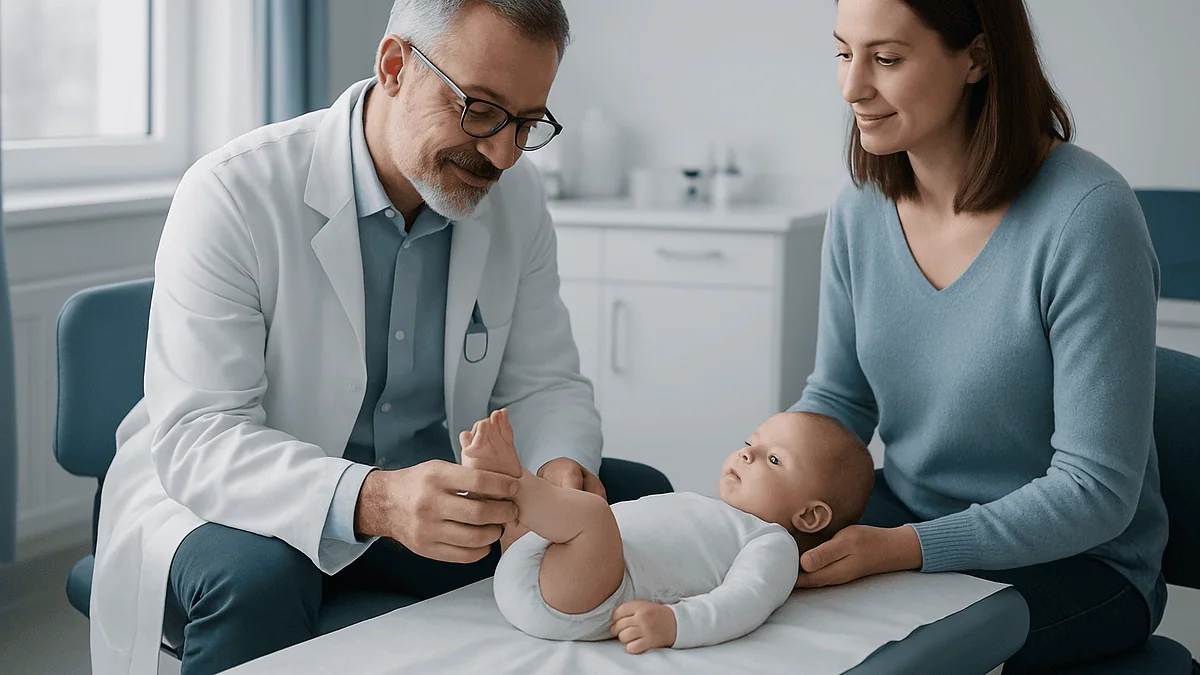
Key Takeaways
- Clubfoot affects roughly 1 in 1,000 babies, occurs about twice as often in boys, and involves both feet in around half of cases.
- Treatment ideally begins within the first one to two weeks of life, when tendons and ligaments are softest, but the same casting approach works when started later in infancy.
- Most feet are corrected after about four to eight weekly casts, and most babies then need a minor procedure to lengthen the Achilles tendon followed by a final cast of about three weeks.
- A flexible foot that a clinician can move into normal position is positional talipes, which usually resolves on its own; a stiff foot is structural clubfoot, which never does.
- Boots-and-bar bracing runs nearly full time for about three months and then during sleep for several years, and skipping it is the leading reason a corrected foot relapses.
- A treated clubfoot may end up slightly smaller with a thinner calf, but children typically walk on time and play sports without restriction.
Clubfoot does not need to be corrected in the delivery room, but treatment ideally begins within the first one to two weeks of life, when a newborn's ligaments and tendons are most pliable. Gentle weekly stretching and casting usually corrects the foot within a few months, and the method still works well when started later in infancy. Untreated clubfoot does not resolve on its own.
The first thing most parents notice is not the foot. It is the nurse’s pause. A hand that lingers a second longer over one tiny leg, then a soft “I’m just going to have the doctor take a look.” By the time anyone says the word clubfoot, a parent has already run through a dozen worst cases in their head, and almost none of them are true.
Here is the part that rarely gets said early enough: this is one of the most correctable conditions a baby can be born with. The foot that turns inward and downward today can, in the large majority of cases, be brought into a flat, flexible, walking position without a major operation. The work happens in plaster, in patience, and in a pair of small boots connected by a bar.
What parents most want to know, once the shock settles, is timing. How fast do we have to move? Is a week’s delay a problem? Does “mild” mean we can wait? Those are fair questions, and the evidence gives fairly clear answers.
How soon does clubfoot need to be corrected?
Soon, but not instantly. Both the NHS and Mayo Clinic describe treatment that typically starts within the first one to two weeks after birth. That window is not arbitrary. A newborn’s ligaments, joint capsules and tendons are unusually soft and stretchable, a leftover of the hormonal environment of pregnancy. Every week that passes, those tissues stiffen a little, and the foot becomes marginally harder to coax into position.
The distinction that matters is between ideal and necessary. Ideal is early. Necessary is a wider window than most parents fear. The same gentle stretch-and-cast approach that works at two weeks also works at two months, and orthopedic teams routinely treat babies whose diagnosis was delayed or whose family had to travel to reach care. Later starts may mean a few more casts and a slightly longer road, not a lost opportunity.
So the honest answer has three parts. First, there is no emergency in the first days; a healthy newborn can go home, feed and settle before casting begins. Second, the first weeks are the sweet spot, and a referral should be made promptly rather than left for a later check-up. Third, if that window has passed for any reason, the door has not closed. What changes with delay is effort and duration, not whether correction is possible.
One more point worth stating plainly: waiting to “see if it improves” is the one strategy the evidence does not support for true clubfoot. The condition does not resolve on its own, and the later a child begins, the more the growing bones adapt to the wrong position.
What clubfoot actually is, and what it isn't
The medical name is congenital talipes equinovarus, a mouthful that describes exactly what the eye sees. Equinus means the foot points down like a horse’s hoof; varus means the heel turns inward. Add a high arch and a forefoot that curves toward the midline, and you have the classic shape: a foot that looks as though it has been rotated inward and tucked under the leg, with the sole facing sideways or even upward.
This is a structural problem, not a positional one. The bones of the foot are present but misaligned, and the soft tissues on the inner and back side of the foot are shortened and tight. According to Mayo Clinic, the affected calf muscles are often underdeveloped, and the foot may be up to about half an inch shorter than the other. It is not painful for the baby, which surprises many parents who watch their infant kick happily with a foot that looks so wrong.
Clubfoot is also common enough that most pediatric orthopedic teams see it every week. NHS figures put it at roughly 1 in every 1,000 babies. Mayo notes it occurs about twice as often in boys and that both feet are affected in around half of cases.
Two things it is not: it is not usually part of a wider condition. Most babies with clubfoot are otherwise healthy, with what clinicians call idiopathic clubfoot, meaning no identifiable cause. In a smaller group, clubfoot accompanies conditions affecting nerves or muscles, such as spina bifida, and the orthopedic team will examine the whole child, not just the foot, to rule those out.
Why would a baby be born with clubfoot?
Nobody has a complete answer, and any source that claims one is overreaching. What the evidence shows is a mix of genetic tendency and environmental influence that adds up during the second trimester, when the foot is forming and rotating into its final position.
Genetics carries real weight. Mayo Clinic lists family history among the clearest risk factors: if one parent or a sibling had clubfoot, the chance rises. Researchers have identified several genes involved in limb development that appear more often in affected families, but no single “clubfoot gene” explains most cases. The pattern looks like many small contributions rather than one switch.
Environment matters too. Smoking during pregnancy is the best-documented modifiable risk factor; Mayo describes the risk as significantly higher when a woman who smokes also has a family history. Very low amniotic fluid, which restricts the space a fetus has to move, has also been associated with clubfoot, and some infections during pregnancy have been studied as possible contributors.
What has been largely dismissed is the old idea that the foot simply got “squashed” in the womb. That explanation fits positional foot deformities, which are flexible and resolve. True clubfoot involves changes in the bones and connective tissue that begin early in development, well before the fetus is large enough to be cramped.
For parents, the practical takeaway is modest: in the great majority of cases, clubfoot happened for reasons no one controlled, and it usually says nothing about the rest of the baby’s health.
Is it my fault my baby has clubfoot?
No. That deserves to be a full sentence on its own, because the question sits under almost every other question parents ask.
The way a mother slept, the exercise she did or did not do, a stressful week at work, one glass of wine before she knew she was pregnant, a tight waistband, a long car journey: none of these have any evidence linking them to clubfoot. The foot’s position is set by processes in early development that operate below the level of daily choices. MedlinePlus summarizes the situation simply: the cause is not known, though a family history makes it more likely.
The one exception people sometimes seize on is smoking. It is true that smoking during pregnancy is associated with a higher risk. It is also true that most babies of parents who smoked do not have clubfoot, and that plenty of babies with clubfoot were born to parents who never smoked a cigarette. Association is a population-level statistic, not a verdict on an individual pregnancy. If smoking is part of your story, the useful response is support to stop now, not guilt about what is already done.
Guilt has a cost beyond the emotional one. Parents who feel responsible sometimes hesitate to ask questions, or push for faster or more aggressive treatment than the foot needs, or blame themselves again if the foot relapses years later. The orthopedic teams who do this work will tell you that relapse is a known feature of the condition, not a parenting failure. Carrying that distinction into the first appointment makes everything after it easier.
Can mild clubfoot correct itself?
True clubfoot, even when it looks mild, does not correct itself. What can correct itself is a different condition that often gets called clubfoot by relatives and, occasionally, by tired new parents at three in the morning.
The NHS draws the line clearly. Positional talipes describes a foot that has been held in an awkward position in the womb and looks turned in at birth, but is flexible: a clinician can gently move it into a normal position with the fingers. It typically improves on its own over weeks, sometimes with the help of simple stretches a physiotherapist demonstrates for parents. No casts, no boots.
Structural clubfoot is stiff. The foot resists being moved into a neutral position, the heel stays tucked in, and the crease on the inside of the foot is deep. Even a “mild” structural clubfoot has these features, and mildness mostly predicts fewer casts rather than a different outcome. Left alone, the shortened tendons and ligaments remain short, and the growing bones remodel around the deformity. A foot that looked only slightly turned at birth can look considerably worse by the time a child is pulling up to stand.
The distinction cannot be made reliably by eye, including by experienced eyes, from across a room or in a photograph. It is made by hand, by someone who examines many infant feet. That is why the right response to “it looks a bit turned in” is a prompt examination, not a wait-and-see plan based on how mild it appears.
How clubfoot is diagnosed, before and after birth
Many families first hear the word in a dimly lit ultrasound room. The NHS notes that clubfoot may be spotted during the routine anomaly scan carried out between 18 and 21 weeks of pregnancy. A prenatal finding cannot be treated before birth, and ultrasound occasionally over- or under-calls it, but knowing early has a practical advantage: parents can meet the orthopedic team, understand the plan and grieve the picture-perfect newborn photos before the baby arrives, so that the first weeks are about action rather than shock.
After birth, diagnosis is clinical. A doctor looks at the shape of the foot and, crucially, feels how it moves: whether the heel can be brought down and out, how the forefoot behaves, how deep the creases are. Mayo Clinic describes this examination as the basis of diagnosis; X-rays are sometimes used but are not required, because much of an infant’s foot is still cartilage that does not show up well.
Clinicians often score the severity using a standardized scale that grades several features of the foot. The score is not a prognosis in itself, but it helps predict the likely number of casts and gives a baseline to track progress week by week.
The examination extends beyond the foot. The team will check the hips, spine and general muscle tone, because clubfoot occasionally travels with other conditions. Finding nothing else is the most common result, and it is worth hearing explicitly.
Serial casting explained: the Ponseti method week by week
The approach used almost everywhere today is deceptively simple. Rather than forcing the foot straight, the clinician stretches it a small amount in a specific sequence and holds the gain in a plaster cast from toes to upper thigh. About a week later, the cast comes off, the foot has softened into its new position, and the process repeats a little further along. Each cast is a single frame in a slow-motion film of the foot rotating outward.
Timing is what makes the method work in infants. According to the NHS, casts are usually changed weekly, and most feet are corrected after roughly four to eight casts. Mayo Clinic describes cast changes once or twice a week over several months. The exact count depends on how stiff the foot was at the start and how much correction each cast achieves.
What surprises parents most is how little the baby minds. The stretching is gentle, done while the infant feeds or sleeps, and the cast is not painful once on. Casting appointments become a rhythm: the soak or saw to remove the old cast, a bath, a few minutes of stretching, fresh plaster, a wriggling baby wrapped back up.
Parents’ job in this phase is observation. Casts should stay dry and snug; toes should stay pink and warm. A cast that slips down the leg or a toe that disappears inside it means a call to the clinic, not a wait until the next appointment. Skin should be checked at the edges. Nothing here requires special skill, but it does require attention, week after week, for the better part of two months.
Does the Achilles tendon have to be released?
Usually, yes, and this is the part of treatment that makes parents sit up. After the casts have rotated the foot outward, one problem typically remains: the heel is still pulled up because the Achilles tendon, the thick cord at the back of the ankle, is too short. Stretching alone rarely fixes it.
The solution is a small procedure called a tenotomy. NHS guidance notes that most babies need this minor operation to loosen the tendon, and Mayo Clinic describes it as part of the standard sequence for most children. The tendon is released through a tiny incision, often under local anesthetic in an outpatient setting, and a final cast is applied to hold the foot in its corrected position for about three weeks while the tendon heals and regrows at its new, longer length.
An infant’s Achilles tendon is remarkably good at this. It reforms across the gap and functions normally afterward, which is one reason the procedure is timed for the first months of life rather than later childhood, when healing is slower and the tendon is less forgiving.
Parents often ask whether the tenotomy can be skipped if the foot “looks fine.” The answer usually lies in what the clinician feels: if the ankle cannot bend upward past a certain point with the heel down, the tendon is the reason, and leaving it tight invites relapse. This is a decision for the treating team, based on the examination, not on appearance.
Boots and bar: the phase that actually decides the outcome
If serial casting is the correction, bracing is the insurance. A corrected clubfoot wants to drift back toward its old position for years, and the only reliable way to prevent that is to hold the feet turned outward in a brace: soft boots attached to a rigid bar, worn on a schedule that shrinks over time.
The schedule is demanding at first and then merciful. Mayo Clinic describes full-time wear, typically about three months, followed by nights and naps for up to about three years. NHS guidance describes wearing the boots roughly 23 hours a day for three months, then at night until the child is about four or five years old. Teams vary in how long they continue the night phase, and the clinician treating your child will set the plan.
| Phase | What happens | Typical timing |
|---|---|---|
| Serial casting | Weekly gentle stretching and plaster casts | About 4 to 8 casts (NHS) |
| Tendon release | Minor procedure to lengthen the Achilles; final cast | Final cast about 3 weeks (NHS, Mayo) |
| Full-time bracing | Boots and bar nearly all day and night | About 3 months (NHS, Mayo) |
| Night bracing | Boots and bar during sleep and naps | Until roughly age 3 to 5 (Mayo, NHS) |
Bracing is where treatment most often falters, and not because it fails. It is because a toddler cries, a grandparent objects, a family goes on holiday, and the boots stay in the bag. Mayo is blunt that relapse is far more likely when the brace is not worn as directed. The foot does not know it was corrected; it only knows what position it spends its nights in. Building the boots into bedtime routine early, when the child has no memory of life without them, is the single most useful thing parents can do.
How long does clubfoot treatment take from start to finish?
Two answers, depending on what “finished” means. The active correction, the part with weekly appointments and plaster, is short: roughly two months of casting followed by a three-week cast after the tendon release. Most babies finish this phase before they are old enough to roll over. Parents who braced themselves for a year of hospital visits are often startled to be handed a pair of boots at three or four months old and told the hard part is done.
Then there is the long tail. The bracing phase runs for years, and follow-up appointments continue through early childhood and often until the foot stops growing. The NHS describes night bracing until age four or five; Mayo Clinic describes up to about three years. During this period the clinician checks that the foot remains flexible, that the heel stays down, and that the child is walking with the sole flat rather than on the outer edge.
Relapse can and does happen, particularly in the toddler years and particularly when bracing has lapsed. It is usually caught early at a follow-up visit and treated with a short return to casting, sometimes with a small procedure to rebalance a tendon that is pulling the foot inward. That is not a treatment failure so much as a known feature of the condition, and one reason follow-up matters even when everything looks perfect.
For planning purposes, then: expect an intense two to three months, a demanding first quarter of bracing, and then a low-effort but non-negotiable routine that lasts until preschool.
What if treatment starts late?
Delays happen for ordinary reasons: a birth far from a pediatric orthopedic service, a diagnosis missed because the baby was swaddled, a family that moved, a pandemic. The good news is that the stretch-and-cast approach was never limited to newborns. Orthopedic teams use it in babies diagnosed at several months and in toddlers who were never treated, and it continues to produce well-corrected feet, though the road is longer.
What changes with age is tissue behavior. An older infant’s ligaments are stiffer, so each cast gains a little less and more casts are needed. Bones have begun to adapt to the deformity, so the correction must overcome shape as well as tightness. By walking age, the foot may have developed calluses or altered skin where the child has been weight-bearing on its outer edge. None of this rules out casting; it simply means the clinician plans for more of it and watches more carefully for the need to add a tendon procedure.
In children who reach school age untreated, or whose earlier correction relapsed and was not addressed, surgery to release or reposition soft tissues, and occasionally to reshape bone, becomes more likely. Mayo Clinic notes that more extensive surgery is generally reserved for severe cases or those that have not responded to nonsurgical treatment, and that surgically treated feet tend to be stiffer over time.
The message for a parent reading this after a missed window is straightforward. You have not run out of options. You have, if anything, an even stronger reason to book the appointment this week rather than next.
What happens if clubfoot is left untreated?
The foot stays turned, and the child learns to walk on it anyway. Children are relentlessly adaptable, and an untreated clubfoot does not stop a toddler from getting around. It does change how. The child bears weight on the outer edge and sometimes the top of the foot, surfaces that were never designed for it.
Over years, that mismatch shows. Mayo Clinic and MedlinePlus describe the consequences: thick calluses and skin breakdown where the wrong part of the foot meets the ground, an awkward gait, trouble fitting shoes, and a growing risk of arthritis as joints are loaded in positions they cannot tolerate. The calf on the affected side remains thin. Pain, absent in infancy, arrives with size and mileage.
The social cost is quieter but real. A child who cannot wear ordinary shoes or who walks noticeably differently faces attention they did not ask for, and physical activity becomes something to avoid rather than enjoy. In parts of the world where treatment is not available, untreated clubfoot remains a significant cause of lifelong disability, which is why global health programs have invested in training local clinicians in casting.
None of this is inevitable. The point of laying it out is not to frighten but to answer the question honestly, because “it’s only cosmetic” is a myth that occasionally delays care. Clubfoot is a functional problem with a well-established solution, and the outcome of treating it early is a foot that works, fits a shoe and does not hurt.
Will my child walk, run and play normally?
In the overwhelming majority of cases, yes. Children treated in infancy with casting and bracing typically walk at the usual age, keep up on playgrounds and sports fields, and grow into adults whose feet do not limit them. Johns Hopkins Medicine and Mayo Clinic both describe the expected outcome of early, well-managed treatment as a functional, nearly normal foot.
“Nearly” is doing honest work in that sentence, and parents deserve the specifics. A corrected clubfoot is often slightly smaller than the other foot, sometimes by a shoe size. The calf on that side is usually thinner and may remain so into adulthood, no matter how many stairs the child climbs. The ankle may bend upward a little less than the other. Mayo notes the affected leg may also be marginally shorter. These differences are visible to a parent who knows where to look and invisible to almost everyone else.
What the evidence does not support is the idea that a treated child is fragile. Running, jumping, dance, football, gymnastics: none are off-limits. Occasionally a child with a corrected clubfoot tires a little faster on long walks or reports stiffness after unusual exertion, and a clinician can advise on stretching if that happens.
The best predictor of a good long-term result is not the severity at birth. It is whether bracing was completed and whether follow-up caught any early drift. Parents cannot control the first; they have enormous influence over the second.
When to see a specialist, and red flags during treatment
Any newborn whose foot appears turned inward or downward, or whose foot cannot easily be moved into a normal position, should be examined by a pediatrician promptly and referred to a pediatric orthopedic specialist if clubfoot is suspected. The referral should happen within days, not at the six-week check. If clubfoot was seen on a prenatal scan, ask to have the orthopedic appointment arranged before delivery so the plan is waiting when the baby arrives.
During casting, contact the treating team the same day if you notice: toes that are pale, blue, dusky or cold; toes that have swollen or disappeared up inside the cast; a cast that has cracked, softened, become wet or slipped down the leg; an unpleasant smell or fluid from the cast; or a baby who is unusually inconsolable and cannot be settled by feeding or comfort. These can signal a cast that is too tight or a skin problem underneath, and they are fixed quickly when caught early.
During the bracing years, book an earlier review if the child begins walking on the outside edge of the foot, if the heel seems to lift or turn in again, if the foot looks stiffer than before, or if the brace no longer fits or causes persistent red marks or blisters. Skin irritation is a reason to adjust the fit, not to stop wearing the boots.
Seek urgent medical attention for any child with a cast who develops fever together with a foul odor, spreading redness above the cast, or severe unrelenting pain. Those are rare, but they are not situations to sleep on.
Frequently asked questions
How soon does clubfoot need to be corrected after birth?
Treatment usually starts within the first one to two weeks of life, according to NHS and Mayo Clinic guidance. There is no need to begin in the first hours, but an early referral takes advantage of how pliable a newborn’s ligaments and tendons are. If the window is missed, the same stretching and casting method still works in older infants, though it may require more casts and a longer course.
Can mild clubfoot correct itself?
True clubfoot, even when mild, does not correct itself, because the tendons and ligaments are structurally shortened. A foot that looks turned in but can be gently moved into a normal position by hand is usually positional talipes, a flexible condition that often resolves on its own with simple stretches. Only an examination by an experienced clinician can tell the two apart, so any turned-in foot should be assessed promptly.
Why would a baby be born with clubfoot?
The exact cause is unknown in most cases. Evidence points to a combination of genetic tendency and environmental influences during early fetal development. Family history raises the risk, and smoking during pregnancy is the best-documented modifiable factor. Low amniotic fluid has also been associated with clubfoot. In a small minority, clubfoot accompanies a condition affecting nerves or muscles, which is why the whole baby is examined.
Is it my fault my baby has clubfoot?
No. Nothing about how a parent slept, exercised, ate or sat during pregnancy causes clubfoot, and most cases have no identifiable cause at all. Smoking during pregnancy is linked to higher risk at a population level, but most babies of parents who smoked do not have clubfoot, and many babies with clubfoot were born to parents who never smoked. Guilt is common and unfounded.
What happens if clubfoot is left untreated?
The foot stays turned, and the child eventually walks on its outer edge or top. Over time this leads to thick calluses, skin problems, an awkward gait, difficulty wearing shoes, a persistently thin calf and an increased risk of arthritis and pain. Untreated clubfoot is a significant cause of disability where treatment is unavailable. Early casting and bracing prevents nearly all of these consequences.
How many casts does clubfoot treatment take?
Most feet are corrected after about four to eight weekly casts, according to the NHS, with the number depending on how stiff the foot was at the start. Mayo Clinic describes cast changes once or twice a week over several months. After casting, most babies have a minor procedure to lengthen the Achilles tendon, followed by a final cast for about three weeks.
Is the Achilles tendon procedure a real surgery?
It is a minor procedure, often done under local anesthetic as an outpatient. A small cut releases the tight Achilles tendon so the heel can drop into position, and a final cast holds the foot while the tendon heals at its new length over about three weeks. Most babies need it, because stretching alone rarely fully corrects the tight heel cord.
How long does a child have to wear the clubfoot boots and bar?
Nearly full time, around 23 hours a day, for about three months, then during sleep and naps for several years. Mayo Clinic describes night bracing for up to about three years; the NHS describes it until about age four or five. The treating clinician sets the schedule. Consistent bracing is the main factor that prevents the foot from drifting back toward its original position.
Will my child walk normally after clubfoot treatment?
In most cases, yes. Children treated in infancy typically walk at the usual age and take part in running, sports and play without restriction. The treated foot is often slightly smaller, the calf thinner, and the ankle a little less flexible than the other side, but these differences rarely affect function. Completing the bracing phase and attending follow-up are the strongest predictors of a good long-term result.
Can clubfoot come back after it has been corrected?
Yes. Relapse is a recognized feature of the condition, most often in the toddler years and especially when bracing has lapsed. It is usually caught at a routine follow-up visit as the heel turning in or the child walking on the outer edge of the foot. Early relapse is typically treated with a short return to casting, sometimes with a small tendon procedure, rather than major surgery.
References
This article is for general information only and is not a substitute for professional medical advice. Please consult a qualified doctor about your individual situation.

More from the Blog

Local, Regional or General? Anesthesia Options for Finger and Toe Surgery Explained
Most finger and toe operations can be done under local anesthesia, usually a digital block that numbs only the affected digit while you stay…

Who May Be a Candidate for Proximal Fibular Osteotomy When Inner-Knee Arthritis Is Mild?
Proximal fibular osteotomy is usually considered for adults whose knee osteoarthritis is confined mainly to the inner (medial) compartment, who have mild to moderate…

Knee Pain Causes: Why Knees Hurt (and Why It Differs in Women)
Knee pain in women most often comes from patellofemoral (front-of-knee) pain, osteoarthritis, tendon or bursa irritation, meniscus wear, or ligament injury. Women see certain…

Shoulder, Elbow, Wrist or Hand: How Upper Limb Surgery Is Planned for Each Joint
Upper limb surgery covers operations on the shoulder, elbow, wrist and hand, from nerve releases and tendon repairs to fracture fixation and joint replacement.…

Life After Adult Reconstruction Surgery: Why High-Impact Sport Is Limited and What Replaces It
After adult reconstruction surgery, the orthopedic term for hip and knee replacement, most care teams advise a long-term shift away from running, jumping and…

How Foot Surgery Is Performed: Bunion Correction, Fusion and Tendon Repair Step by Step
Foot surgery is usually performed as a day procedure under a nerve block, spinal or general anesthetic. The surgeon makes one or more small…