

Hyperparathyroidism Work-Up: Calcium and PTH Tests, Bone Density and Kidney Checks Explained
Key Takeaways
- Primary hyperparathyroidism is diagnosed from blood, not scans: high calcium paired with a PTH that is raised or fails to drop is the hallmark pattern.
- A PTH in the normal range is abnormal when calcium is high, because healthy glands suppress hormone output almost completely in that setting.
- In roughly 8 out of 10 cases the cause is a single benign adenoma, and parathyroid cancer is rare, according to the National Institute of Diabetes and Digestive and Kidney Diseases.
- A 24-hour urine calcium is the test that separates true hyperparathyroidism from familial hypocalciuric hypercalcemia, an inherited mimic that does not need surgery.
- Bone density testing for this condition should include the forearm, because PTH preferentially thins cortical bone that hip and spine scans can miss.
- Sestamibi, ultrasound and 4D CT are ordered to locate a gland before surgery, and a negative scan does not rule the disease out.
A hyperparathyroidism work-up starts with blood tests for calcium and parathyroid hormone (PTH), usually repeated to confirm the pattern of raised or non-suppressed PTH alongside high calcium. Doctors then add vitamin D, phosphate, kidney function, a 24-hour urine calcium, a bone density scan and often kidney imaging. Localization scans such as ultrasound or sestamibi come only after the diagnosis is secure, to plan possible surgery.
The phone call usually comes about something else. A routine panel before a knee operation, a check-up for tiredness that never quite lifted, a follow-up after a kidney stone. Tucked into the results is one line the reader did not expect: calcium, slightly high. Then a second request for blood, then a mention of a hormone most people have never heard of, and suddenly there is a diagnosis on the table with a name that sounds like a thyroid problem but is not one.
That gap between “your calcium is up” and “here is what we think is happening” is what hyperparathyroidism workup tests are designed to close. The work-up is more methodical than dramatic. It is a sequence of blood and urine measurements, a bone scan, a look at the kidneys, and only later, if at all, an imaging study to find the gland responsible.
This article walks through that sequence in the order a careful endocrinology team would run it, explains what each result can and cannot tell you, and separates the questions a test answers from the decisions that belong to you and your clinicians.
What hyperparathyroidism workup tests are actually measuring
Behind the thyroid, usually four glands the size of a lentil sit and do one job: keep blood calcium inside a narrow band. They do it by releasing parathyroid hormone (PTH), a messenger that tells bone to release calcium, tells the kidneys to hold on to it, and nudges the kidneys to activate vitamin D so the gut absorbs more. When calcium rises, healthy glands go quiet. When it falls, they speak up.
Hyperparathyroidism means one or more of those glands keeps talking when it should be silent. In primary hyperparathyroidism, the problem lives in the gland itself. According to the National Institute of Diabetes and Digestive and Kidney Diseases, in roughly 8 out of 10 cases the cause is a single benign growth called an adenoma; in most of the remainder, several glands have enlarged. Cancer of a parathyroid gland is rare.
The work-up exploits the feedback loop. A normal gland faced with high calcium suppresses PTH toward the bottom of its range. A misbehaving gland does not. So the tests are less about any single number and more about whether the two numbers make sense together. Calcium high and PTH low points away from the parathyroids and toward another cause, such as cancer-related hypercalcemia or excess vitamin D. Calcium high and PTH high, or even PTH sitting in the upper-normal range when it ought to be suppressed, is the signature of a gland that has lost its off switch.
Every downstream test, from vitamin D to bone density to kidney imaging, either sharpens that inference or measures what years of extra calcium and PTH may have done to bone and kidney. Holding that logic in mind makes the rest of the pathway feel far less like a maze.
The two most common lab findings: high calcium with an unsuppressed PTH
Ask an endocrinologist for the hallmark of primary hyperparathyroidism and the answer is a pairing, not a value. Blood calcium above the reference range, together with PTH that is either above range or in the upper part of normal at a moment when it should be low. Mayo Clinic describes exactly this combination as the basis of diagnosis: elevated or high-normal PTH in the presence of high calcium.

Why does the PTH not have to be frankly high? Because the reference range for PTH is built from people whose calcium is normal. A PTH value in the middle of that range is entirely appropriate for someone with normal calcium and entirely inappropriate for someone whose calcium is elevated. Laboratories cannot flag that mismatch automatically; a clinician reading both results side by side can.
A few details matter in the blood draw itself. Calcium fluctuates modestly through the day and with hydration, so most teams repeat the measurement rather than acting on one result. The National Institute of Diabetes and Digestive and Kidney Diseases notes that high calcium is often discovered incidentally on routine bloodwork, which means the first abnormal value may have been drawn without the patient fasting or in the middle of an illness. A second sample taken under calmer conditions, ideally with PTH measured from the same draw, is far more informative.
Modern PTH assays measure the intact hormone, which avoids confusion from fragments that linger in the blood, particularly in people with reduced kidney function. Your report may label it “intact PTH” for this reason.
Two numbers, drawn together, interpreted together: that is the core of primary hyperparathyroidism diagnosis. Everything else in the work-up either rules out imitators or maps the consequences.
Is there a gold standard test for diagnosing hyperparathyroidism?
People searching this question often expect the answer to be a scan. It is not. The diagnosis of primary hyperparathyroidism is biochemical: it rests on the calcium and PTH relationship described above, confirmed on repeat testing and supported by the exclusion of other explanations. Mayo Clinic and the National Institute of Diabetes and Digestive and Kidney Diseases both frame blood tests, not imaging, as the way the condition is identified.
This surprises patients because imaging feels more definitive. A picture of a swollen gland seems harder to argue with than a hormone level. Yet scans miss small adenomas, occasionally highlight thyroid nodules that have nothing to do with the parathyroids, and cannot tell whether a gland that looks enlarged is actually overproducing hormone. A negative scan does not rule the condition out, and a positive scan without the matching bloodwork does not rule it in.
So the phrase clinicians use is “biochemically confirmed.” That confirmation typically involves:
- At least two calcium measurements above range, or persistently high-normal calcium with a clearly raised PTH.
- An intact PTH that is elevated or not appropriately suppressed.
- A vitamin D level to exclude deficiency, which can raise PTH on its own.
- Kidney function to exclude the secondary pattern seen in chronic kidney disease.
- A urine calcium measurement to exclude a rare inherited mimic.
Only when those pieces line up does imaging enter the conversation, and then for a different purpose: locating the gland, not proving it is overactive. Keeping the two roles separate saves patients from an unhelpful experience in which a normal-looking scan is mistaken for reassurance, or an incidental finding is mistaken for a diagnosis.
Repeat, corrected and ionized calcium: why one result is never enough
Roughly half the calcium in blood is bound to a protein called albumin, and only the free, or ionized, portion is biologically active. That single fact explains most of the fuss around repeat calcium testing. If albumin is low, as it can be in illness, poor nutrition or liver disease, total calcium reads lower than the active fraction really is. If albumin is high, usually from dehydration or a tight tourniquet, total calcium reads falsely high.

Laboratories address this in two ways. The first is a calculated adjustment, often reported as “corrected” or “adjusted” calcium, which estimates what the total would be at a standard albumin. The second is a direct measurement of ionized calcium from a specially handled sample. Ionized calcium avoids the estimate altogether but is more sensitive to how the blood is collected and transported, so not every laboratory offers it routinely.
Why repeat at all? Because the decision that follows a confirmed diagnosis, whether that is surgery or years of monitoring, is a consequential one, and because calcium moves. Hydration, a recent meal, and some medicines can shift a borderline value across the threshold and back. Certain diuretics, lithium, and high-dose calcium or vitamin D supplements can raise blood calcium; a clinician will usually ask about these before repeating the test, and may ask you to hold a supplement for a period before the redraw. That is a decision for the prescribing team, never one to make alone.
The practical upshot for patients: a second, and sometimes third, blood draw is standard practice, not a sign that something was missed. Bring a list of everything you take, including over-the-counter antacids and supplements, and mention any recent illness or dehydration. It shortens the path to a clear answer.
Vitamin D, phosphate and kidney function: the supporting blood tests
Once calcium and PTH have declared themselves, three further blood tests do the quiet work of confirming that the pattern truly comes from the parathyroids.
25-hydroxyvitamin D is the storage form of vitamin D and the best marker of a person’s overall supply. Deficiency matters here for two reasons. It raises PTH by itself, because the glands respond to the drop in absorbed calcium, which can muddy the interpretation in someone whose calcium is only borderline. And in genuine primary hyperparathyroidism, low vitamin D can mask how high calcium would otherwise be. Guidelines summarized by the National Institutes of Health Office of Dietary Supplements note that PTH rises as vitamin D status falls, which is why teams want the level documented before drawing conclusions.
Phosphate tends to run low or low-normal in primary hyperparathyroidism because PTH pushes the kidneys to excrete it. A low phosphate alongside high calcium adds weight to the diagnosis. High phosphate with high PTH points instead toward kidney disease.
Creatinine and estimated glomerular filtration rate (eGFR) measure how well the kidneys filter. This matters in both directions. Chronic kidney disease causes a secondary rise in PTH as the kidneys lose the ability to activate vitamin D and clear phosphate, so reduced eGFR changes the whole interpretation. Long-standing primary hyperparathyroidism can also impair kidney function over time, which is one of the factors that shifts a team from monitoring toward recommending surgery.
A fourth marker, alkaline phosphatase, sometimes appears on the panel. It rises when bone is turning over rapidly and can hint at how much skeletal effect the excess PTH is having. None of these tests is diagnostic alone. Together they turn a suggestive calcium and PTH pair into a coherent story.
What disease mimics hyperparathyroidism? The 24-hour urine calcium and FHH
There is one condition that produces almost the same bloodwork as primary hyperparathyroidism and yet needs no surgery at all. It is called familial hypocalciuric hypercalcemia (FHH), an inherited change in the calcium-sensing receptor that resets the body’s thermostat so it tolerates a higher calcium as normal. People with FHH have mildly raised calcium and normal or mildly raised PTH from childhood, usually without symptoms or complications.
The test that separates the two is a 24-hour urine collection for calcium and creatinine. In primary hyperparathyroidism the kidneys typically spill a normal or increased amount of calcium into the urine, because there is simply more of it to filter. In FHH the kidneys reabsorb calcium avidly, so urinary calcium is low relative to what the blood level would predict. Clinicians express this as a calcium-to-creatinine clearance ratio; a low ratio raises suspicion for FHH and may prompt genetic testing, particularly if relatives share the pattern.
The collection itself is unglamorous but important. You are given a container, asked to discard the first morning urine, then collect every void for the following 24 hours, including the first urine of the next morning. Missing even one void distorts the result, and vitamin D deficiency can lower urinary calcium in a way that mimics FHH, which is another reason the vitamin D level is checked first.
The urine test carries a second purpose. Very high urinary calcium is one of the findings that guideline groups treat as a reason to lean toward surgery in someone otherwise free of symptoms, because it signals higher stone risk. So the same collection that rules out an imitator also helps stage the real condition.
Getting this test right avoids an operation nobody needed, which is why experienced teams rarely skip it.
Primary, secondary, tertiary and normocalcemic: a table of lab patterns
The word hyperparathyroidism covers several different situations, and the laboratory pattern is what tells them apart. Secondary hyperparathyroidism is the glands doing their job in response to a problem elsewhere, most often vitamin D deficiency or chronic kidney disease. Tertiary hyperparathyroidism describes glands that have been stimulated for so long, usually by kidney failure, that they become autonomous and keep overproducing even after the original trigger is addressed. Normocalcemic primary hyperparathyroidism is a newer category in which PTH is persistently high, calcium stays within range, and every secondary cause has been excluded.
| Pattern | Blood calcium | PTH | Phosphate | Typical driver |
|---|---|---|---|---|
| Primary | High | High or inappropriately normal | Low or low-normal | Adenoma or gland hyperplasia |
| Secondary | Low or normal | High | Variable; high in kidney disease | Vitamin D deficiency, chronic kidney disease |
| Tertiary | High | Very high | Often high | Long-standing kidney failure |
| Normocalcemic primary | Normal, repeatedly | Persistently high | Normal | Gland dysfunction, secondary causes excluded |
| FHH (mimic) | Mildly high | Normal or mildly high | Normal | Inherited receptor change |
The table is a guide, not a verdict. Real patients straddle categories, particularly when kidney function is borderline or vitamin D has recently been replaced. What the table does show is why the supporting tests are non-negotiable: without a vitamin D level and an eGFR, a secondary pattern can be mistaken for a primary one, and the treatment paths for those two diagnoses are entirely different.
Normocalcemic disease deserves a special note. Because calcium is normal, it is often found only when PTH is measured for another reason, such as a low bone density result. Guideline groups generally ask for the pattern to be confirmed on several occasions and for all secondary causes to be excluded before the label is applied.
Bone density testing in hyperparathyroidism: what the DXA scan looks for
Extra PTH, year after year, asks bone to release calcium it would rather keep. The bone density scan measures the cumulative bill. The test is a dual-energy X-ray absorptiometry (DXA) scan, a low-dose X-ray that estimates the mineral content of bone at set sites and reports it as a T-score, the number of standard deviations from the average for a healthy young adult of the same sex.
The World Health Organization classification, used across most guidelines, defines osteoporosis as a T-score of minus 2.5 or lower, and low bone mass, sometimes called osteopenia, as a T-score between minus 1.0 and minus 2.5. In a hyperparathyroidism work-up, the scan is not just a screening exercise. A T-score in the osteoporosis range at any measured site, or a fragility fracture on imaging, is one of the criteria that most guideline groups treat as a reason to recommend surgery even when the patient feels well.
One detail distinguishes this DXA from a routine one: the forearm. Standard osteoporosis screening measures the hip and spine. Primary hyperparathyroidism has a particular appetite for cortical bone, the dense outer shell, and the distal third of the forearm is rich in it. Teams working up parathyroid disease therefore usually ask for the forearm to be scanned as well, because it can show loss that the hip and spine have not yet registered.
The scan itself takes a few minutes, involves lying still on a padded table, and exposes you to a fraction of the radiation of a standard chest X-ray. Metal jewelry and belt buckles come off; most clothing stays on. Results arrive as a report your clinician interprets alongside the blood and urine findings, and if you are placed under monitoring rather than treated surgically, the DXA becomes a recurring measure of whether bone is holding steady.
Kidney checks: stones, eGFR and why imaging the kidneys matters
The kidneys sit at the receiving end of high blood calcium. They filter it, they try to reabsorb what the body needs, and they excrete the surplus into urine that is now more prone to forming crystals. Two kinds of damage follow. Stones, which announce themselves loudly, and a slower decline in filtering capacity, which does not.
That is why a complete hyperparathyroidism work-up includes both a functional and a structural kidney assessment. The functional part is the creatinine and eGFR already on the blood panel. National Institute of Diabetes and Digestive and Kidney Diseases guidance defines chronic kidney disease as an eGFR below 60 that persists for three months or more, and most parathyroid guidelines use that same threshold as a marker of kidney involvement significant enough to favor surgical treatment.
The structural part is imaging. Kidney ultrasound is the usual first choice: no radiation, no contrast, and good at finding stones or nephrocalcinosis, the fine calcium deposits within kidney tissue that indicate longer exposure. Where ultrasound is inconclusive, or where a stone is suspected but not seen, a low-dose CT of the abdomen may follow. Some teams image every patient with confirmed primary hyperparathyroidism; others reserve it for people with a history of stones, high urinary calcium, or symptoms. Mayo Clinic lists kidney stones among the recognized complications of untreated disease, and finding a silent stone can change the management plan for someone who considered themselves symptom-free.
There is a practical thread to pull here. If you have ever passed a stone, had a stone analyzed, or been told you have “gravel” on a scan, say so at the first visit. A prior stone history is one of the clearest signals that the kidneys are already being affected, and it may shorten the work-up by moving the conversation about definitive treatment forward.
Sestamibi scan, ultrasound and 4D CT: locating the gland, not diagnosing the disease
Once the biochemistry is settled and the team is considering an operation, the question changes from “is there a problem” to “where is it.” This is where parathyroid imaging earns its place. The purpose is to help a surgeon plan a focused approach rather than explore all four glands, and Mayo Clinic is explicit that these scans are used to locate an overactive gland before surgery, not to make the diagnosis.
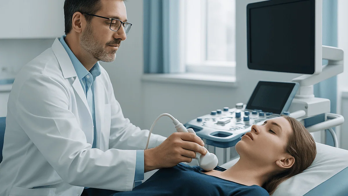
Three studies dominate. Neck ultrasound uses sound waves to look behind the thyroid for an enlarged gland. It is quick, radiation-free and often the first step, though it can struggle with glands tucked behind the breastbone or hidden by thyroid nodules. A sestamibi scan involves an injection of a small amount of a radioactive tracer that overactive parathyroid tissue tends to hold on to longer than surrounding tissue; a gamma camera then pictures where the tracer lingers. It is frequently combined with a CT for anatomical detail. Four-dimensional CT, the fourth dimension being time as contrast washes in and out, can pick up small or unusually placed glands that the other two miss, at the cost of more radiation and the use of iodine contrast.
Each has blind spots. Multiple enlarged glands are harder to spot than a single adenoma. Thyroid nodules can mimic parathyroid tissue on ultrasound and occasionally on sestamibi. A negative study does not mean there is no disease; it means the gland was not confidently located, and experienced surgeons operate successfully in that situation using intraoperative PTH measurement to confirm the culprit has been removed.
If your work-up stops short of imaging because you and your team have chosen monitoring, nothing has been skipped. Localization is a surgical planning tool, and ordering it before that decision is made adds cost, radiation and sometimes confusion without adding certainty.
Who usually gets the full hyperparathyroidism workup tests, and who is asked to wait
Almost everyone with a confirmed high calcium and unsuppressed PTH receives the core panel: repeat calcium, intact PTH, vitamin D, phosphate, creatinine and eGFR, and a 24-hour urine collection. The bone density scan and kidney imaging follow in most people with primary disease, because their results directly feed the decision between surgery and monitoring. Where practice varies is in how quickly the localization scans are ordered, and whether some patients are placed under observation without them.
Guideline groups broadly agree that surgery is the definitive treatment and that it is usually recommended when the disease is clearly affecting bone or kidney, when calcium is well above range, or when the person is younger, on the reasoning that decades of exposure lie ahead. Mayo Clinic describes a watchful-waiting approach as reasonable for people with mildly raised calcium, normal kidney function and normal bone density who have no symptoms, with regular blood tests and periodic bone density checks. The National Institute of Diabetes and Digestive and Kidney Diseases notes that the condition most often affects people over 60 and is more common in women, particularly after menopause, which is also the group in whom asymptomatic, incidentally found disease is most common.
People are commonly asked to wait, or to complete more testing first, in three situations: when vitamin D deficiency has just been found and needs to be addressed before the calcium and PTH are reinterpreted; when calcium is only borderline and a second or third measurement is needed; and when pregnancy is possible, since some imaging is deferred.
Where medicines enter the picture, for example a calcimimetic that quiets the gland’s calcium sensor, or a bone-protecting agent, they are options a specialist may raise for people who cannot or prefer not to have surgery. What they do, how long they take to act and whether they suit you are questions for the prescribing team.
What the following weeks usually look like
The rhythm of a parathyroid work-up is stop-start by design, because so much of it is confirmation. A typical path, described in Mayo Clinic and NHS patient information and mirrored in most endocrine clinics, runs something like this.
The first visit follows the incidental finding. Expect a detailed history: stones, fractures, fatigue, low mood, bone or joint aches, digestive complaints, a family history of calcium problems or endocrine tumors, and a full medicine and supplement review. Blood is usually redrawn the same day for calcium, albumin, intact PTH, vitamin D, phosphate and kidney function. You may leave with the 24-hour urine container.
Over the next two to four weeks, the repeat bloods and urine come back. If vitamin D is low, your clinician may address that first and re-test afterward, which can add several weeks. The DXA scan and kidney ultrasound are typically scheduled in this window; both are outpatient studies requiring no preparation beyond removing metal.
By the second visit, the team can usually say whether the pattern is primary hyperparathyroidism, a secondary form, or something needing genetic follow-up. If the findings favor surgery and you are inclined toward it, localization imaging is ordered and a surgical referral made. If the findings favor observation, a monitoring schedule is agreed, usually involving regular calcium and kidney function tests and periodic bone density scans, on intervals your team sets.
The NHS describes primary hyperparathyroidism as often mild and slowly progressive, which is why this measured pace is appropriate for most people. Very high calcium with symptoms is the exception, and in that case the timeline compresses sharply, sometimes to a same-day assessment.
Between appointments, hydration is the one piece of self-care almost every guideline mentions, because dehydration concentrates calcium in the blood. Beyond that, avoid starting or stopping supplements without asking.
What people often get wrong about the hyperparathyroidism work-up
“It is a thyroid problem.” The parathyroids sit behind the thyroid and share the name, but they are separate glands with a separate hormone. Thyroid function tests are normal in hyperparathyroidism unless a second, unrelated condition is present.
“My PTH is in the normal range, so it cannot be that.” A normal PTH in the presence of high calcium is abnormal. The gland should be near-silent. This is the most common reason the diagnosis is missed, and it is why the two results must be read together.
“The scan will tell us if I have it.” Imaging locates; bloodwork diagnoses. A clear sestamibi scan in someone with confirmed biochemistry does not undo the diagnosis, and an incidental bright spot in someone with normal calcium does not create one.
“I feel fine, so nothing is happening.” Bone loss and early kidney change are silent. The DXA scan and eGFR exist because the body does not always send a signal. Several guideline groups have moved away from the word “asymptomatic” for exactly this reason.
“I should stop taking calcium.” Cutting dietary calcium sharply can push PTH higher still and worsen bone loss. Most guidelines advise normal dietary intake rather than restriction, and any change to supplements should go through your clinician.
“One high calcium means surgery.” A single result means a repeat. Even confirmed disease does not automatically mean an operation; the bone, kidney and urine findings are what tip the balance, and the decision is shared.
“Vitamin D will make my calcium dangerous.” In someone with primary hyperparathyroidism and true deficiency, cautious repletion under supervision is standard, because low vitamin D itself drives PTH and bone loss. The key word is supervised; the team monitors calcium as they do it.
Questions to ask your care team about your results
A work-up produces a lot of numbers. These questions help turn them into a plan you understand and agree with.
- Has my calcium been confirmed as high on more than one occasion, and was it corrected for albumin or measured as ionized calcium?
- Was my PTH drawn at the same time as the high calcium, and does the combination fit primary hyperparathyroidism or a secondary cause?
- What was my vitamin D level, and does it need to be addressed before we interpret the other results?
- What did the 24-hour urine show, and has familial hypocalciuric hypercalcemia been excluded?
- What are my T-scores at the spine, hip and forearm, and do any of them meet the threshold that guidelines use to recommend surgery?
- What is my eGFR, and did the kidney imaging show stones or calcium deposits?
- Based on all of this, do you see me as someone who would usually be offered surgery, or someone for whom monitoring is reasonable? What would change that?
- If we choose monitoring, which tests will be repeated, how often, and what result would prompt a rethink?
- If we move toward surgery, which localization scan do you recommend and why, and what happens if the gland is not found on imaging?
- Are there medicines that could be discussed if surgery is not an option for me, and what do they do?
- Should any of my relatives have their calcium checked?
- Is there anything in my current medicines or supplements that could be affecting these results?
Bring a written list, and ask for a copy of your results so you can track them over time. A monitoring plan works best when the person being monitored can see the same trend line their clinician sees.
When to call your doctor
Most people going through a parathyroid work-up have mildly raised calcium and no urgent symptoms, and the pace described above is appropriate. A small number develop a marked rise in calcium, sometimes triggered by dehydration, illness or a new medicine, and that situation needs prompt assessment.
Contact your care team the same day, or seek urgent care, if you notice:
- Severe or worsening thirst with passing large volumes of urine, particularly alongside nausea or vomiting that stops you keeping fluids down.
- New confusion, unusual drowsiness or difficulty staying awake, which Mayo Clinic and MedlinePlus both list among the features of significantly elevated calcium.
- Sudden severe pain in the flank or lower back, blood in the urine, or fever with urinary symptoms, which may indicate a kidney stone or infection.
- Palpitations, an irregular heartbeat, fainting or chest pain.
- A fall or minor knock that produces bone pain out of proportion to the injury, raising concern for fracture.
- Persistent vomiting or severe abdominal pain, since high calcium can occasionally be associated with pancreatitis.
Call within a few days, rather than urgently, if you develop steadily increasing tiredness, low mood, muscle weakness, constipation or bone aches during the work-up, or if a new medicine or supplement has been started since your last calcium test. Mention it before your next draw so the result can be interpreted correctly.
Between visits, keep drinking fluids unless you have been told otherwise, do not stop or start calcium or vitamin D on your own, and let any other clinician you see know that your calcium is under investigation. Every decision about testing intervals, imaging and treatment sits with your treating team; the purpose of this guide is to make those conversations clearer, not to replace them.
Frequently asked questions
What is the gold standard for diagnosing hyperparathyroidism?
The gold standard is biochemical: repeated blood measurements showing high calcium together with a PTH level that is elevated or not appropriately suppressed, after excluding vitamin D deficiency, kidney disease and familial hypocalciuric hypercalcemia. No imaging test can make the diagnosis on its own. Scans such as ultrasound or sestamibi are used later to locate the overactive gland when surgery is being planned, not to confirm that the condition is present.
What is the hallmark of hyperparathyroidism?
The hallmark of primary hyperparathyroidism is the combination of raised blood calcium with a PTH that has not switched off. In a healthy person, high calcium drives PTH toward the bottom of its range. When PTH stays high or even mid-normal despite high calcium, the feedback loop has been lost, which points to a gland producing hormone independently. Low phosphate often accompanies the pattern and adds supporting weight.
What are the two most common lab findings of primary hyperparathyroidism?
The two defining findings are elevated serum calcium and an elevated or inappropriately normal intact PTH, drawn at the same time. Mayo Clinic describes this pairing as the basis of the diagnosis. Supporting findings frequently include low or low-normal phosphate, a normal or raised 24-hour urine calcium, and sometimes a raised alkaline phosphatase reflecting increased bone turnover, but the calcium and PTH relationship is what defines the condition.
What disease mimics hyperparathyroidism?
Familial hypocalciuric hypercalcemia is the closest mimic. It is an inherited change in the calcium-sensing receptor that makes the body tolerate mildly high calcium as normal, producing bloodwork that looks like mild primary hyperparathyroidism. The difference shows in a 24-hour urine collection, where calcium excretion is low relative to blood levels. Cancer-related hypercalcemia and excess vitamin D also raise calcium, but they suppress PTH, which separates them from parathyroid disease.
How are PTH and calcium levels interpreted together in primary hyperparathyroidism diagnosis?
Clinicians read the two values as a pair rather than against their individual reference ranges. Calcium is first corrected for albumin or measured as ionized calcium. If it is high, the question becomes whether PTH is suppressed as it should be. A PTH that is above range, or sitting in the upper part of normal, is inappropriate in that context and supports the diagnosis. A low PTH with high calcium points to a non-parathyroid cause.
Why do I need a 24-hour urine test for hyperparathyroidism?
The 24-hour urine collection serves two purposes. It measures calcium excretion relative to creatinine, which distinguishes primary hyperparathyroidism from familial hypocalciuric hypercalcemia, a condition that does not require surgery. It also identifies people with very high urinary calcium, who face a greater risk of kidney stones; most guidelines treat that finding as a reason to lean toward surgical treatment even in someone without symptoms. Collecting every void for the full day is essential for an accurate result.
What does a sestamibi scan for the parathyroid show, and does a normal scan mean I do not have the condition?
A sestamibi scan uses a small amount of radioactive tracer that overactive parathyroid tissue retains longer than surrounding tissue, so a gamma camera can picture where the tracer lingers and help a surgeon plan a targeted operation. A normal scan does not exclude hyperparathyroidism. Small adenomas and multiple enlarged glands are often missed, and the diagnosis rests on blood tests. Mayo Clinic describes these scans as tools for locating a gland before surgery rather than for diagnosis.
Why is the forearm included in a bone density scan for hyperparathyroidism?
Parathyroid hormone thins cortical bone, the dense outer layer, faster than the spongy bone that dominates the spine. The distal third of the forearm is rich in cortical bone, so it often shows loss before the hip or spine does. Adding the forearm to a standard DXA scan gives a truer picture of how much the condition has affected the skeleton, and a T-score of minus 2.5 or lower at any site is a finding most guidelines treat as favoring surgery.
Can vitamin D deficiency cause a high PTH without hyperparathyroidism?
Yes. Low vitamin D reduces calcium absorption from the gut, and the parathyroid glands respond by raising PTH to keep blood calcium stable. This is called secondary hyperparathyroidism and is a normal response, not a gland disorder. Calcium is typically normal or low in this setting rather than high. Because deficiency can also mask how high calcium would otherwise be in true primary disease, teams check a 25-hydroxyvitamin D level before finalizing the interpretation.
How often are tests repeated if I am being monitored instead of having surgery?
Mayo Clinic describes monitoring as regular blood tests for calcium and kidney function alongside periodic bone density scans, with intervals set by the treating team based on how stable the results are. Many people are seen at least yearly for bloodwork, with DXA repeated less frequently. A rise in calcium, a fall in eGFR, a new kidney stone, a fracture or a drop in bone density are the findings that typically prompt a rethink of the plan.
References
- Primary Hyperparathyroidism, National Institute of Diabetes and Digestive and Kidney Diseases (NIH)
- Hyperparathyroidism, MedlinePlus Medical Encyclopedia
- Hyperparathyroidism, NHS
- Hyperparathyroidism, Cleveland Clinic
This article is for general information only and is not a substitute for professional medical advice. Please consult a qualified doctor about your individual situation.

More from the Blog

Living With Hashimoto Thyroiditis: Monitoring Routines, Fatigue and Symptoms to Report
Living with Hashimoto thyroiditis usually means a steady routine rather than a crisis: taking thyroid hormone replacement consistently if your clinician prescribes it, having…

Before Your Thyroid Appointment: Previous Tests, Ultrasound Reports and Medicine Lists to Bring
For a thyroid appointment, bring every previous thyroid blood test you can find (with dates and the lab's reference ranges), the written report from…

What Do Blood Gases, Ketones and Electrolytes Tell the Team During DKA Treatment?
During DKA treatment, blood gases show how acidic the blood is and whether that acid is clearing; blood ketones show whether insulin has switched…

Traveling With Type 1 Diabetes: Time Zones, Supplies and Planning Care Abroad
Traveling with type 1 diabetes is usually safe with planning: see your diabetes team several weeks ahead, carry all insulin and supplies in hand…

Radioactive Iodine vs Thyroid Surgery for Hyperthyroidism: What Each Path Involves
Radioactive iodine and thyroid surgery are both definitive treatments for hyperthyroidism, meaning each aims to permanently reduce or remove overactive thyroid tissue. Radioactive iodine…

Can Hyperthyroidism Treatment Swing to Hypothyroidism? Why Long-Term Monitoring Continues
Yes. Treatment for an overactive thyroid can swing thyroid hormone levels too low, producing hypothyroidism. After radioactive iodine or total thyroid removal this is…