

Intraoral Scanning vs Traditional Impressions: What Digital Dentistry Changes for the Patient
Key Takeaways
- For single crowns and short bridges, systematic reviews on PubMed find crowns from digital scans fit as well as those from conventional impressions, so fit alone rarely decides the method.
- Intraoral scanner accuracy tends to fall as the scan path lengthens, which is why full-arch implant bridges are often verified with a physical model or a conventional impression.
- The camera can only record what it can see, so a bleeding gum or a margin deep below the gumline is the most common reason a dentist switches from scanner to tray.
- Complete dentures still rely on conventional impressions because the material captures how gums compress under load, something a surface scan cannot record.
- Impression material can lift a loose or poorly cemented crown, which is why temporaries are placed after the impression and why scanning removes that particular risk.
- Whichever record is taken, Cleveland Clinic describes a typical wait of about two to three weeks in a temporary crown while the laboratory makes the permanent one.
An intraoral scanner captures a digital 3D map of the teeth with a wand-shaped camera, while a traditional impression records their shape in a tray of setting material. For single crowns and short spans, systematic reviews find both produce clinically acceptable fit; scanning is usually quicker and easier to tolerate, while conventional impressions remain standard for many full-arch, denture and deep-margin cases. The treating dentist chooses based on the individual mouth.
The tray comes toward your mouth loaded with something that looks like pink putty, and you already know the drill: bite, breathe through your nose, count the ceiling tiles until the dentist says you can relax. For decades that was the only way to copy a tooth precisely enough to build a crown on it. Then a colleague mentioned her dentist simply “took a video” of her teeth. No tray, no taste, done in minutes.
That gap between two very different experiences is why so many people search for intraoral scanner vs impressions before a crown, bridge or dental implant appointment. Is the newer method really better, or just newer? Does the old way still have a place?
The honest answer is more interesting than a simple upgrade story. Both techniques are trying to solve the same problem, each has a weak spot, and the choice should follow the tooth, not the technology.
What actually happens when your teeth are scanned
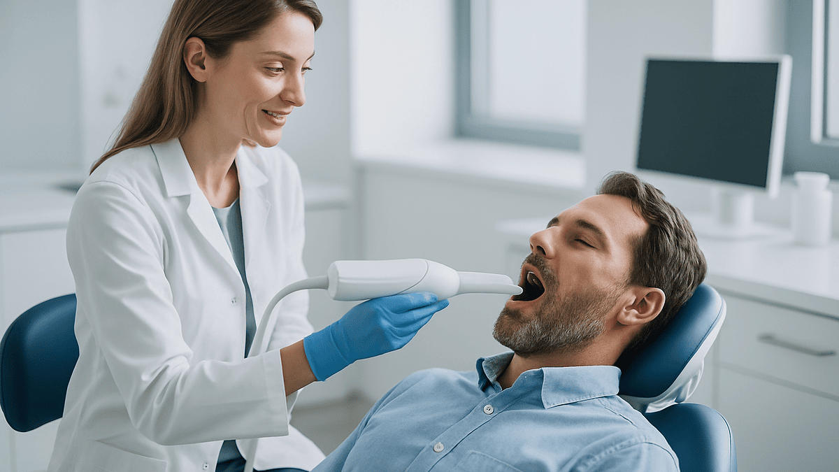
An intraoral scanner is a handheld wand, about the size of a large electric toothbrush, with a small camera and a light source at its tip. The dentist or assistant moves it slowly over the teeth while software stitches thousands of tiny images into one three-dimensional model that appears on a screen in real time. You may be asked to open wide, turn your head, or bite together so the scanner can record how the upper and lower teeth meet.
Nothing sets, nothing hardens, and there is no tray. Saliva and blood are the main enemies, because the camera cannot read a surface it cannot see, so the tooth is dried with air and sometimes a cotton roll or a retraction cord placed at the gumline is used to lift the gum away from the edge of the preparation. A tooth prepared for a crown has a fine line, the margin, where the crown will finish; capturing that line sharply is the whole point.
Once the model is complete, the software flags any gaps or blurred zones and the operator re-scans those spots. The finished file can be sent electronically to a dental laboratory, or in some practices used to mill a restoration in the office. Some clinicians still make a physical printed model from the scan for checking fit.
Two terms come up repeatedly in the research on this topic. Trueness is how close the digital model is to the real tooth. Precision is how consistently repeated scans agree with one another. A scanner can be precise but not true, which matters when a crown must seat within a hair’s breadth, and it is why studies test both.
How traditional impressions work, step by step
A conventional impression records the shape of your teeth as a negative, the way a footprint records a shoe. A U-shaped tray is filled with impression material, most commonly a silicone-type elastomer or, for less demanding work, a seaweed-derived material called alginate. The loaded tray is seated over the teeth and held still while the material sets, usually a few minutes, then eased off in one motion.
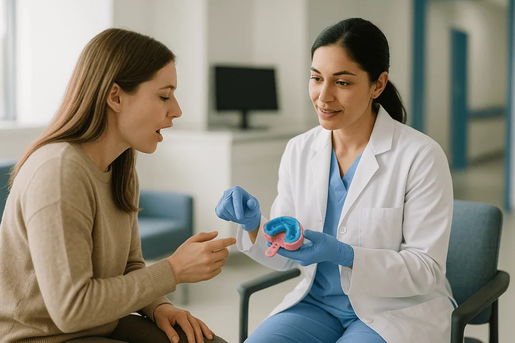
The impression is disinfected, checked under light for pulls, bubbles or voids around the margin, and sent to the laboratory. There a technician pours dental stone into it to create a positive model, mounts upper and lower models together using a bite record, and fabricates the crown, bridge or denture on that model. Many laboratories now scan the stone model into a computer, so a “traditional” impression often becomes a digital workflow partway through.
Where the method shines is in its physics. A well-handled elastomer flows into narrow spaces, slips under the gum edge, and copies soft tissue under gentle pressure. For a full denture, that compressibility is the goal: the impression captures how the gums behave under load, not only where they sit at rest.
Where it falters is human. Materials can be mixed inconsistently, trays can move during setting, and the impression can distort if it is stored too warm or poured too late. Each of those errors is invisible to the patient and often to the dentist until the crown does not fit. Understanding this explains why the digital alternative arrived, and why it did not sweep the older method aside.
Intraoral scanner vs impressions: what the accuracy evidence shows
The most useful question is not which method is more accurate in a laboratory, but whether the finished restoration fits well enough in a mouth. The marker researchers use is the marginal gap, the tiny space between the edge of a crown and the tooth. Larger gaps can trap plaque and invite decay under the crown.
A systematic review and meta-analysis of fixed prosthodontic studies indexed on PubMed found that crowns made from digital impressions had marginal fit comparable to, and in some pooled analyses tighter than, crowns from conventional impressions, with both falling inside ranges clinicians consider acceptable; the authors noted that many included studies were small and of mixed quality (see references). For a single crown or a short bridge, in other words, the evidence does not support choosing one method purely on the grounds of fit.
| Aspect | Intraoral scan | Traditional impression |
|---|---|---|
| Single crown, short bridge | Clinically acceptable fit in reviews | Clinically acceptable fit in reviews |
| Full arch, many teeth | Accuracy tends to fall as span grows | Generally more consistent across the arch |
| Deep or bleeding margins | Camera cannot see what it cannot see | Material can flow under the gum |
| Edentulous ridges, dentures | Few landmarks; limited evidence | Established standard |
| Comfort and gagging | No tray or setting material | Tray and material may trigger gagging |
| Storage and re-use | File retained; can be re-sent | Physical model may distort or be discarded |
Notice that the table is not a scorecard. It is a map of where each technique is on home ground, and the treating team reads that map against your particular tooth.
Are intraoral scanners accurate enough for digital impressions for dental implants?
Dental implants raise the stakes. A crown sits on a tooth that has a little natural give in its ligament; an implant is fused to bone and does not move. If a bridge spanning several implants is even slightly out of position, the metal or ceramic can be under permanent strain, so implant records demand a level of accuracy that a single tooth does not.
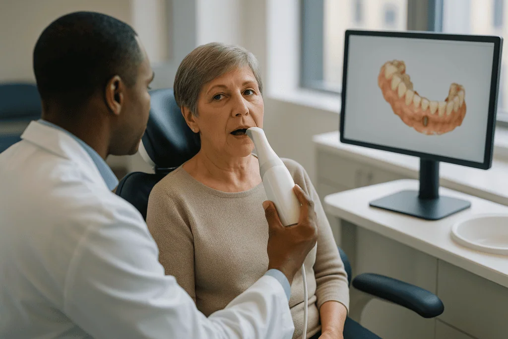
To scan an implant, the dentist screws a small marker called a scan body onto the implant. The scanner reads the marker’s shape and orientation, and the software works out exactly where the implant sits beneath the gum. For one or two implants close together, published studies and the meta-analysis referenced below generally find digital records perform comparably to conventional ones.
Across a full arch of implants the picture is more mixed. Bench studies repeatedly show that as the scanner travels farther from its starting point, small stitching errors accumulate, a bit like drawing a long straight line freehand across a page. Many laboratories and surgeons therefore verify a full-arch digital record with a physical check, or still take a conventional implant impression using a rigid tray and splinted components for the definitive restoration. That is not old-fashioned caution; it reflects what the evidence currently supports.
For the patient, the practical difference is modest. Scan bodies are small, the scan itself is quick, and a conventional implant impression adds a few minutes with a tray. Both approaches are compatible with the healing timeline Mayo Clinic describes for implants, in which the bone grows around the post over several months before the final restoration is made.
Dental impression gag reflex: does scanning really help comfort?
For a meaningful minority of people, the worst part of dentistry is not the drill but the tray. The gag reflex is a protective response triggered when something touches the back of the tongue, soft palate or throat; impression material, which flows toward exactly those areas and then must be held still while it sets, is an efficient trigger. Some patients avoid needed crowns for years because of it.
Here scanning has a genuine, patient-centered advantage. The wand touches only the front of the mouth or hovers just above the teeth, there is no bulky tray behind the molars, and the operator can pause at any moment without spoiling anything. Systematic reviews of patient-reported outcomes summarized on PubMed consistently find that people rate digital impressions as more comfortable and less stressful than conventional ones, and that chairside time is often shorter, though the size of that difference varies between studies and operators.
Scanning is not effortless for everyone. Keeping the mouth open for several minutes can tire the jaw, and people with a small mouth or limited opening may find the wand tip intrusive at the back. Cheek retractors are sometimes used, which is uncomfortable for some. The scanner’s light is bright but not harmful.
If gagging is a known problem for you, say so before the appointment. Dentists use positioning, breathing techniques, distraction and, for conventional impressions, faster-setting materials and smaller trays. A strong gag reflex is one of the more common reasons a clinician reaches for the scanner when either method would otherwise do, and it is a perfectly good reason to ask about it.
Do dentists still use impressions?
Yes, widely, and not only in practices that have yet to buy a scanner. The two techniques coexist because they excel at different jobs, and many dentists who scan every crown still keep impression material on the shelf.
Complete dentures are the clearest example. A denture rests on gums, not teeth, and its stability depends on recording how those tissues compress and how the muscles of the cheeks and tongue shape the border. Impression material under gentle pressure captures that behavior; a camera records only the surface at rest. The NHS lists dentures among routine dental treatments and the conventional workflow remains the reference standard for them, even though digital denture design is developing.
Deep margins are another. When a crown edge finishes well below the gum, especially with bleeding or a bulky gum, the scanner cannot see the line no matter how skilled the operator. A silicone impression displaces the gum and flows to the margin. Some dentists scan, inspect, and switch to a tray if the software shows a blurred edge.
Retention matters too. A stored digital file can be re-sent if a crown is lost or a lab error occurs, which is convenient. A physical impression or model, by contrast, is bulky, can distort, and is often discarded, but it can be poured and checked with the naked eye, something many technicians still value.
So the honest reading of intraoral scanner vs impressions is not a replacement story. It is a story of the toolbox getting larger. Hybrid workflows, where an impression is taken and then scanned in the laboratory, are extremely common and give the patient the tactile advantages of the tray with the design advantages of software.
What are the downsides of using an intraoral scanner?
Marketing tends to present scanning as pure upside, so it is worth listing where the method genuinely struggles, because these are the situations in which your dentist may decline to use one.
- Line of sight. The camera reads only what it can see. Blood, saliva, a rolled gum or a margin tucked far below the gumline produce holes or fuzz in the model.
- Span length. Small stitching errors add up over a long scan path, so accuracy is less reliable across a whole arch than across three or four teeth.
- Featureless surfaces. Smooth, shiny ceramic, metal or wide edentulous ridges give the software few landmarks to match, which can cause the model to drift.
- Operator skill. Scan strategy, the order and angle in which the wand moves, changes results. A scanner in inexperienced hands can produce a worse record than a well-made impression.
- Motion and moisture. Tongue movement, a fogged lens or a patient who cannot hold still lengthens the scan and increases the chance of errors.
- Verification. Because nothing physical exists, errors may not be discovered until the crown arrives, unless a model is printed for checking.
None of these is a reason to fear scanning. They are the reasons the systematic reviews referenced here draw a line between short-span work, where digital and conventional records perform comparably, and long-span or soft-tissue work, where conventional methods remain the benchmark. A good clinician knows the limits of the tool in hand, and a good patient explanation includes them.
Intraoral scanner vs impressions: who is scanned, and who is usually asked to wait
There is no waiting list for a scan in the sense of a queue, but there are situations in which a dentist postpones or bypasses the digital record, and it helps to know why.
Scanning is commonly used for single crowns, inlays and onlays, short bridges, orthodontic records for clear aligners, whitening trays, night guards, implant crowns on one or two fixtures, and monitoring tooth wear over time. In these cases the evidence supports equivalent fit, and the comfort and speed advantages are real.
The people more often asked to have a conventional impression, or to wait before scanning, include those whose gum has not yet healed after surgery, so the margin is bleeding or swollen; those needing a complete denture or a large implant-supported bridge across the arch; those whose crown preparation finishes deep below the gum; and children or adults who cannot keep the mouth open long enough for a full scan. Someone with a fresh extraction socket adjacent to the tooth may also be asked to return once the tissue settles, since a swollen ridge today will not match a healed ridge in three weeks.
A few individuals are steered the other way. Anyone with a severe gag reflex, a strong aversion to the taste or feel of impression material, or a history of impression-related distress is a natural candidate for scanning when the case allows.
The decision belongs to the treating team, who weigh the tooth, the tissue, the laboratory’s requirements and your comfort together. If you have a preference, voice it; if the dentist explains why the other method suits your case, that explanation is usually rooted in one of the limits above.
What the following days and weeks usually look like
Whichever record is taken, the days afterward follow a similar rhythm, and it is the laboratory rather than the scanner that sets the pace.
Same day, the tooth is usually covered with a temporary crown, a plastic or resin placeholder that protects the prepared tooth and keeps the neighbors from drifting. Cleveland Clinic describes wearing a temporary for roughly two to three weeks while the permanent crown is made, though laboratory turnaround varies and some practices with in-office milling fit a permanent crown the same day for suitable cases.
During that interval the tooth may be sensitive to cold or pressure, the gum may be tender where a cord was placed, and the temporary can feel rough or slightly high. Sticky or very hard foods are typically avoided on that side. If a temporary comes loose, keep it and contact the practice rather than attempting to glue it yourself, since incorrect seating can change how the final crown fits.
At the fitting appointment the dentist tries in the restoration, checks the margin with an explorer and radiographs where needed, adjusts the bite, and cements or screws it into place. Digital records make small adjustments quicker because the laboratory can compare the delivered crown to the original file, but they do not remove the need for this check.
Dental implants run on a longer clock. Mayo Clinic notes that the jawbone grows into the implant surface over several months before a permanent tooth is attached, so the scan or impression for the final crown is often taken weeks or months after surgery, once the gum has matured around a healing cap. The waiting is biology, not technology.
Can dental impressions pull crowns out? In-chair worries answered
It is a reasonable fear: a rubbery material grips your teeth and is then tugged free. Can it take a crown with it?
Occasionally, yes, and dentists know it. Impression material can lift a crown, bridge or filling that was already loose, poorly cemented, or held in mainly by friction, particularly an older restoration on a short tooth. A temporary crown is deliberately held with weak cement and is the most likely casualty; that is why the temporary is normally placed after the impression, not before. If a permanent crown comes away in an impression, it is generally a sign that its cement had already failed, and the event reveals a problem rather than causing one. The dentist can usually clean and re-cement it, or advise replacement if decay is found underneath.
Scanning removes this risk entirely, since nothing grips the teeth. It also avoids two rarer conventional-impression events: material breaking off and lodging between teeth or under the gum, and, very uncommonly, a fragment being inhaled or swallowed. Skilled technique, a well-fitting tray and blocking large undercuts keep these rare.
Other common worries deserve short answers. The scanner’s light is a structured visible light or laser at low intensity, not ionizing radiation, so there is no X-ray exposure from the scan itself. Impression materials are generally well tolerated; true allergic reactions are rare, but mention any known latex or chemical sensitivities beforehand, since some tray adhesives and materials contain substances to which a small number of people react. Neither method is painful, though a retraction cord at the gumline can sting briefly and is sometimes placed with local anesthetic already on board.
Which intraoral scanner is considered the most accurate?
This is one of the most-searched questions on the topic, and the honest answer is that independent evidence does not crown a winner, and a patient explainer should not try to.
Laboratory comparisons published in dental journals do test scanners side by side, and their rankings shift from study to study depending on what is scanned, how the model is measured, which software version was used and who held the wand. A device that leads on a single-tooth trial may trail on a full-arch one. Reviews summarizing these comparisons tend to conclude that most current scanners achieve clinically acceptable trueness and precision for short-span work, and that differences between devices are frequently smaller than differences between operators.
That last point deserves emphasis. Scan strategy, meaning the order and angle in which the surfaces are captured, drying technique, retraction of the gum, and knowing when to stop and re-scan a blurred zone all influence the final model more than the badge on the handle. A meticulous clinician with a modest scanner will typically produce a better record than a hurried one with the latest model.
For you as a patient, the useful questions are therefore not about manufacturers. They are: is the laboratory receiving a file it can work with, has the operator confirmed the margin is captured, and is the method suited to the type of restoration planned. The answers to those questions determine fit.
This article deliberately names no devices or companies. Product performance is a moving target, marketing claims are not the same as peer-reviewed evidence, and the choice of equipment sits with the practice, not with a magazine.
What people often get wrong about digital impressions vs traditional impressions
Myths gather around any new technology, and a few about scanning are worth correcting because they can shape decisions.
“Digital is always more accurate.” Not as a blanket statement. For single crowns and short bridges, reviews find the two methods comparably accurate. For full arches and soft-tissue work, conventional impressions remain the benchmark. Accuracy depends on the case.
“If my dentist uses putty, the practice is behind the times.” Many highly experienced clinicians choose impressions for specific cases precisely because they understand where scanners fall short. Using a tray for a denture or a deep margin is a judgment call, not a lag.
“Scanning means my crown will be ready the same day.” Some practices mill certain crowns in the office, but many scan and send the file to a laboratory, with a turnaround similar to a conventional impression. Cleveland Clinic’s two-to-three-week temporary period still applies to most cases.
“The scanner replaces X-rays.” A surface scan shows the crowns of teeth and the gums. It cannot show decay under an old filling, the root, the bone or the nerve. Radiographs remain a separate tool for a separate purpose.
“A digital file means no fitting adjustments.” Every restoration is checked in the mouth and adjusted as needed. Digital records reduce some lab errors but do not eliminate the try-in.
“Impressions are dangerous.” They are routine, well tolerated and used safely in enormous numbers. The rare events described earlier are managed by technique, and scanning is chosen for comfort more often than for safety.
Questions to ask your care team
A short conversation before the record is taken can make the appointment easier and the result better. Bring whatever applies.
- Which method are you planning for my case, and what about my tooth or gum led you to that choice?
- If I have a strong gag reflex or anxiety about the tray, are there alternatives or comfort measures you can offer?
- Will the record be sent to a laboratory or used to make the restoration here, and roughly how long should I expect to wear a temporary?
- If my crown margin is below the gum, how will you make sure it is captured?
- For an implant bridge, will you verify the digital record with a physical model or a conventional impression?
- Do you keep my scan file, and could it be reused if the restoration is lost or damaged?
- Are there any materials in the impression or tray adhesive I should know about, given my allergies?
- What should I do if the temporary comes loose or the tooth becomes very sensitive before the fitting?
You are not expected to understand trueness and precision or to weigh full-arch stitching error. You are entitled to hear, in plain words, why one method suits your mouth and what the plan is if the first record is not good enough. Dentists generally welcome these questions, because a patient who knows what a retraction cord is for, or why the mouth must stay dry for a moment, is a patient who can help the record come out right the first time.
When to call your doctor
Neither a scan nor an impression is likely to cause harm, and most discomfort afterward is mild and short-lived. A few signs, though, mean the practice or an emergency service should hear from you promptly rather than at the next scheduled visit.
Contact your dentist the same day if a temporary crown comes off, cracks or feels high enough to stop your teeth meeting normally; if a piece of impression material seems stuck between teeth or under the gum and you cannot remove it gently with floss; if the prepared tooth becomes severely sensitive to heat or throbs on its own, which can signal an inflamed nerve; or if bleeding from the gumline where a cord was placed does not settle with gentle pressure.
Seek urgent care, calling emergency services if needed, for swelling that spreads to the face, jaw or neck, fever with facial swelling, difficulty swallowing or breathing, or a sudden rash, wheeze or throat tightness after an impression, which could indicate a rare allergic reaction. The NHS notes that a spreading dental infection can become serious quickly and should not wait for a routine appointment. If you believe a fragment of material was inhaled and you are coughing or short of breath, that also needs immediate assessment.
For dental implants, Mayo Clinic advises reporting pain that worsens rather than eases in the days after surgery, persistent numbness of the lip or chin, or a healing cap that loosens. Anything that feels wrong in the mouth is worth a phone call; the treating team would rather hear about a small worry early than a large one late.
Frequently asked questions
Do dentists still use impressions?
Yes. Conventional impressions remain routine for complete dentures, deep subgingival crown margins, many full-arch implant bridges and cases where bleeding or swelling obscures the tooth. Many practices use both methods and choose per case, and some take a physical impression that is later scanned in the laboratory, combining the strengths of each.
Are intraoral scanners accurate?
For single crowns and short bridges, systematic reviews find scanner-based restorations achieve marginal fit comparable to conventional impressions, within clinically accepted ranges. Accuracy becomes less consistent across a full arch or over featureless surfaces, and it depends heavily on the operator’s technique, drying of the field and retraction of the gum.
What are the downsides of using an intraoral scanner?
The main limitations are line of sight, span length and operator skill. Blood, saliva or a margin hidden under the gum produce gaps in the model; errors accumulate over long scans; smooth ceramic or wide toothless ridges give the software few landmarks. Because no physical model exists, some errors surface only when the restoration arrives.
Can dental impressions pull crowns out?
Occasionally, if a crown or filling was already loose or poorly cemented, impression material can lift it out. Temporaries are especially vulnerable, which is why they are placed after the impression. A permanent crown removed this way usually had failed cement already, and the dentist can typically clean and re-cement it or assess for decay beneath.
Which intraoral scanner is considered the most accurate?
No independent evidence consistently ranks one device above the rest. Laboratory comparisons shift with the case type, software version and operator, and reviews find most current scanners acceptable for short-span work. Differences between operators are often larger than differences between devices, so technique and case selection matter more than the badge on the wand.
Does an intraoral scan help with a dental impression gag reflex?
Usually, yes. The wand touches only the teeth and can be paused at any time, and there is no tray or setting material against the palate or throat. Patient-reported studies consistently rate scanning as more comfortable than conventional impressions. Tell your dentist beforehand so the method and comfort measures can be planned.
Are digital impressions used for dental implants?
Yes, using a small marker called a scan body screwed onto the implant so software can locate it. For one or two implants the evidence supports comparable accuracy to conventional records. For long implant bridges across the arch, many clinicians verify the digital record with a physical model or take a conventional splinted impression.
How long does a digital impression take compared with a traditional one?
Studies summarized on PubMed generally report shorter chairside time for scanning, though the difference varies with the operator and the case. A conventional impression must be mixed, seated and held until it sets over a few minutes, then inspected. Either way, the laboratory turnaround, not the record, determines when the crown is ready.
Does the scanner expose me to radiation?
No. Intraoral scanners use low-intensity visible or laser light to capture surface shape, not ionizing radiation. They show the outer surfaces of teeth and gums only and cannot reveal decay under fillings, roots or bone, so radiographs remain a separate and necessary tool when your dentist needs that information.
Will I need a temporary crown whether I am scanned or have an impression?
In most cases, yes. Unless the practice mills a suitable crown in the office the same day, the prepared tooth is protected with a temporary while the laboratory works. Cleveland Clinic describes a typical wait of about two to three weeks. Sensitivity and mild gum tenderness are common during this period.
References
This article is for general information only and is not a substitute for professional medical advice. Please consult a qualified doctor about your individual situation.

More from the Blog

All On 4 for International Patients: How Long to Stay, the Healing Gap and the Second Trip
All on 4 international patients usually need two separate trips. The first covers surgery and a fixed temporary bridge, with most surgeons asking patients…

Who Is a Candidate for Zygomatic Implants? Severe Upper Jaw Bone Loss and the Alternatives
A candidate for zygomatic implants is usually an adult who has lost most or all upper teeth and whose upper jaw has too little…
How to Clean All On 6 Teeth: Water Flossers, Superfloss and Professional Maintenance Visits
To clean All-on-6 teeth, brush the bridge and the gum line twice a day with a soft brush and fluoride toothpaste, then clean beneath…
The Dental Implant Process: Every Stage From First Scan to Final Crown
The dental implant process typically unfolds over three to nine months. It begins with a 3D scan and treatment planning, followed by any needed…
Why an ENT Check May Come Before a Sinus Lift When You Have Chronic Sinusitis or Allergies
An ENT (ear, nose and throat) check often comes before a sinus lift because the graft is placed directly beneath the maxillary sinus lining,…

Sinus Lift Healing Timeline: From Graft Placement to the Day Your Implants Can Be Planned
A sinus lift usually needs several months to heal before implants can be planned. Cleveland Clinic and Mayo Clinic describe bone graft maturation as…