

Mole Removal: How Dermatologists Do It and What to Expect
Key Takeaways
- Most adults carry between 10 and 40 moles, and a typical mole has a natural life cycle of roughly 50 years, often fading on its own in later life.
- Safe mole removal takes about 30 minutes in a clinic under local anesthetic: the numbing injection stings for seconds, and the procedure itself is painless.
- Mole-removal creams work by chemically burning skin, frequently scar worse than surgery, and destroy the tissue a lab would need to rule out melanoma.
- Dermatologists avoid lasers and freezing for true pigmented moles because those methods leave no specimen to test and can cause confusing partial regrowth.
- A shave-removed mole can partially regrow as flat pigment since deeper melanocytes survive; any pigment returning in a scar should be shown to a clinician.
- The ABCDE changes, asymmetry, irregular border, multiple colors, diameter over 6 millimeters, and evolution, are the specific signs that turn a mole from cosmetic question into medical appointment.
The only safe way to get rid of a skin mole is professional removal. A dermatologist typically uses shave excision or surgical excision under local anesthetic; the visit usually takes about 30 minutes and the wound heals over two to three weeks. Home remedies, mole-removal creams, and cutting or freezing moles yourself are unsafe and can hide an early skin cancer.
It usually starts in front of a mirror. A mole on the jaw that catches the razor every third morning. One on the shoulder that a bra strap has been arguing with for years. Or the one on your back that a partner glanced at last week and said, quietly, “Has that always looked like that?”
Those three little scenarios, annoyance, friction, and doubt, cover almost every reason people end up in a dermatology chair asking for a mole to come off. And the good news is that removal is one of the most routine, most predictable procedures in medicine: a numbing injection, a few minutes of work, a small dressing, done.
What deserves more attention is everything around those few minutes, why the internet’s shortcuts are genuinely risky, what the pathology lab does with your mole afterward, and which changes in a mole should move you from “someday” to “this month.”
First, which kind of mole did you mean?
A confession from the editing desk: type “how to get rid of moles” into a search engine and most of what comes back involves lawns, tunnels, and exasperated gardeners. The English language gave the same name to a burrowing mammal and a spot of pigment on your skin, and search engines have never quite recovered.
This article is about the skin kind: the small brown, tan, or pink spots that dermatologists call nevi. If you’re battling the velvet-coated kind that raises ridges across your backyard, you’ll want a garden or wildlife resource instead; stomping tunnels and folk deterrents are questions for an extension service, not a hospital magazine.
The mix-up is worth pausing on for one serious reason. Some of the aggressive advice written for yard moles, burn them out, poison them, dig them up, has a disturbing echo in what people attempt on their own skin. Mole-removal pastes, kitchen acids, freezing kits, even razor blades all circulate online as “fast fixes.” None of them belong anywhere near a pigmented spot on your body, for reasons we’ll unpack below.
So: skin moles from here on. What they are, why they show up, how a dermatologist actually takes one off, what healing looks like week by week, and, most important of all, which moles should never be removed casually because they first need to be examined properly.
What is a mole, and why do you have so many?
A mole is simply a cluster of melanocytes, the cells that produce your skin’s pigment, growing together instead of spreading out evenly. When those cells bunch up, you see a small brown, tan, black, or sometimes pink spot, flat or raised, smooth or slightly bumpy.
They are remarkably common. Most people have somewhere between 10 and 40 moles by adulthood, according to the Mayo Clinic, and the great majority appear before age 40. About 1 in 100 babies is born with a mole already in place, per the Cleveland Clinic; these congenital moles are usually harmless but tend to be monitored more closely over a lifetime.
Here’s the fact that surprises almost everyone: moles are not permanent fixtures. A typical mole has a life cycle of roughly 50 years, the Cleveland Clinic notes. It emerges, often darkens or rises slightly through your twenties and thirties, then slowly flattens and fades, sometimes disappearing entirely in older age. Hormonal shifts during adolescence and pregnancy can make moles darker or more prominent for a while, which is normal and expected.
Sun exposure matters too. Ultraviolet light both encourages new moles and raises the long-term risk that pigment cells behave badly, which is why people with heavy childhood sun exposure often carry more moles, and why sun protection is part of any honest conversation about them.
The practical upshot: a mole is ordinary tissue, not a foreign object, and “getting rid of it” is a minor surgical question, not a cleansing, drawing-out, or dissolving one, whatever the internet suggests.
Can you get rid of moles at home? Please don't
No home method removes a mole safely, and several can do real harm. It’s worth being specific about what’s actually being sold and shared online, because the marketing is persuasive and the risks are not printed on the label.
- Mole-removal creams and pastes work, when they “work” at all, by chemically burning through skin. The result is often a sunken, pale scar considerably uglier than the mole, and sometimes a mole that’s only partially destroyed, leaving pigment cells behind under scar tissue where they’re harder to monitor.
- Home cutting or shaving risks bleeding that’s hard to control, infection, and a jagged wound. A dermatologist controls depth, bleeding, and sterility; a bathroom does not.
- Freezing kits and cautery pens sold for warts and skin tags aren’t designed for pigmented lesions and destroy tissue indiscriminately.
- Kitchen acidsvinegar, garlic, citrus, cause chemical irritation and burns with no meaningful effect on the mole’s cells.
The scarring alone would be reason enough to refuse. But the deeper problem is diagnostic, and it’s the single most important idea in this article: a small percentage of things that look like moles are early melanomas, the most serious form of skin cancer. When a dermatologist removes a suspicious mole, the tissue goes to a laboratory and gets a definitive answer. When you burn or cut one off at home, you destroy the evidence, and if it was a melanoma, the remaining cells continue growing under a scar, invisible, until the cancer is far more advanced. Mayo Clinic and NHS guidance is unambiguous on this point: mole removal is a clinician’s job.
What happens before removal: how a dermatologist reads a mole
Removal never comes first. Every reputable clinician starts by deciding what the mole most likely is, because that answer determines which technique to use, or whether removal should be a formal biopsy instead of a cosmetic tidy-up.
The screening framework most dermatologists use is the ABCDE checklist, described by the Mayo Clinic: Asymmetry (one half unlike the other), Border irregularity (scalloped, blurred edges), Color variation (multiple shades within one mole), Diameter (larger than about 6 millimeters, roughly a pencil eraser), and Evolution (any change in size, shape, color, or symptoms like itching or bleeding). None of these features means a mole is cancerous; each one raises the bar for a closer look.
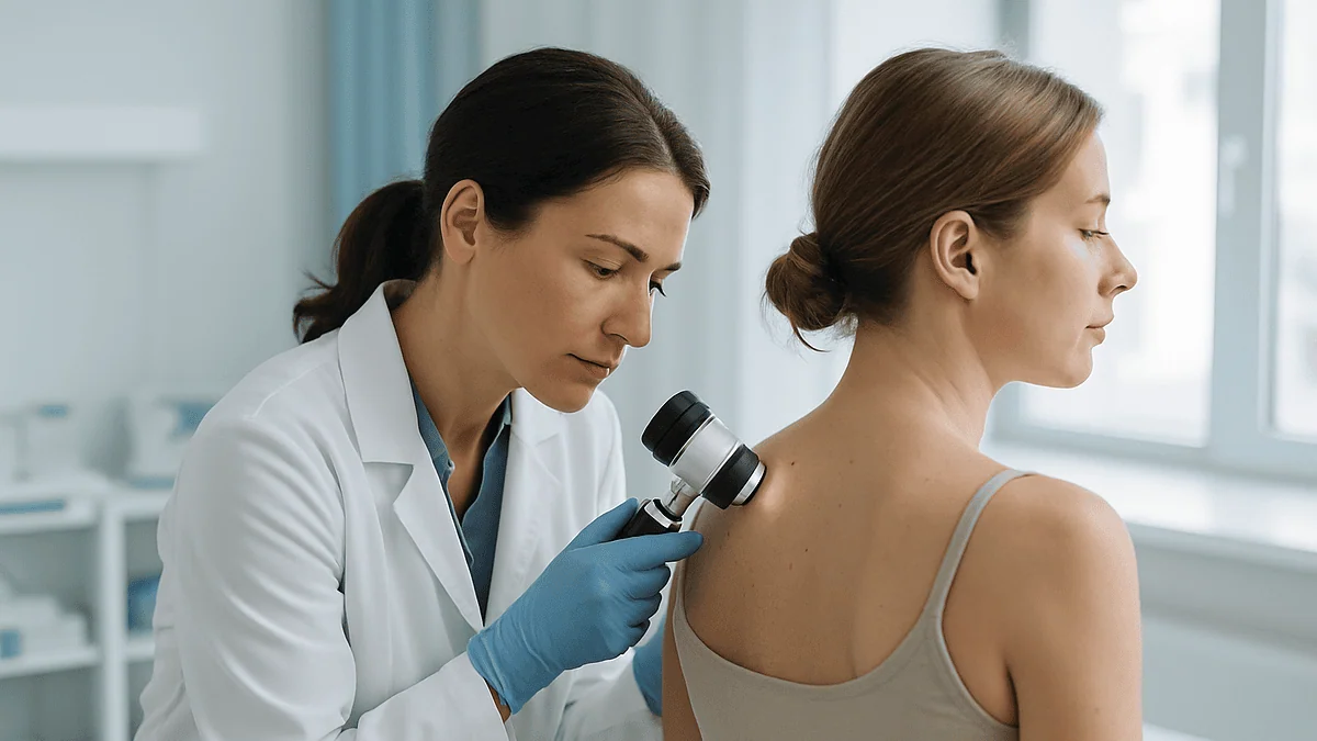
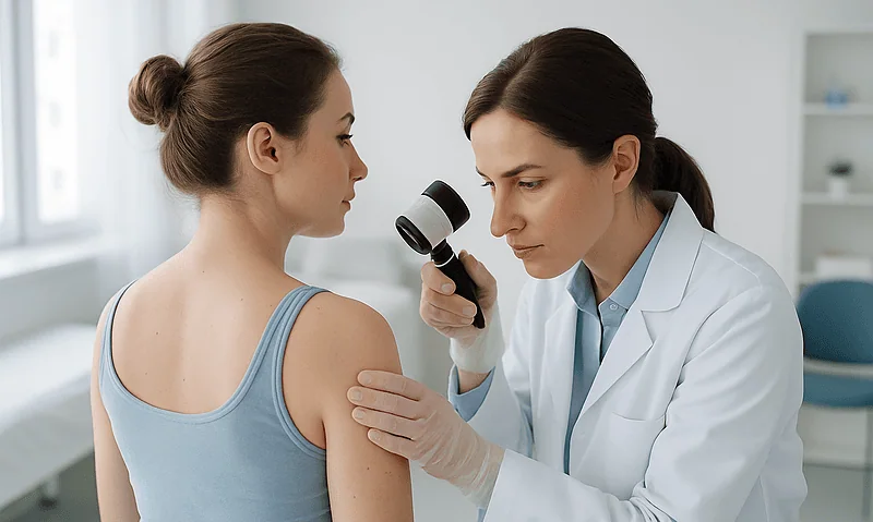
That closer look usually involves a dermatoscope: a handheld magnifier with polarized light that reveals pigment patterns beneath the surface invisible to the naked eye. Trained eyes can distinguish the orderly network of a benign mole from the chaotic structures that warrant a biopsy, which meaningfully reduces both missed cancers and unnecessary removals.
Then comes the sorting decision. A clearly benign mole being removed because it snags or bothers you cosmetically can be taken off with the simplest technique. A mole with any suspicious feature gets removed in a way that preserves the whole lesion for the pathologist, usually a deeper excision with a margin of normal skin. The examination shapes the surgery, which is precisely what no cream, kit, or kitchen remedy can replicate.
Shave excision: the quick option for raised moles
For a raised, benign-appearing mole, shave excision is the workhorse. After cleaning the area, the clinician injects a small amount of local anesthetic just under the mole: a brief sting, then numbness within a minute or two. A small, flexible blade then shaves the mole flush with, or slightly below, the surrounding skin surface. The whole thing typically takes a few minutes of actual cutting; the NHS notes that a mole-removal visit overall usually runs about 30 minutes, most of which is preparation and paperwork.
There are no stitches. Bleeding is controlled with light pressure and, often, gentle cauterization or a chemical styptic applied to the base. What’s left behind looks and heals much like a shallow graze: a small pink circle that crusts, then re-epithelializes over one to three weeks, gradually blending toward your normal skin tone over the following months.
Two honest caveats. First, because a shave removes the visible portion of the mole but not necessarily every pigment cell in the deeper skin, some moles partially regrow, usually as a flat, lighter patch of pigment. That regrowth is typically harmless but should always be shown to a clinician, since regrowth can occasionally mimic worrying change. Second, shave excision is reserved for moles the dermatologist is confident are benign; anything ambiguous deserves the deeper technique described next.
The shaved tissue, by the way, doesn’t go in the trash. Responsible practice sends it to pathology, which is your quiet insurance policy that “obviously benign” really was.
Surgical excision: removing the mole and everything under it
When a mole is flat, deep, large, or carries any whisper of suspicion, dermatologists reach for full surgical excision. The principle is simple: remove the entire mole through the full thickness of the skin, along with a small rim, a margin, of normal-looking skin around it, so the pathologist can confirm nothing was left behind.
The experience from the patient’s side: local anesthetic numbs the area, the surgeon cuts an ellipse (a lens shape) around the mole, lifts the tissue out, and closes the wound with stitches. An ellipse rather than a circle, because straight-line closures lie flatter and heal into finer scars. Depending on the site, stitches on the face may come out in about a week, while stitches on the back or legs, where skin is under more tension, often stay closer to two weeks, in line with NHS and Cleveland Clinic guidance on minor skin surgery.
Excision trades a slightly longer scar for near-certainty. Because the entire lesion comes out with margins, regrowth is rare, and the pathology report examines the mole from top to bottom. If the report shows atypical cells or, uncommonly, an early melanoma, the fact that everything was removed with a margin becomes enormously valuable, sometimes it’s the entire treatment.
Aftercare is modest: keep the wound dry for the first day or two, avoid heavy lifting or stretching across the site while stitches are in, and expect a fine line that starts pink and fades over the better part of a year. Your clinician will tailor the specifics to the location.
What about lasers, freezing, or burning a mole off?
Reasonable question, lasers sound tidy, and freezing works fine for warts. But for true melanocytic moles, most dermatologists decline both, and the reasoning is worth understanding rather than just accepting.
The core problem is the missing specimen. Lasers vaporize tissue; cryotherapy destroys it in place; electrocautery chars it. All three eliminate the possibility of pathological examination. Since even experienced dermatologists occasionally get a surprise from the lab, a mole that looked innocent but showed atypical cells under the microscope, destroying the tissue means accepting a small but real chance of missing a diagnosis that mattered. NHS and Mayo Clinic guidance consistently favors techniques that preserve tissue for testing when a pigmented lesion is removed.
There’s a second issue specific to pigmented lesions: incomplete destruction. Laser and freezing treatments may remove surface pigment while leaving melanocytes alive in the deeper dermis. Those cells can regrow in scattered, irregular patterns within scar tissue: a phenomenon that can look alarmingly like melanoma on later examination and sometimes forces a biopsy that would never otherwise have been needed.
Where do these tools legitimately shine? On things that merely resemble moles. Seborrheic keratoses (waxy, stuck-on age spots), skin tags, and certain benign vascular spots are routinely and appropriately treated with freezing or cautery once a clinician has confidently identified them. That identification step is the hinge: destructive methods are fine for lesions that don’t need testing, and wrong for lesions that might.
If a clinic leads with laser for a pigmented mole without discussing biopsy, that’s a reasonable moment to ask more questions.
Does mole removal hurt?
Far less than most people fear, and the honest breakdown goes like this.
The only genuinely uncomfortable moment is the anesthetic injection: a needle prick followed by a stinging, pressure-like sensation as the numbing medication spreads under the mole. It lasts perhaps five to ten seconds. Many patients compare it to a bee sting that quits early. Some clinicians ice or vibrate the skin first, or buffer the injection, specifically to soften this step.
After that, the area is numb, fully. You may feel pressure, tugging, or movement, which the brain finds strange but not painful. Patients routinely chat through the procedure. If you feel anything sharp, say so; a small top-up of anesthetic fixes it immediately.
Once the numbness wears off a few hours later, expect the wound to feel like what it is: a small graze (after a shave) or a stitched cut (after an excision). Most people describe soreness or tenderness for a day or two, managed comfortably with the simple measures your clinician recommends. Wounds over joints or on the back, anywhere skin stretches with movement, tend to complain slightly longer.
What about itching? A healing wound often itches around days five through ten. That’s usually a normal part of repair, not a problem, though spreading redness, warmth, or discharge is a different matter (more on infection signs in the aftercare section).
One more reassurance: because everything happens under local anesthetic, you drive yourself home, return to desk work the same day, and skip the fasting, sedation, and recovery-room rituals of bigger procedures entirely.
Which method fits which mole? A side-by-side look
Technique selection is a judgment call your dermatologist makes based on the mole’s depth, location, appearance, and any suspicion of atypia, but seeing the options side by side demystifies the conversation you’ll have in the exam room.
| Method | Best suited for | Stitches? | Tissue tested? | Typical initial healing |
|---|---|---|---|---|
| Shave excision | Raised, benign-appearing moles | No | Yes, specimen sent to lab | About 1–3 weeks, like a graze |
| Surgical excision | Flat, deep, large, or suspicious moles | Yes | Yes, full lesion with margin | Stitches out in ~1–2 weeks; scar matures over months |
| Punch excision | Small moles needing full-depth removal | Usually 1–2 | Yes | About 1–2 weeks |
| Laser / freezing / cautery | Non-mole lookalikes (skin tags, seborrheic keratoses) after confident diagnosis | No | No, tissue destroyed | Days to ~2 weeks |
Notice the pattern running down the “tissue tested” column. The three techniques dermatologists actually use for true moles all preserve the specimen; the destructive methods are reserved for lesions that don’t need a microscope’s opinion. Healing timelines above reflect general NHS and Cleveland Clinic guidance for minor skin procedures: your own wound may run a little faster or slower depending on the site, your age, and habits like smoking, which measurably slows skin repair.
Cost and coverage vary with the reason for removal: lesions removed because of medical concern are handled differently from purely cosmetic requests, so ask the practice to clarify before the procedure rather than after.
Aftercare: what the first two weeks actually look like
Mole-removal aftercare is blessedly boring, and boring is the goal. Here’s the realistic timeline.
Day 1: Keep the dressing on and the area dry. A little oozing or pinpoint bleeding through the bandage is normal; firm pressure for ten minutes settles most of it. Expect mild soreness as the anesthetic fades.
Days 2–7: Most clinicians have you gently wash the site with mild soap and water once daily, pat dry, apply a thin layer of whatever ointment they’ve recommended, and re-cover it. Moist, covered wounds heal faster and scar less than wounds left to dry and crust: a well-established principle in wound care. Skip swimming pools, hot tubs, and heavy workouts that stretch the area, especially if you have stitches.
Week 2: Shave sites are usually filling in nicely, pink and flat. Excision stitches come out around now, roughly one week on the face, closer to two on the trunk and limbs. Itching is common and usually benign.
Weeks 3 onward: The wound is closed; the scar’s slow makeover begins. New scars are pink or reddish and gradually fade over many months.
Two non-negotiables. First, protect the healing site from sun, fresh scars pigment easily, and a broad-spectrum sunscreen (the CDC recommends SPF 15 or higher for general use; many dermatologists suggest more) or simple covering keeps the final scar as invisible as possible. Second, know the infection signs: spreading redness, increasing rather than decreasing pain, warmth, swelling, pus, or fever. Any of those warrants a same-week call to the clinic.
Will there be a scar, and can the mole come back?
Yes to a scar, honestly stated: any procedure that removes skin leaves one. The real questions are how visible it will be and how it compares with the mole you started with, and here the news is genuinely good.
A shave excision typically leaves a flat, pale mark roughly the size of the original mole, often barely noticeable within a year. A surgical excision leaves a fine line somewhat longer than the mole was wide (that lens-shaped closure again), which starts pink, may feel firm for a few months, and matures into a thin white line. Scars keep improving for a full year or more, per Cleveland Clinic guidance on skin healing, so judge the result at twelve months, not at six weeks.
Your body has a vote, too. People prone to thick or keloid scarring, and wounds on high-tension areas like the chest, shoulders, and upper back, tend to scar more visibly. If that’s you, say so beforehand; technique, closure choices, and early scar care can all be adjusted.
As for regrowth: after a full excision with margins, recurrence is rare. After a shave, some pigment can return at the site because melanocytes deeper in the skin survived, usually appearing as a flat, evenly colored patch. That’s typically harmless, but there’s an important rule attached: any pigment returning in a removal scar should be evaluated by a clinician, because regrowth within scar tissue can be difficult to distinguish from something more serious on appearance alone. Bring the original pathology report if you have it; it makes the assessment far easier.
Why every removed mole should visit the lab
Here’s the step patients rarely see, and it may be the most valuable part of the entire process: the specimen jar.
After removal, the mole travels to a pathology laboratory, where it’s preserved, sliced thinner than paper, stained, and examined under a microscope by a pathologist: a physician who specializes in reading tissue. Within days to a couple of weeks, a report comes back classifying exactly what the lesion was: an ordinary benign nevus, a mildly atypical one, or, uncommonly, something requiring further treatment.
Why bother when the dermatologist already said it looked fine? Because visual inspection, even expert visual inspection with a dermatoscope, is an estimate: an extremely good one, but an estimate. The microscope is the answer key. Occasionally a mole removed for purely cosmetic reasons turns out to harbor atypical cells, and that finding changes the plan: perhaps a wider excision of the site, perhaps closer surveillance of your other moles going forward. Discovering that on a pathology slide, while the lesion is thin and fully removed, is the best-case version of an unwelcome surprise. Skin cancers caught early are far more manageable than those found late: the entire logic of the CDC’s skin cancer messaging rests on that asymmetry.
Practical advice: ask two questions at your appointment. “Will this be sent to pathology?” (for any true mole, the answer should be yes) and “How will I get the result?” Then actually collect the result, a quick call or portal message, rather than assuming silence means all clear. Keep a copy. Future clinicians examining that patch of skin will thank you.
When to see a doctor about a mole
Most moles never need anything from you but a passing glance. A specific short list of changes, though, should prompt an appointment, promptly, not eventually.
See a clinician soon if a mole shows any of these red flags: it’s changing in size, shape, or color; it has become asymmetric or developed irregular, blurred borders; it contains multiple colors or an area of black; it’s larger than about 6 millimeters (a pencil eraser) and growing; it itches persistently, bleeds, crusts, or won’t heal; or it simply looks different from all your other moles, dermatologists call this the “ugly duckling” sign. A brand-new pigmented spot appearing after age 40 also deserves professional eyes, since new moles become less common with age while melanoma risk rises. These criteria track the ABCDE framework described by the Mayo Clinic and echoed across NHS and CDC guidance.
Don’t let reassuring details talk you out of it. Melanoma can arise in skin that was never sunburned, in people with dark skin tones (often on palms, soles, or under nails), and in spots that don’t hurt at all. Pain is a famously unreliable alarm for skin cancer.
Two calibrating truths so this lands as information rather than fear. First, the overwhelming majority of moles, including many odd-looking ones, are benign, and most changes people notice turn out to be innocent. Second, when melanoma is found early, while still thin, treatment is typically a straightforward excision. The entire point of watching your moles isn’t to live anxiously; it’s to convert a potentially serious disease into a minor procedure by showing up a few months sooner.
Living with the moles you keep: checks, sun, and common sense
Removal is a fine answer for one troublesome mole. For the other 10 to 40, the strategy is stewardship, and it costs almost nothing.
Start with a monthly self-check: the same week each month makes it a habit. In good light, work systematically: face and scalp (a hair dryer on cool helps part the hair), neck, chest, arms including palms and between fingers, torso front and back with a hand mirror or a partner’s help, legs, and finally the soles of your feet and between your toes. You’re not memorizing every spot; you’re building enough familiarity that change stands out. Photographs help enormously: a quick phone picture of a mole next to a ruler or coin, dated, turns “I think it’s bigger?” into an answerable question.
Then, sun protection, which is less about the moles you have than the skin around them. UV exposure drives most skin cancers, and the protective playbook from the CDC is short: broad-spectrum sunscreen (SPF 15 or higher, reapplied every two hours and after swimming or sweating), shade during peak hours from roughly 10 a.m. to 4 p.m., hats and clothing that cover, and no tanning beds, full stop. People with many moles, fair skin, a history of blistering sunburns, or a family history of melanoma benefit most from this diligence, and are exactly the people who should ask a clinician whether a periodic professional skin exam makes sense for them.
None of this is burdensome. Ten minutes a month and a tube of sunscreen buy you the two things that matter most in skin health: early detection and less damage to detect.
Frequently asked questions
How do you get rid of moles quickly?
The fastest safe option is an in-office shave excision by a dermatologist: after a brief numbing injection, a raised benign mole comes off in minutes, with the whole visit typically lasting about 30 minutes. There is no legitimately faster route, creams, kits, and home cutting are slower to heal, scar worse, and destroy tissue that should be tested. Booking one clinic appointment is genuinely the quick way.
Can I remove a mole at home with cream or apple cider vinegar?
No, neither is safe or effective. Removal creams act by burning through skin, often leaving a sunken scar with mole cells still alive underneath, and kitchen acids cause chemical irritation without reliably removing anything. Worse, both destroy the tissue a pathologist would examine, so if the spot was an early melanoma, that diagnosis is lost while the cancer keeps growing under scar tissue. Professional removal preserves the specimen and your safety.
Why should you never cut off a mole yourself?
Three reasons: bleeding and infection risk, worse scarring, and, most critically, the loss of diagnosis. Dermatologists send removed moles to a laboratory to confirm they’re benign; a small fraction of mole-like spots are early melanomas, and home removal destroys or discards that evidence. If cancerous cells remain in the skin, they continue growing invisibly beneath a scar. What looks like a shortcut can convert a curable early cancer into a late one.
How long do moles last?
Around 50 years, on average, moles have a natural life cycle, according to Cleveland Clinic guidance. A mole typically appears in childhood or early adulthood, may darken or rise slightly through the middle years, then gradually flattens, lightens, and often disappears entirely in older age. Gradual, symmetrical fading over years is normal; rapid change over weeks or months in either direction is what deserves a clinician’s look.
Does mole removal hurt?
Only briefly. The local anesthetic injection stings for about five to ten seconds; after that the area is completely numb, and you’ll feel pressure or tugging at most during the procedure. Once the numbness fades a few hours later, expect mild soreness for a day or two, comparable to a small cut or graze. Wounds on high-movement areas like the back or shoulders may stay tender slightly longer.
Can a mole grow back after removal?
Sometimes, depending on the technique. After a shave excision, pigment cells deeper in the skin can survive and regrow as a flat, lighter patch, usually harmless, but it should always be evaluated because regrowth in scar tissue can mimic worrying change. After a full surgical excision with a margin of normal skin, recurrence is rare. Keep your pathology report; it helps any future clinician interpret the site correctly.
Will mole removal leave a scar?
Yes, every removal leaves some mark, though usually a subtle one. Shave excisions typically heal to a flat, pale spot about the size of the original mole; surgical excisions leave a thin line that fades from pink to faint white over a year or more. Location matters: chest, shoulder, and upper-back skin scars more visibly. Sun protection on the healing site and following your clinician’s wound-care instructions noticeably improve the final result.
Are removed moles always tested for cancer?
They should be. Standard practice is to send any true melanocytic mole to a pathology laboratory, where it’s examined under a microscope to confirm it’s benign, even when it was removed for purely cosmetic reasons. Occasionally an innocent-looking mole shows atypical cells, and finding that early changes the follow-up plan. Ask two questions at your appointment: whether the specimen will go to pathology, and how you’ll receive the result.
Can a normal-looking mole still be melanoma?
Rarely, yes, which is why change over time matters more than appearance at a single moment. Melanoma can begin looking fairly ordinary and reveal itself through evolution: growing, developing new colors, itching, or bleeding. It can also arise on skin that was never sunburned and in people with dark skin tones, often on palms, soles, or under nails. Any mole that’s changing, or that looks unlike all your others, warrants professional evaluation.
Why won't my dermatologist use a laser on my mole?
Because lasers vaporize the tissue, leaving nothing for a pathologist to examine, and preserving the specimen is a core safety principle for pigmented lesions. Lasers can also destroy surface pigment while leaving deeper melanocytes alive, causing patchy regrowth within scar tissue that can later mimic melanoma and force unnecessary biopsies. Lasers and freezing remain perfectly appropriate for confidently diagnosed non-mole lesions like skin tags and seborrheic keratoses.
References
This article is for general information only and is not a substitute for professional medical advice. Please consult a qualified doctor about your individual situation.

More from the Blog

What a Congenital Nevus Check Involves: Dermoscopy, Photo Mapping and Tracking Change
A congenital nevus check is a skin examination of a birthmark mole. A clinician inspects the whole nevus and the surrounding skin, uses a…
How Does Phototherapy Help Atopic Dermatitis? What Sessions Involve and Who May Be Offered Them
Phototherapy for atopic dermatitis exposes affected skin to carefully measured ultraviolet light, usually narrowband UVB, in a clinic cabinet several times a week for…
A Daily Routine That Supports Atopic Dermatitis Treatment: Bathing, Moisturizing and Nail Care
A supportive atopic dermatitis daily routine rests on three anchors: a short, lukewarm bath or shower with a gentle, fragrance-free cleanser; a generous layer…

Is Coconut Oil Good for Your Hair? The One Oil That Penetrates
Coconut oil can genuinely help hair, and the reason is unusual: its main fatty acid, lauric acid, is small enough to penetrate the hair…
Peptides in Skincare: What Signal Peptides Do and What the Studies Show
Peptides in skincare are short chains of amino acids applied topically to signal skin cells, most often to encourage collagen production or soften expression…
How to Get Clear Skin: What Actually Works, According to Dermatology Trials
Dermatology trials show that clear skin comes mainly from treating acne consistently: a gentle cleanser, benzoyl peroxide, an adapalene retinoid, daily sunscreen, and patience…