

Physical Therapy After Selective Dorsal Rhizotomy: Why Daily Home Exercises Matter So Much

Key Takeaways
- SDR cuts a portion of the sensory nerve rootlets that drive spasticity in the legs, so the stiffness reduction is permanent, but the operation adds no strength on its own.
- The CDC estimates cerebral palsy affects about 1 in 345 children, with spastic forms making up roughly 80% of cases, and SDR targets only the lower-limb spasticity in a selected subgroup.
- Children often stand less steadily in the first weeks after surgery because weakness that stiffness was masking becomes visible, which is expected rather than a setback.
- Program descriptions from Johns Hopkins Medicine and the NHS place the most intensive rehabilitation in the first several months, with walking gains typically tracked across the first year.
- Spasticity does not return after SDR, but tightness from growth spurts and under-use can, which is why stretching, orthotic reviews and hip and spine surveillance continue for years.
- Progress is measured against a child's own baseline using tools such as the Gross Motor Function Measure and gait analysis, not against videos of other children online.
Physical therapy after selective dorsal rhizotomy (SDR) is considered an essential part of the treatment rather than an optional follow-up. The surgery reduces spasticity by cutting a portion of overactive sensory nerve rootlets in the lower spine, which often reveals underlying muscle weakness and unfamiliar movement patterns. Months of intensive therapy, supported by daily home exercises, help children build strength, balance and new motor skills on the treating team's plan.
A father sits on the floor of a hospital playroom, counting out loud while his six-year-old lifts one knee toward the ceiling, wobbles, laughs, and tries again. The scar on her lower back is only two weeks old. The hard work, as the family is learning, started the moment the operation ended.
Families who research selective dorsal rhizotomy usually spend months weighing the surgery itself: the risks, the eligibility criteria, the rare permanence of cutting nerve tissue. Far fewer arrive prepared for what comes next. Physical therapy after SDR is not an afterthought tacked onto a neurosurgical procedure. It is the second half of the treatment, and in many ways the longer half.
This explainer walks through what the surgery changes in the body, why therapists describe the months that follow as rebuilding rather than recovering, and why the exercises done at the kitchen table on an ordinary Tuesday may matter as much as any session in a clinic gym.
What is selective dorsal rhizotomy and what actually happens in the operation?
Selective dorsal rhizotomy, usually shortened to SDR, is a spinal operation designed to reduce spasticity in the legs. Spasticity is a form of muscle stiffness in which muscles contract involuntarily because the signals that normally dampen reflexes are impaired. It is the hallmark of spastic cerebral palsy, which the CDC describes as the most common form, affecting about 80% of people with the condition.
The logic of the operation rests on a reflex loop. Sensory nerve fibers carry stretch information from the leg muscles into the spinal cord through the dorsal roots, the bundles that enter the back of the cord. In a child with spasticity, that incoming traffic triggers an exaggerated outgoing command to contract. During SDR, a neurosurgeon opens a small window in the bone of the lower spine, identifies the sensory roots that supply the legs, and divides each one into smaller strands called rootlets. Each rootlet is stimulated electrically while the team watches the muscles respond. Rootlets that produce an abnormal, spreading or sustained response are cut; those with a normal response are left intact.
Only a portion of the sensory rootlets is divided, which is what makes the procedure selective. The motor roots, which carry commands out to the muscles, are not touched. The result, as described by Johns Hopkins Medicine, is a permanent reduction in the abnormal reflex activity driving the stiffness, while the ability to move the legs is preserved.
Two features of this design shape everything that follows. The change is lasting, since nerve rootlets do not regrow. And the operation removes stiffness without adding strength. A leg that has been held rigid for years, often relying on that rigidity to stand, is suddenly asked to work in a new way. That gap is exactly what physical therapy after SDR exists to fill.
Why is physical therapy after SDR treated as part of the surgery, not a follow-up?
Ask a therapist who works with these children and the phrase you will hear is that SDR removes a barrier. Spasticity acts like a brake jammed on while the engine is running. Release it and the car can finally move freely, but the driver still has to learn to steer.
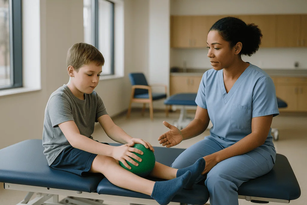
Many children with spastic diplegia, the pattern in which both legs are affected more than the arms, have used stiffness as a scaffold. Locked knees and tight calves can hold a body upright even when the muscles beneath are weak. The NHS describes SDR as a way to improve walking and ease pain, yet guidance from the same source is clear that children need months of physiotherapy afterward to relearn how to use their legs. Once the brake is released, weakness that was always there becomes visible. Children sometimes stand less confidently in the first weeks after surgery than they did before it, which can alarm parents who were not warned.
There is a second reason therapy is inseparable from the operation. The developing brain is remarkably adaptable, a property called neuroplasticity, meaning the nervous system reorganizes itself in response to repeated experience. Movement patterns are learned through repetition, and a child who has walked on their toes for years has a deeply practiced habit. Changing that pattern requires thousands of correct repetitions, not a single surgical event.
This is why centers that perform SDR, according to Johns Hopkins Medicine, typically build in an intensive rehabilitation program lasting many months, and why the decision to operate is often made jointly by neurosurgeons and rehabilitation specialists. A child who cannot access regular therapy afterward, for whatever reason, may be a less suitable candidate, not because the surgery would fail technically but because its purpose would be left incomplete.
Who is SDR usually for, and who is usually asked to wait?
SDR is not a treatment for cerebral palsy itself. Cerebral palsy is a group of lifelong conditions affecting movement and posture, caused by injury to or abnormal development of the brain before, during or shortly after birth, and the CDC estimates it affects about 1 in 345 children in the United States. The surgery addresses one feature of the condition, lower-limb spasticity, in a specific group of children.
According to Johns Hopkins Medicine and the NHS, the children who tend to be considered have a particular profile:
- Spastic diplegia, with spasticity concentrated in the legs and reasonable arm and trunk control.
- Enough underlying strength to walk, with or without a walker or crutches, which is often expressed using the Gross Motor Function Classification System. The GMFCS is a five-level scale describing everyday mobility, and children at levels II and III are the group most commonly described as candidates.
- Good selective motor control, meaning the ability to move one joint without the whole leg moving along with it.
- Spasticity as the dominant problem, rather than dystonia, which is involuntary twisting movement, or fixed contractures, which are permanent shortening of muscles and tendons.
- A family and school environment able to support months of frequent therapy.
Age matters as well. Many programs operate within an early-to-middle-childhood window, before the growing skeleton has developed severe bony deformities and while the nervous system remains highly plastic. Exact age ranges vary between programs and health systems.
Children are often asked to wait, or guided toward other options, when weakness rather than stiffness limits their walking, when dystonia is mixed in, when contractures already need orthopedic correction, or when there is no realistic plan for the rehabilitation phase. Adults with cerebral palsy are sometimes evaluated too, though the evidence base is smaller. Every one of these judgments rests with the treating team, drawing on examination, gait analysis and often video review.
What does physical therapy after SDR involve in the first days?
The first week sets the tone, though it looks nothing like the exercise program families picture. Children typically remain in hospital for several days after the operation, as described by Johns Hopkins Medicine, with the earliest priorities being pain control, protecting the healing incision and the gradual return to sitting and standing.
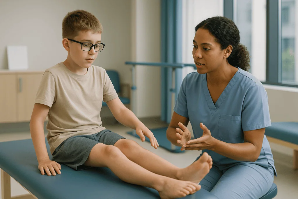
Pain management in children is planned entirely by the medical team, often combining medicines given through a vein or by mouth with positioning and comfort measures. Doses and schedules are individualized and are not something a family should adjust. What parents can expect is that discomfort usually eases over the first days and that the team will set rules about lying flat, how much to sit up, and when it is safe to bend or twist the lower back. Those rules protect the muscles and membranes around the spinal cord as they heal.
Therapy in this window is gentle and deliberate. A physical therapist will typically begin with:
- Positioning the legs to encourage straight hips and knees, since the muscles no longer pull them into flexion.
- Passive range-of-motion movements, in which the therapist moves the joint while the child relaxes, to keep the newly available movement available.
- Breathing, trunk and head control work done lying down.
- Progression to supported sitting, then standing in a frame or with assistance, once the surgical team gives clearance.
Many children feel wobbly and surprisingly weak when they first stand. Parents describe legs that feel like soft noodles compared with the firm resistance they knew. That sensation is expected and is the clearest early sign that the spasticity has reduced. It is also the first lesson in why the months ahead matter: the stiffness is gone, and now the real work of building strength and control can begin.
SDR rehabilitation timeline: what the weeks and months usually look like
No two children follow the same path, and the ranges below are typical descriptions drawn from program outlines published by Johns Hopkins Medicine and the NHS rather than promises. They are a map, not a schedule, and the treating team will adjust it week by week.
| Phase | Typical timing | Main focus | Where therapy happens |
|---|---|---|---|
| Hospital phase | First several days | Comfort, incision care, safe positioning, first supported sitting and standing | Inpatient ward |
| Intensive early phase | Roughly the first 4–6 weeks | Range of motion, early strengthening, trunk control, relearning standing balance | Inpatient rehabilitation or daily outpatient visits, with home exercises |
| Building phase | Roughly weeks 6 through month 6 | Progressive strengthening, gait retraining, stairs, transfers, endurance | Several outpatient sessions a week plus daily home program |
| Consolidation phase | Months 6 through 12 and beyond | Refining walking pattern, higher-level skills, sport and play participation | Reduced clinic frequency, home program continues |
| Long-term follow-up | Years, through growth spurts | Monitoring for contractures, hip and spine changes, adjusting orthotics | Periodic specialist review |
A pattern worth noticing in that table is how the balance shifts. Early on, the therapist does most of the work and the child is largely a participant. By the building phase, the home program carries a growing share of the total practice, and by the consolidation phase it is the main vehicle. Johns Hopkins Medicine notes that intensive therapy is usually recommended for at least several months, with the greatest gains in walking often reported over the first year.
Growth complicates the picture. A child may be walking better than ever at age seven and then hit a growth spurt at eleven in which bones lengthen faster than muscles. Spasticity itself does not return, since the divided rootlets do not regrow, but tightness from growth is a separate issue. Long-term follow-up exists to catch exactly that.
Why daily home exercises after rhizotomy matter so much
Here is the arithmetic that shapes everything. Suppose a child attends physical therapy three times a week for an hour each session. Over a year, that is roughly 150 hours of supervised practice. The same child is awake for perhaps 5,000 hours in that year. The clinic is where the plan is designed and corrected; the rest of the week is where it is actually practiced.
Three mechanisms explain why that practice has to be daily rather than occasional.
The first is strength. Muscles adapt to repeated loading by growing stronger, but the adaptation fades if loading stops. Children after SDR often have years of accumulated weakness in the hip extensors, the muscles at the back of the hip that keep the body upright, and in the muscles that lift the foot. Rebuilding those muscles follows the same rules as any strength training, and consistency over weeks is what produces measurable change, as general guidance from Mayo Clinic on physical therapy for cerebral palsy emphasizes.
The second is motor learning. Walking is a skill, and a skill is encoded in the brain through repetition. A child who practices heel-first steps for twenty minutes in clinic and then toe-walks for the remaining fifteen hours of the day has, in effect, rehearsed the old pattern far more than the new one. Daily home exercises tip that ratio.
The third is range of motion. Newly released joints will drift back toward tightness if they are not moved through their full range regularly, particularly during growth. Stretching and positioning at home maintain what the surgery made possible.
None of this requires a home gym. It requires minutes, repeated, and a family that understands why those minutes are not optional. Therapists often say that the parents are the primary therapists after SDR and the clinic staff are the coaches. The evidence on motor learning supports that framing.
What does a typical home exercise program include?
Home programs are written individually, and any list here is descriptive rather than instructional. The treating therapist decides what a particular child should do, how often, and when to progress. What follows is the shape such programs commonly take, so that families know what to expect and what to ask about.
- Stretching and positioning. Sustained holds for the calf muscles, hamstrings and hip flexors, and time spent in positions that encourage straight hips, such as lying on the stomach while reading or watching a screen.
- Strengthening. Bridges, sit-to-stand repetitions from a chair, heel raises, step-ups onto a low stair, and later squats or resisted work. The emphasis is usually on the hip and knee extensors and the ankle muscles.
- Balance and trunk control. Standing on one foot while brushing teeth, reaching across the body for toys, kneeling and half-kneeling games that challenge stability.
- Gait practice. Deliberate walking with attention to heel contact, step length and arm swing, often in front of a mirror or recorded on a phone so the child can see the pattern.
- Functional tasks. Stairs, getting up from the floor, walking on uneven ground, carrying objects, all of which translate strength into daily life.
The progression matters as much as the content. Early programs may be short and gentle; by mid-rehabilitation they are often longer and more demanding. A good program also changes with the child’s interests. For a six-year-old, strengthening framed as a game lasts longer than strengthening framed as homework.
Standing frames, exercise bikes and treadmills appear in some programs, usually introduced by the therapist and sometimes borrowed or loaned through the clinic. Pool-based therapy is popular because water supports body weight and lets children practice movements they cannot yet manage on land. Hippotherapy, which uses the movement of a horse to challenge balance, is offered in some places, though the research base for it remains limited and it is best regarded as a supplement to, not a replacement for, the core program.
How do families actually fit daily exercises into real life?
The honest answer is: imperfectly, and that is fine. Families who sustain a home program for a year rarely describe heroic discipline. They describe habits.
The most reliable strategy therapists recommend is attaching exercises to things that already happen. Stretches before bed, bridges during a favorite television show, single-leg standing at the bathroom sink, sit-to-stand practice every time the child gets off the couch. Spreading the work through the day often suits children better than one long block, and it mirrors how motor skills are learned in the first place, through distributed practice.
School is the other half of the equation. Many children spend more waking hours at school than at home, and a short conversation between the family, the school physical therapist and the classroom teacher can turn transitions, physical education and even seating into practice opportunities. The NHS notes that children with cerebral palsy may be entitled to support plans at school, and a post-SDR rehabilitation period is a reasonable moment to review one.
Siblings and friends can help more than families expect. A child who refuses to do heel raises for a parent will frequently do them to beat an older brother in a contest. Charts, stickers and small rewards work for younger children; for older ones, seeing their own progress on a phone video from month one next to month four is often more motivating than any sticker.
Rest days are not failure. Illness, exhaustion and simple bad days happen. The research on motor learning rewards consistency across months, not perfection across days. What tends to undermine progress is the slow drift in which a week of skipped sessions becomes a month, which is why regular check-ins with the therapist, even brief ones, matter. They reset the plan and remind everyone why they started.
Caregiver fatigue is real. Parents who feel they are failing because the program slipped should say so to the team. Adjusting the program is routine; silently abandoning it is the outcome everyone is trying to avoid.
What else runs alongside physical therapy after SDR?
Physical therapy is the central thread, but it rarely runs alone.
Orthotics, which are braces worn on the lower legs and feet, are almost universal in the months after surgery. Ankle-foot orthoses hold the ankle in a stable position, support the arch and help the weakened muscles that lift the foot while they regain strength. Because the leg changes shape and function during rehabilitation, orthotics are typically reassessed and often remade during the first year. A brace that fit at week six may be wrong by month six.
Occupational therapy addresses the practical business of daily life: dressing, toileting, getting into a car, managing a school day. Children whose arms are mildly affected may also work on fine motor skills. Where speech or swallowing is involved, speech and language therapy continues as before, since SDR does not alter those functions.
Medicines sometimes remain part of the picture even after spasticity in the legs has been reduced. Oral medicines that act on the central nervous system to reduce muscle tone are sometimes continued or tapered, and botulinum toxin injections, which temporarily weaken specific overactive muscles, are occasionally used for residual tightness in muscles the surgery did not fully address or in the upper limbs. Any change to these medicines, whether starting, stopping or adjusting, is a decision for the prescribing clinician, and families should not alter them on their own after surgery, even if the legs feel looser.
Orthopedic monitoring continues through childhood. Hip displacement and spinal curvature are known risks in cerebral palsy regardless of SDR, and the NHS and Mayo Clinic both describe regular surveillance as standard. Some children later need orthopedic procedures to lengthen tendons or correct bone alignment; the aim of good rehabilitation is to reduce that need where possible, not to guarantee its absence.
Psychological support is often underused. A year of intensive therapy is demanding for a child and a family, and a psychologist or social worker attached to the rehabilitation team can make the difference between a program that survives and one that quietly collapses.
How do therapists measure whether rehabilitation is working?
Progress after SDR is slow enough that families can lose sight of it. Formal measurement exists precisely to make invisible change visible.
The most widely used tool is the Gross Motor Function Measure, or GMFM, a standardized assessment in which a therapist scores a child’s ability to perform tasks such as rolling, sitting, crawling, standing and walking. It is usually repeated at set intervals, often before surgery and then at several points during the first year or two afterward, so that scores can be compared. A rise in the standing and walking sections is the pattern teams hope to see.
Instrumented gait analysis offers a more detailed picture. In a gait laboratory, reflective markers are placed on the legs, cameras record walking from several angles, force plates in the floor measure how weight is applied, and small electrodes record which muscles are active and when. The result is a frame-by-frame account of joint angles and muscle timing. Johns Hopkins Medicine describes gait analysis as part of both candidate selection and follow-up, because it can show changes a clinician’s eye cannot catch.
Simpler measures matter too. Spasticity itself is graded on scales that rate resistance to passive movement. Range of motion at each joint is recorded with a goniometer, a protractor for joints. Walking speed, step length and the distance a child can walk in a set time are recorded, and so are the aids a child needs. Moving from a walker to crutches, or from crutches to independent walking indoors, is a functional milestone that often matters more to families than any numerical score.
What these measures cannot do is predict an individual child’s result. Published research describes group trends, and children within those groups vary widely. The honest use of measurement is to track the child in front of the team against their own baseline, and to adjust the program when a particular area stalls.
What are the risks, and what are the alternatives to SDR?
No operation on the spinal cord is trivial, and families deserve a plain account of both the risks and the other options.
Risks described by Johns Hopkins Medicine and the NHS include infection, leakage of cerebrospinal fluid from the surgical site, bladder or bowel disturbance that is usually temporary, altered sensation in the legs and feet, back pain, and, rarely, more lasting weakness or sensory loss. Because sensory rootlets are cut, some change in sensation is expected; the goal of intraoperative monitoring is to limit that change to what is necessary. Over the longer term, the known concerns are hip displacement and spinal deformity, though these also occur in children with cerebral palsy who never have surgery, which makes their relationship to SDR difficult to untangle. Surveillance is the response.
The alternatives sit on a spectrum from reversible to permanent:
- Oral antispasticity medicines, which act throughout the nervous system and are fully reversible but may cause drowsiness and affect the whole body rather than the legs alone.
- Botulinum toxin injections, which weaken targeted muscles for a period of months and are repeated over time.
- Intrathecal baclofen, in which a small pump implanted under the skin delivers medicine directly into the fluid around the spinal cord; it is adjustable and reversible but involves an implanted device that needs refilling and can malfunction.
- Orthopedic surgery, which lengthens tendons or realigns bones but does not address the neurological cause of spasticity.
- Ongoing therapy, orthotics and casting, without any procedure.
Each has a place, and the NHS and Mayo Clinic describe them as options chosen according to the pattern and severity of a child’s spasticity rather than as a ranking. SDR is distinctive in being permanent and in targeting the reflex loop itself, which is why candidate selection is so careful. The decision is made by the treating team with the family, and a second opinion is a reasonable thing to ask for.
What people often get wrong about recovery after selective dorsal rhizotomy
Several misunderstandings surface again and again in clinic conversations.
“The surgery makes the legs stronger.” It does the opposite in the short term. SDR reduces stiffness, and stiffness was often doing the job that strength should have done. Weakness becomes more apparent, not less, until therapy rebuilds the muscles. Families who expect a child to walk better the week after surgery are frequently disappointed; families who expect a wobbly start and a gradual climb tend to cope far better.
“Spasticity will come back.” The divided rootlets do not regrow, so the reflex overactivity they carried does not return. What can return is tightness from growth, from under-use, or from contractures that were already forming. Those are different problems with different solutions, mostly involving stretching, orthotics and sometimes orthopedic care.
“Intensive therapy means more is always better.” Recovery has a dose, but it also has a ceiling for fatigue. An exhausted child practices poorly and resents the program. The evidence on motor learning favors consistent, well-targeted practice over marathon sessions.
“If we miss sessions, the surgery was wasted.” Missed days are not catastrophic. Missed months are a problem. The difference is whether the family returns to the plan.
“SDR treats cerebral palsy.” It treats one feature of it. The underlying brain injury is unchanged, and the condition remains lifelong. Honest teams are careful to say that the surgery aims to manage spasticity and improve function, not to remove the condition.
“Children in online videos represent typical results.” Videos that circulate tend to show the most striking changes. Published evidence describes a range of outcomes, and the NHS is explicit that improvement varies from child to child. A family’s own baseline measures are the right comparison, not a stranger’s highlight reel.
Questions to ask your care team before and after SDR
Good questions do more than gather information. They signal to the team what a family cares about and open conversations that might otherwise not happen. The following are worth bringing to appointments, ideally written down.
Before surgery:
- What specifically about my child’s spasticity, strength and motor control makes them a candidate, and what would make you hesitate?
- Which GMFCS level is my child, and what does your experience suggest children at that level typically work toward after SDR?
- What does your rehabilitation program look like in the first six weeks, and in the first year? How much of it is inpatient, outpatient and home-based?
- How will the home program be taught, and who do we contact when we have questions between visits?
- What are the alternatives for my child, and why do you favor this option?
- What sensory changes should we expect, and how will bladder and bowel function be monitored?
During rehabilitation:
- How will progress be measured, and when will we see the scores?
- Which exercises matter most right now, and which can we drop if time is short?
- How will we know when orthotics need adjusting?
- What should school be doing, and can you communicate directly with the school therapist?
- Should any of my child’s medicines change, and who makes that decision?
- What are the signs that something is wrong, and who do we call, including out of hours?
Long term:
- How often will hips and spine be checked, and with what imaging?
- What should we watch for during growth spurts?
- At what point does the formal program end, and what does maintenance look like afterward?
Teams generally welcome these questions. A family that understands the plan is far more likely to carry it out, and every clinician involved knows that the home program is where results are made.
When to call your doctor after SDR
Most of the rehabilitation months are uneventful, but families should know the signs that need prompt medical attention rather than waiting for the next scheduled appointment. The list below reflects complications described by Johns Hopkins Medicine and general post-spinal-surgery guidance; the team will give its own written instructions, which take precedence.
Contact the surgical or rehabilitation team the same day, or seek urgent care, if you notice:
- Fever, or redness, swelling, warmth or discharge at the incision, which can indicate infection.
- Clear fluid leaking from the wound, or a soft swelling under the skin near the scar, which may suggest a cerebrospinal fluid leak.
- A severe headache that is worse when sitting or standing and eases lying flat, another possible sign of fluid leak.
- New or worsening weakness in the legs, or loss of movement that was previously present.
- New numbness, tingling or loss of sensation, particularly spreading or affecting the groin or buttocks.
- Difficulty passing urine, loss of bladder or bowel control, or no bowel movement for several days accompanied by abdominal discomfort.
- Severe back pain not controlled by the comfort measures the team prescribed.
- Calf pain, swelling or redness in one leg, or sudden breathlessness or chest pain, which can signal a blood clot and need emergency assessment.
- Any sudden change in alertness, persistent vomiting or a stiff neck.
Less urgent but still worth a call within a few days: a brace causing skin redness that does not fade within about half an hour of removal, a plateau or backward step in walking that lasts more than a couple of weeks, pain during specific exercises, or a child who has become persistently distressed by the program. These are not emergencies, but they are reasons to adjust the plan rather than push through.
When in doubt, call. Rehabilitation teams expect these calls and would far rather hear about a concern early than discover it at a routine review. Every decision about investigation and treatment rests with the clinicians who know the child.
Frequently asked questions
How long does selective dorsal rhizotomy recovery take?
Recovery is usually described in months rather than weeks. Hospital stays last several days, intensive therapy commonly fills the first weeks, and program outlines from Johns Hopkins Medicine describe rehabilitation continuing for at least several months, with walking changes tracked over the first year. Children vary widely, and the treating team sets the pace based on measured progress.
What does physiotherapy after SDR surgery focus on?
It focuses on three things: keeping the newly released joints moving through their full range, rebuilding strength in muscles that were weak beneath the stiffness, and retraining the walking pattern itself. Early sessions are gentle and supervised; later ones progress to gait training, stairs, balance and endurance, with a daily home program carrying a growing share of the practice.
Why do children seem weaker right after SDR?
Because spasticity was doing part of the job of strength. Many children with spastic diplegia stand on locked, stiff legs rather than on active muscle. Once the surgery reduces the reflex overactivity, the underlying weakness is exposed. Physical therapy exists to rebuild that strength, and the wobbly early phase is expected rather than a sign that something went wrong.
Can spasticity come back after rhizotomy?
The spasticity carried by the divided rootlets does not return, because cut sensory rootlets do not regrow. What can develop is tightness from rapid growth, under-use or contractures that were already forming. These are separate problems, managed with stretching, orthotics and sometimes orthopedic care, which is why long-term follow-up through growth spurts is routine.
How often should home exercises after rhizotomy be done?
The treating therapist sets the schedule for each child, so there is no universal answer. Most programs ask for some practice every day, often spread across short bursts rather than one long block, because motor skills and strength both respond to consistent repetition over months. Missing an occasional day is not harmful; drifting away for weeks is what undermines progress.
Is SDR a treatment for cerebral palsy itself?
No. Cerebral palsy is a lifelong condition caused by injury to or abnormal development of the developing brain, and SDR does not change that. It treats one feature of the condition, spasticity in the legs, in children whose stiffness rather than weakness is the main limit on walking. Other aspects of cerebral palsy continue to need their own management.
Who decides whether a child is ready for SDR?
A multidisciplinary team, usually including a pediatric neurosurgeon, a rehabilitation physician or neurologist, physical therapists and often an orthopedic surgeon, reviews examination findings, GMFCS level, gait analysis and the family’s capacity to support months of therapy. The decision is made jointly with the family, and asking for a second opinion is reasonable and common.
Will my child still need leg braces after SDR?
Usually yes, at least for a period. Ankle-foot orthoses support the ankle and help the muscles that lift the foot while they regain strength. Because the legs change during rehabilitation, braces are typically reassessed and often remade in the first year. Whether and when bracing is reduced is a decision for the therapy and orthotics team based on measured function.
What is the Gross Motor Function Measure used for?
The GMFM is a standardized assessment in which a therapist scores a child’s ability to perform tasks across lying, sitting, crawling, standing and walking. It is repeated at intervals before and after SDR so that a child’s progress can be compared against their own earlier scores. It measures function, not spasticity, which makes it useful for showing whether therapy is translating into real-world movement.
Can adults have SDR and the same rehabilitation?
Some adults with spastic cerebral palsy are evaluated for SDR, and rehabilitation follows the same principles of range of motion, strengthening and gait retraining. The evidence base in adults is smaller than in children, and factors such as established contractures, joint wear and the lower plasticity of the adult nervous system shape expectations. Candidacy and planning rest with the treating team.
References
- NHS: Cerebral palsy, Treatment
- CDC: About Cerebral Palsy
- NIH National Institute of Neurological Disorders and Stroke: Cerebral Palsy
- MedlinePlus: Cerebral Palsy
This article is for general information only and is not a substitute for professional medical advice. Please consult a qualified doctor about your individual situation.

More from the Blog

Living With a Baclofen Pump: Incision Care, Bending Limits and Programming Visits
Living with a baclofen pump means caring for two healing incisions for several weeks, avoiding bending, twisting and heavy lifting while the device settles,…
Video-EEG, MRI and Neuropsychological Testing: How the Epilepsy Surgery Target Is Defined
Before epilepsy surgery, specialists combine several tests because no single one can both locate where seizures begin and show what nearby brain tissue does.…

Tethered Cord Syndrome: Why the Spinal Cord Gets Stretched and When Release Surgery Is Advised
Tethered cord syndrome develops when scar tissue, a fatty mass or an abnormally thick band anchors the lower spinal cord so it cannot glide…

When Does a Chiari Malformation Need Surgery? How Neurosurgeons Weigh Watchful Waiting
A Chiari malformation usually needs surgery only when it is causing symptoms that clearly affect daily life, when MRI shows a fluid-filled cavity in…

How Does Tethered Cord Surgery Differ for Adults and Children? Symptoms, Timing and Recovery
Tethered cord surgery in adults and children uses the same core operation, releasing the spinal cord from tissue that anchors it, but the reasons…

Chiari Malformation Surgery Recovery: Neck Stiffness, Wound Checks and Returning to Driving
Recovery after Chiari decompression surgery usually means a hospital stay of a few days followed by several weeks of gradual healing, with neck stiffness…