

Preparing for a Urology CT or MRI: Creatinine Checks, Contrast Allergies and Implants

Key Takeaways
- A CT urogram is usually acquired in three timed passes, non-contrast, nephrographic and excretory, so a single visit can last about an hour even though each scan takes seconds.
- The creatinine blood test estimates how well your kidneys filter, and departments use it to decide whether iodinated contrast is safe, needs a smaller volume, or should be swapped for a different scan.
- Iodinated CT dye and gadolinium MRI dye are chemically unrelated, so a reaction to one does not predict a reaction to the other, though both belong in your record.
- Shellfish allergy has no proven link to contrast reactions; the seafood proteins that trigger allergy are unrelated to the iodine in contrast dye.
- Most joint replacements and ureteral stents are compatible with MRI, while certain pacemakers, cochlear implants, older aneurysm clips and stray metal fragments are the devices that most often stop a scan.
- Staying hydrated before a contrast CT helps the kidneys clear the dye and brightens the excretory images, so fluids are restricted only when the department specifically says so.
Preparing for a urology CT or MRI usually means three things: a recent creatinine blood test to confirm your kidneys can handle contrast dye, an honest account of any past reaction to contrast so the team can plan around it, and a full list of implants or metal in your body, which matters most for MRI. Your radiology team confirms the exact fasting, drinking and medicine instructions.
The letter arrives with the appointment, and the checklist on the back is longer than expected. A blood test first. A question about allergies to “contrast.” A whole paragraph about pacemakers, metal fragments and anything a surgeon may once have left inside you. Most people booked for a kidney or bladder scan have never thought about any of this, and the scan itself suddenly feels like the easy part.
It helps to know what each item on that list is protecting you from. Preparing for a urology CT scan, or an MRI of the urinary tract, is less about rules and more about three safety questions: can your kidneys clear the dye, has your body reacted to dye before, and is there anything inside you that a strong magnet or an X-ray beam needs to know about.
Answer those well and the day of the scan is usually quiet, brief and, by most accounts, a little anticlimactic. This guide walks through what is checked, why it is checked, and what to ask when something on the form does not fit your situation.
Why would a urologist order a CT urogram or urinary tract MRI?
Urine is made in the kidneys, travels down two thin tubes called ureters, collects in the bladder and leaves through the urethra. Any part of that route can hide a stone, a blockage, a cyst or, less often, a growth. Ultrasound sees some of it well, but a good deal of the plumbing sits behind bone and bowel gas where sound waves struggle.
That is where cross-sectional imaging comes in. A CT scan, short for computed tomography, uses a rotating X-ray beam and a computer to build slice-by-slice pictures of the body. An MRI, or magnetic resonance imaging, uses a strong magnet and radio waves rather than radiation to do something similar, and it is particularly good at telling soft tissues apart.
A urologist typically reaches for one of these when a question needs a detailed answer. The commonest triggers, according to Mayo Clinic’s overview of the CT urogram, are blood in the urine (haematuria), suspected stones, recurrent urinary infections without an obvious cause, and follow-up of a known abnormality in the kidney or bladder. Trauma to the flank and unexplained flank pain are other frequent reasons.
The word “urogram” simply means the scan is timed to show the urine-carrying system while contrast dye is passing through it. Dye given into a vein is filtered by the kidneys and lands in the urine within minutes, lighting up the collecting system so that a stone, a narrowing or a filling defect stands out against bright white urine.
Knowing the reason for your particular scan matters for preparation, because a stone protocol may need no dye at all while a haematuria workup nearly always does. Your referral letter or patient portal usually names the protocol; if it does not, that is a fair first question for the booking team.
What is a CT urogram, and what are its three phases?
Lie on a narrow table, hold your breath for a few seconds, and the table slides through a ring the shape of a large doughnut. That is the whole physical experience of a CT scan, repeated two or three times. The cleverness is in the timing.
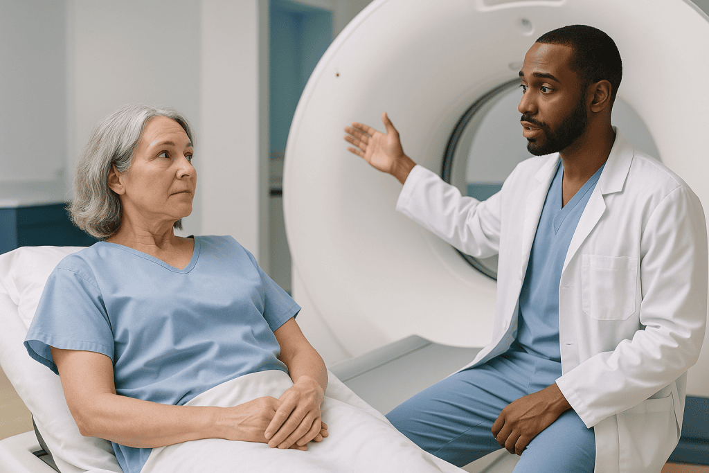
A full CT urogram is usually acquired in three passes, each catching the contrast dye at a different stage of its journey:
- Non-contrast phase. A first scan before any dye is given. Stones are dense and show up brilliantly on their own, and this phase gives the radiologist a baseline to compare with later images.
- Nephrographic phase. Roughly a minute or two after the dye is injected, the kidney tissue itself soaks it up and glows uniformly. Anything that does not glow the same way, such as a cyst or a solid mass, becomes visible.
- Excretory (or delayed) phase. Several minutes later, the kidneys have filtered the dye into the urine. Now the collecting systems, ureters and bladder appear bright white, and the radiologist looks for narrowings, blockages or irregular outlines.
Some departments merge the second and third phases with a split injection to reduce radiation, and some skip phases entirely when the clinical question is narrow. A scan looking only for stones, for example, is often a single non-contrast pass.
Mayo Clinic notes that the appointment as a whole, including preparation and the wait between phases, commonly runs about an hour, while each individual scan takes only seconds to a few minutes. Expect a warm flush, a metallic taste and sometimes a fleeting urge to urinate when the dye goes in. Those sensations are normal and pass within a minute or two.
What organs show on a CT urogram?
Patients often imagine the scan zooms in on the kidneys alone. In practice the field of view runs from the top of the kidneys, tucked just under the ribs, down to the pelvic floor, and everything in that column is recorded whether or not it was the target.
The organs the radiologist is specifically asked to assess are the two kidneys, both ureters, the bladder and, in men, the prostate where it sits beneath the bladder. The adrenal glands, perched on top of each kidney, appear as a matter of course.
Everything else in the abdomen and pelvis is in the frame too: liver, spleen, pancreas, gallbladder, loops of bowel, the aorta and its branches, the lymph nodes along the spine, the uterus and ovaries, and the bones of the lower spine and pelvis. Radiologists are trained to read the whole image, not just the requested organ, which is why reports occasionally mention an “incidental finding” somewhere unexpected.
Most incidental findings are benign. Small liver cysts, harmless adrenal nodules and simple kidney cysts are common in people past midlife and are usually noted, sometimes recommended for a follow-up scan, and then left alone. It can be unsettling to read about a finding nobody was looking for, which is one good reason to go over the report with the clinician who ordered it rather than reading it cold on a portal late at night.
An MRI of the urinary tract covers a similar territory but is often more targeted, for instance a dedicated kidney protocol or a prostate protocol, so the list of what appears may be shorter. Johns Hopkins Medicine’s guide to kidney CT describes the range of structures typically included and why the whole region is imaged rather than one organ in isolation.
How urinary tract MRI differs, and when it is chosen instead of CT
Where CT is fast and superb at spotting stones and calcium, MRI is slower and better at character. It separates fluid from solid tissue, shows the layers of the bladder wall and the zones of the prostate, and does all this without ionising radiation. For a young person who may need many scans over a lifetime, or for someone in whom iodine dye is off the table, those advantages carry real weight.

The experience is different too. You lie inside a tunnel rather than passing through a ring, the machine knocks and hums loudly enough that ear protection is standard, and stillness matters more because each sequence takes minutes rather than seconds. The NHS notes that an MRI can last from 15 to 90 minutes depending on the area and the number of images required.
Contrast is optional. Many kidney and prostate MRI protocols use a gadolinium-based agent, a dye built around the metal gadolinium that changes how tissue behaves in a magnetic field. It is chemically unrelated to iodinated CT dye, so a past reaction to one does not predict a reaction to the other, although both are recorded and considered.
MRI also has its own specific kidney concern, discussed later, and its own hard limits around implants and metal. A stone that would take a CT scanner seconds to find can be surprisingly hard to see on MRI, because stones contain no water and simply appear as a dark gap.
The choice between the two is a clinical judgement that balances the question being asked, your kidney function, any allergy history, radiation exposure and what is inside your body. Your urologist and the radiologist make that call together; the preparation you are asked to do follows from it.
Preparing for a urology CT scan: why the creatinine check comes first
Creatinine is a waste product made steadily by your muscles and cleared almost entirely by your kidneys. When the kidneys slow down, creatinine builds up in the blood, which makes it a convenient, if imperfect, gauge of filtering power. The laboratory usually converts the number into an estimated glomerular filtration rate, or eGFR, an estimate of how many millilitres of blood the kidneys filter each minute.
Radiology departments want that figure before giving iodinated contrast because the dye leaves the body through the very organs being tested. In people whose kidneys already work at reduced capacity, contrast can occasionally cause a further temporary dip in function, sometimes called contrast-associated acute kidney injury. Most healthy kidneys shrug the dye off. The blood test identifies the minority who need a different plan.
MedlinePlus explains that the creatinine test is a simple venous blood draw with no special preparation; the sample can be taken at your family practice, a hospital laboratory or sometimes at the radiology desk on the day. Many departments accept a result from within the past few weeks to a few months if you have no known kidney disease, and want a fresher one if you do. Ask what window your department uses.
Not everyone needs the test. Departments commonly reserve it for people with known kidney disease, diabetes, high blood pressure, heart failure, a single kidney, age past midlife, or a history of kidney surgery, while others test everyone as routine. A non-contrast stone protocol may need no bloodwork at all.
If your eGFR is lower than the department’s threshold, the scan is rarely cancelled outright. More often the team hydrates you, reduces the dye volume, switches to a non-contrast protocol or an ultrasound, or chooses MRI with careful thought about the gadolinium question. That decision sits with the radiologist and your referring clinician.
Kidney function and contrast: iodine dye and gadolinium are not the same risk
The two families of contrast agent behave differently in kidneys that are struggling, and the difference shapes which scan you are offered.
Iodinated contrast, used in CT, is water-soluble and passes out through the kidneys within hours in most people. The concern is a short-term fall in kidney function in the days afterwards, most likely in those who start with a low eGFR, are dehydrated, or receive a large volume of dye. In the great majority of cases the kidneys recover on their own; the aim of screening is to avoid tipping an already fragile kidney over an edge.
Gadolinium-based contrast, used in MRI, was once considered the safer alternative for kidney patients. That changed when a rare scarring condition called nephrogenic systemic fibrosis was linked to certain older gadolinium agents given to people with severe kidney failure or on dialysis. Modern agents are structured to hold the gadolinium far more tightly, and the NHS describes such reactions as rare, but departments still check kidney function before gadolinium in anyone at risk and avoid it entirely in some.
A separate observation is that trace amounts of gadolinium can remain in brain and other tissues for months or years after an injection. No health effect from this retention has been established, and regulators worldwide continue to monitor it; it is reasonable to ask whether a contrast-free MRI would answer your question equally well.
For people on dialysis, timing sometimes matters: some units arrange a dialysis session soon after gadolinium. Those arrangements are made by the kidney team, and anyone on dialysis should make sure the radiology department knows before the appointment rather than on arrival.
Neither dye is off-limits for everyone with kidney disease. The eGFR number, the urgency of the question and the alternatives available are weighed case by case.
Contrast allergies: what counts, what does not, and what the team can do
“Are you allergic to contrast?” trips people up because most have never had contrast, or have had it and cannot remember whether the warm flush counted as a reaction. It did not. Warmth, a metallic taste, a brief feeling of needing to urinate and mild nausea are expected effects of the dye entering the bloodstream, not allergy.
A true contrast reaction ranges from mild (hives, itching, a few sneezes) through moderate (widespread rash, wheeze, facial swelling) to severe (difficulty breathing, a drop in blood pressure, collapse). Mayo Clinic describes serious reactions as uncommon, but any past reaction, however mild, is worth reporting because the risk of a repeat is higher than in someone with no history.
Bring detail if you have it: which type of scan, roughly when, what happened and how it was treated. “I came out in hives after a heart scan” tells the radiologist far more than a tick in a box.
With that information the team has several options. They can choose a different iodinated agent, since reactions are often specific to one product. They can plan a premedication course, typically a corticosteroid combined with an antihistamine started in the hours before the scan, a regimen that is prescribed and timed by the radiology or referring team, never self-arranged. Or they can avoid iodine altogether and use MRI, because gadolinium is a different molecule and cross-reaction is not expected, though a history of any contrast reaction is still noted.
Two widely held beliefs deserve a direct answer. Shellfish allergy has no proven link to contrast reactions; the iodine-in-shellfish theory has not held up, and the proteins that cause seafood allergy are unrelated to contrast dye. Likewise, a skin reaction to topical iodine antiseptic does not predict a reaction to intravenous contrast. Mention both anyway; the team would rather know.
MRI safety and implants: what the screening form is really asking
The magnet in an MRI scanner is switched on around the clock, and it is powerful enough to turn a loose steel object into a projectile. That is why the screening form asks about every implant, device, fragment and piercing with such persistence, and why staff repeat the questions at the door even when you filled the form in at home.
Implants fall broadly into three groups. MR-safe items pose no known hazard in any MRI environment. MR-conditional items can be scanned only under specific conditions, such as a particular magnet strength or a device programmed into a special mode beforehand. MR-unsafe items cannot go in the scanner. Manufacturers assign these labels, and the implant card you were given at surgery usually carries the relevant details.
Items of particular interest in a urology setting include:
- Cardiac pacemakers and defibrillators, many newer models of which are MR-conditional but need programming and monitoring by cardiac staff on the day.
- Sacral nerve stimulators used for bladder control, which have model-specific rules and may need to be switched off.
- Artificial urinary sphincters and inflatable penile implants, most of which are MR-conditional; the card matters.
- Cochlear implants, older aneurysm clips, insulin pumps and some drug-infusion pumps, several of which are unsafe or must be removed.
- Metal fragments in the eye or body from grinding, welding, shrapnel or old injuries; an X-ray may be taken first to check.
Ureteral stents, most orthopaedic joints and modern surgical clips are generally fine, though staff still want to know. Tattoos rarely cause a problem beyond occasional warmth; some transdermal medicine patches contain metal backing and are removed before the scan on the team’s instruction.
For CT, implants are far less of a safety issue. Metal simply produces streaks on the image, so knowing about a hip replacement helps the radiologist interpret the pictures rather than keep you out of the room.
Who is usually scanned straight away, and who is usually asked to wait
Most adults referred for a urinary tract CT or MRI are booked without special hurdles. A few situations prompt a pause, a change of protocol or a conversation first.
Pregnancy. CT uses X-rays, and the developing baby is more sensitive to radiation than an adult. The NHS advises telling the department if there is any chance you are pregnant; a CT is not automatically ruled out when the clinical need is pressing, but ultrasound or MRI without contrast are often preferred. Gadolinium is generally avoided in pregnancy unless the benefit clearly outweighs uncertainty.
Breastfeeding. Only a tiny fraction of either contrast agent reaches breast milk and even less is absorbed by the infant. Mainstream guidance allows feeding to continue; anyone who prefers to pause can discuss a short interruption with the team.
Recent contrast. If you had a contrast CT in the past day or two, the department may prefer to space the studies apart to give the kidneys a rest, particularly if your eGFR is borderline.
Low eGFR or dialysis. As described above, this usually changes the protocol rather than cancelling the scan.
Untreated overactive thyroid. Iodine load can worsen hyperthyroidism, so the thyroid team may want to be involved first. Planned radioactive iodine treatment is another reason to delay iodinated contrast, because the dye can block iodine uptake for weeks.
Acute illness or severe dehydration. Dehydrated kidneys are more vulnerable to contrast, so someone admitted with vomiting or poor intake is often rehydrated before dye is given.
Claustrophobia or inability to lie still. This affects MRI more than CT. Wider-bore scanners, a mirror, music and, occasionally, a mild sedative arranged in advance by the clinical team can make the difference between a completed scan and an abandoned one.
None of these is a permanent barrier. They are reasons for the team to plan, and the earlier they know, the smoother the plan.
CT urogram preparation versus MRI preparation at a glance
Departments vary in detail, so treat this table as a map of what is commonly asked rather than a substitute for your own instructions.
| Preparation item | Urology CT (with iodinated contrast) | Urinary tract MRI (with or without gadolinium) |
|---|---|---|
| Blood test for kidney function | Commonly required, especially with risk factors | Required if gadolinium planned and kidney risk present |
| Contrast allergy history | Any past iodinated contrast reaction; premedication or alternative agent possible | Any past gadolinium reaction; separate from iodine history |
| Implant screening | Noted for image quality; rarely a safety barrier | Mandatory safety screen; MR-conditional devices need documentation |
| Fasting | Often a few hours without solid food; water usually encouraged | Frequently none for kidney or bladder protocols; some prostate protocols vary |
| Bladder | Often asked to arrive well hydrated; empty or fill only as instructed | Comfortably full or empty depending on protocol |
| Metal and clothing | Belts, zips and jewellery over the abdomen removed | All metal removed; gown usually provided |
| Radiation | Yes, X-ray based | None |
| Typical time in the room | Seconds per scan; about an hour for the full visit | 15–90 minutes depending on sequences |
| Diabetes medicines | Some, notably metformin, may be paused after contrast on the team’s instruction | Rarely affected unless kidney function is a concern |
The time figures reflect the NHS and Mayo Clinic descriptions cited below. The fasting row is the one most often misremembered: a CT urogram does not usually call for an overnight fast, and going without fluids is counterproductive because well-hydrated kidneys handle contrast better and produce the bright urine the excretory phase depends on.
Can I pee before a CT urogram? Eating, drinking and medicines on the day
The single most searched question about this scan has a slightly annoying answer: it depends on the protocol, so follow the instruction you were given and, if none was given, ask when you arrive.
Here is why it varies. The excretory phase needs contrast-laden urine sitting in the collecting system and bladder, so some departments ask you to drink water in the hour before and not to empty your bladder until the delayed images are done. Others give the dye, wait, then ask you to empty your bladder part way through so that the ureters fill and drain on the images. A few give a diuretic, a medicine that briefly increases urine flow, to speed the dye through. Each approach has a bladder instruction attached, and the technologist will tell you which applies. Going to the toilet just before you are called, without asking, can occasionally mean a longer wait for the delayed phase.
Food is simpler. Mayo Clinic notes many centres ask for a few hours without solid food before contrast, chiefly to reduce nausea, while clear fluids are usually welcome. Water is the drink of choice; plain coffee or tea is often acceptable, but check.
Take your usual morning medicines with a sip of water unless told otherwise. The exception people most often hear about is metformin, a common diabetes tablet cleared by the kidneys. Because a temporary contrast-related dip in kidney function could let it accumulate, some departments ask patients with reduced kidney function to pause it after the scan for a period the team specifies. That instruction comes from the radiology or prescribing team; do not stop it on your own initiative.
Wear loose clothing without metal fastenings, leave valuables at home, and bring your implant cards, medicine list and any previous imaging reports. Allow for the visit to last longer than the scan itself.
What the following hours and days usually look like
Most people walk out of the department and get on with their day. A cannula site may be a little bruised; the warm flush is long gone; the main lingering effect is mild fatigue from an early start and a lot of water.
Iodinated contrast is cleared by the kidneys within a day in people with normal function. Drinking normally over the following 24 hours supports that, though there is no need to force fluids beyond comfort. Gadolinium follows a similar route. Neither dye changes the colour of urine noticeably, and neither leaves you “radioactive”: a CT scan delivers its X-ray dose during the scan and nothing remains afterwards.
Delayed contrast reactions, most often a rash appearing hours to a couple of days after iodinated dye, are uncommon but recognised. They are usually mild and settle on their own, and are worth reporting so the reaction is recorded for future scans. Anything more than a rash falls under the red-flag section below.
If you were told to pause a diabetes medicine, restart only when the team says so; some departments repeat the creatinine test first in higher-risk patients. If you had a sedative for MRI, someone should drive you home and you should not drive or make significant decisions for the rest of the day.
Results are the part that actually takes time. The images are read by a radiologist, who writes a report for the clinician who ordered the scan. The NHS advises that this typically takes from a few days to a couple of weeks, and that urgent findings are communicated faster. Your urologist then explains the findings in the context of your symptoms and decides on any next step, which may be nothing at all, a repeat scan in months, or a further test.
Reading the report before that conversation is your right, and portals make it easy. Just remember that radiology language is written for another doctor and often lists observations that are not concerning.
What people often get wrong about urology scan preparation
Some of the most common assumptions are backwards, and a few could compromise the scan.
“I should stop drinking so my bladder is empty.” Almost the opposite. Well-hydrated kidneys clear contrast more safely and produce clearer excretory images. Restrict fluids only if specifically told to.
“I am allergic to shellfish, so I cannot have contrast.” There is no established connection. Seafood allergy is a reaction to muscle proteins, not iodine. Report it anyway, but expect it to change nothing about your dye.
“A metal hip means no MRI.” Most modern joint replacements are MR-conditional or safe. The devices that truly stop an MRI are a shorter list: certain pacemakers, cochlear implants, some aneurysm clips, some pumps and stray metal fragments. The implant card, not the assumption, decides.
“I had CT dye once with no problem, so MRI dye is fine too.” Probably, but for a different reason: the two agents are unrelated, so one predicts little about the other in either direction.
“The scan itself will hurt.” The needle for the cannula is the only sharp part. The dye produces warmth, not pain, and the scanners are silent (CT) or loud but painless (MRI).
“I should stop my blood pressure or heart tablets.” Routine medicines are almost always continued. Only specific drugs, chiefly some diabetes medicines and, for MRI, certain patches or pumps, are adjusted, and only on instruction.
“The radiation from one CT is dangerous.” A single abdominal CT delivers a dose measured in the low millisieverts, comparable to a few years of natural background radiation according to MedlinePlus. The risk is small and real, which is why clinicians avoid unnecessary scans, but it is not a reason to refuse a scan that could find a stone blocking a kidney.
“A normal creatinine means my kidneys are perfect.” Creatinine is an estimate that varies with muscle mass, age and hydration. It is a screening tool for contrast safety, not a full kidney assessment.
Questions to ask your care team before a urology CT or MRI
Ten minutes of questions at the booking stage often prevents a rescheduled appointment. These are the ones worth putting to the urology team or the radiology department.
- Which scan exactly is planned, and does it use contrast dye? Knowing whether it is a non-contrast stone CT, a full three-phase urogram or an MRI changes every instruction that follows.
- Do I need a creatinine blood test, and how recent does it have to be? Ask where the sample can be taken and whether the result will reach the department automatically.
- I once reacted to contrast: how will that be handled? Bring whatever details you have and ask whether premedication, a different agent or a different scan is the plan.
- I have this implant: is it safe for MRI, and do you need the card or a device check first? For pacemakers and stimulators, ask who arranges the programming on the day.
- Should I eat and drink normally, and what do you want my bladder to be like when I arrive?
- Which of my medicines, if any, need to be paused, and for how long afterwards? Ask specifically about diabetes tablets if you take them.
- Could I be pregnant, or am I breastfeeding: does that change the plan?
- I am worried about the tunnel: what options exist for claustrophobia, and does anything need arranging in advance?
- How long should I allow for the whole visit, and can I drive afterwards?
- When and how will I get the results, and who will explain them to me?
- Is there a reasonable alternative, such as ultrasound or a contrast-free protocol, and what would we lose by choosing it?
Write the answers down or ask for them by message. Instructions given verbally at a busy clinic desk have a way of evaporating by the morning of the scan, and the radiology team would far rather answer the same question twice than turn you away at the door.
When to call your doctor: red flags before and after the scan
Contrast scans are safe for the overwhelming majority of people, and most of what happens afterwards is nothing at all. A short list of symptoms should not be waited out.
Call emergency services or go to the nearest emergency department if, during or in the hours after the scan, you develop:
- Difficulty breathing, wheeze, or a feeling of the throat closing.
- Swelling of the face, lips or tongue.
- Faintness, collapse or a racing heartbeat with light-headedness.
- Widespread hives spreading rapidly.
These are signs of a severe contrast reaction. They almost always begin within minutes, which is why you are watched in the department, but a late-starting severe reaction is treated in exactly the same way.
Contact the clinic, your family doctor or an out-of-hours service the same day if you notice:
- A new rash, itching or mild hives appearing hours to two days after the scan.
- Passing much less urine than usual over the following day, or swelling of the ankles, which can signal a temporary drop in kidney function.
- Spreading redness, warmth, hardness or worsening pain at the injection site, particularly if the arm felt tight during the injection; a small amount of dye can leak under the skin.
- Fever, chills or new flank pain in the days after the scan.
- A pacemaker or stimulator that seems to behave differently after an MRI.
Before the scan, call the department rather than turning up if you develop vomiting, a high fever or an acute illness, if you learn you are pregnant, if you discover an implant you had forgotten, or if your kidney team has changed your treatment since the referral. Each of these can alter the protocol, and a quick phone call is far simpler than a wasted journey.
Whatever the symptom, the people who ordered and performed the scan want to hear about it. They hold the details of what was given and when, and they are the right team to decide what, if anything, needs doing next.
Frequently asked questions
Can I pee before a CT urogram?
Only if the technologist says so, because the excretory phase depends on contrast-filled urine being in the collecting system and bladder at the right moment. Some protocols ask you to hold urine until the delayed images are done; others ask you to empty part way through so the ureters can be seen filling and draining. Ask when you check in rather than using the toilet just before you are called.
What is a CT urogram and how is it different from a plain CT of the abdomen?
A CT urogram is an abdominal and pelvic CT timed around an injection of iodinated contrast so that the kidneys, ureters and bladder appear bright while urine carrying the dye passes through them. A plain abdominal CT may use contrast too, but it is not timed for the delayed excretory phase, so the collecting system and ureters are far less clearly shown.
Why is a creatinine test needed before CT contrast?
Iodinated contrast is cleared by the kidneys and can cause a temporary dip in function in people whose kidneys are already working at reduced capacity. Creatinine, converted to an eGFR, estimates that capacity. A result within the department’s accepted window lets the team decide whether to proceed normally, reduce the dye volume, hydrate you first, or choose a non-contrast or MRI alternative.
What are the three phases of a CT urogram?
The non-contrast phase is a baseline scan that shows stones and calcium. The nephrographic phase follows a minute or two after injection, when kidney tissue absorbs dye evenly and masses or cysts stand out. The excretory or delayed phase comes several minutes later, when filtered dye fills the collecting systems, ureters and bladder, revealing narrowings, blockages or irregular outlines.
What organs show on a CT urogram?
The kidneys, adrenal glands, ureters, bladder and, in men, the prostate are the target organs. Because the scan covers the whole abdomen and pelvis, the liver, spleen, pancreas, gallbladder, bowel, major blood vessels, lymph nodes, reproductive organs and spine appear as well. Radiologists report everything visible, which is why incidental findings unrelated to the urinary tract are sometimes mentioned.
Why would a urologist order a CT urogram?
The commonest reasons are blood in the urine, suspected kidney or ureteric stones, recurring urinary infections without a clear cause, unexplained flank pain, trauma to the kidney region, and follow-up of a previously seen abnormality. The scan answers whether there is a stone, blockage, cyst or growth anywhere along the route urine travels, with more detail than ultrasound can usually provide.
Does a shellfish or iodine antiseptic allergy mean I cannot have contrast dye?
No established link exists between seafood allergy and contrast reactions, because seafood allergy is caused by muscle proteins rather than iodine. A skin reaction to topical iodine antiseptic also does not predict a reaction to intravenous contrast. Mention both on your form so the team has the full picture, but expect them to change nothing about the dye you receive.
Which implants stop you having an MRI, and what about MRI safety with a pacemaker?
Devices that most often prevent or complicate MRI are certain pacemakers and defibrillators, cochlear implants, older aneurysm clips, some insulin or drug pumps, sacral nerve stimulators and loose metal fragments. Many modern pacemakers are MR-conditional and can be scanned once cardiac staff reprogram them for the session. The implant card and manufacturer label decide, so bring the card to booking.
Do I need to stop metformin or other medicines before a urology CT?
Most routine medicines continue as normal. Metformin is the exception people hear about most: because it is cleared by the kidneys, some departments ask patients with reduced kidney function to pause it after contrast for a period the team specifies, sometimes with a repeat creatinine test before restarting. Never stop or restart a prescribed medicine for a scan without an explicit instruction from your clinical team.
How long does a urology MRI take and can I have one if I am claustrophobic?
The NHS notes an MRI can last from 15 to 90 minutes depending on how many sequences are needed. Claustrophobia is common and manageable: wider-bore scanners, going in feet first where the protocol allows, a mirror to see out, music through headphones and a companion in the room all help. A mild sedative can sometimes be arranged in advance by the clinical team, in which case someone must drive you home.
References
This article is for general information only and is not a substitute for professional medical advice. Please consult a qualified doctor about your individual situation.

More from the Blog

Reading a Bladder Disease Treatment Plan: Questions to Ask Your Urologist Before You Decide
Before agreeing to a bladder disease treatment plan, ask your urologist which specific condition is being treated, what test results support that diagnosis, why…

Is Tiredness After Hemodialysis Normal? Cramps, Low Blood Pressure and What to Report
Yes, feeling tired after hemodialysis is common and expected for many people, because a session removes fluid and waste quickly and shifts salts and…

Shockwave Therapy or Endoscopic Removal: Which Kidney Stone Treatment Fits Your Stone?
Shock wave lithotripsy breaks a kidney stone from outside the body using focused sound waves, while ureteroscopy passes a thin scope up the urinary…

Orchiopexy Recovery for Children: Swelling, Incision Care and When Rough Play Can Resume
Most children recover from orchiopexy, the operation that moves an undescended testicle into the scrotum, over roughly two to four weeks. Expect scrotal swelling…

UTI Warning Signs: Fever, Flank Pain and the Moment to Seek Urgent Care
A UTI that brings fever, chills, or pain in the side or back (flank pain) may mean the infection has reached a kidney. Seek…

Who Is a Candidate for Robotic Urology? Prior Surgery, Body Weight and Heart Health Considered
A candidate for robotic urologic surgery is usually someone who needs an operation on the prostate, kidney, bladder or ureter, is fit enough for…