

Proton Therapy Planning: The Simulation CT, Your Immobilization Device and Bladder Prep
Key Takeaways
- A proton therapy simulation delivers no treatment; it is a CT-based planning visit that records your position and anatomy as the template for every session.
- Protons release most of their energy at the Bragg peak and stop, so any change in tissue density along the beam path can shift where the dose lands.
- Thermoplastic masks are perforated mesh softened in warm water and molded to your face in minutes; they hold position rather than restrict breathing.
- For pelvic sites, a comfortably full bladder lifts small bowel out of the field and stabilizes the target, and daily imaging checks that the volume matches planning day.
- Bowel gas lets protons travel farther than planned, which is why some teams ask patients to avoid gas-forming foods and check the daily image before treating.
- The NHS notes that radiotherapy usually starts a few days to a few weeks after planning, with proton plans often needing the longer end for range calculations and physics checks.
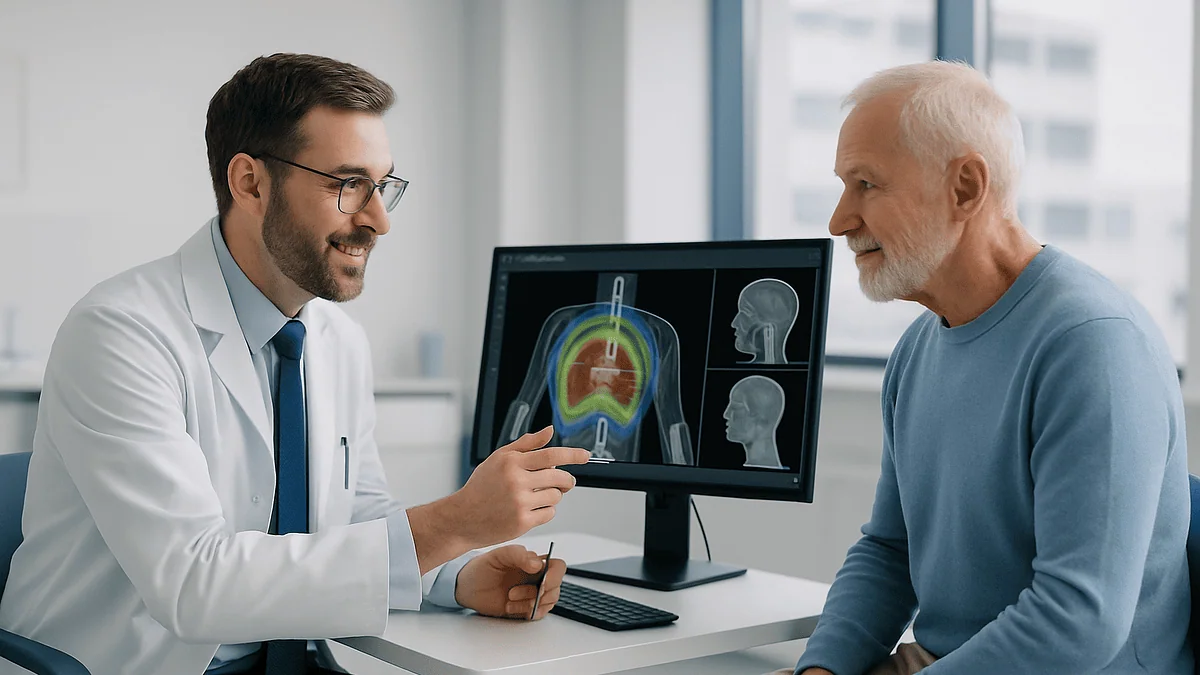
A proton therapy simulation is the planning appointment held before treatment starts. You lie in the exact position you will use for every session, a custom immobilization device such as a mask or body mold is made, and a CT scan maps your anatomy for the physics team. For pelvic and some abdominal cancers, you may be asked to arrive with a comfortably full bladder so that your organs sit in the same place each day.
The appointment letter says “simulation,” and the word lands oddly. Nothing about cancer treatment feels like a rehearsal. Yet that is exactly what a proton therapy simulation is: a dress rehearsal in which no radiation is delivered, a mold takes the shape of your shoulders or the back of your head, and a scanner records the way your body actually sits on the table.
People arrive with a small cluster of worries. Will the mask feel tight? What if I cannot hold my bladder? Why is a second scan needed when I have already had so many? The answers are mostly reassuring, and they share one theme. Protons stop inside the body at a depth that depends on what they pass through, so the planning team needs you positioned, and your organs arranged, the same way on the planning day and on every treatment day that follows.
This explainer walks through the three parts most patients ask about: the planning CT, the immobilization device, and bladder preparation.
What actually happens at a proton therapy simulation
The simulation is a planning visit, not a treatment. According to the NHS, radiotherapy planning involves a CT scan in the treatment position, and the therapy itself starts a few days or weeks later once the plan has been calculated and checked. A CT (computed tomography) scan is an X-ray study that builds cross-sectional pictures of the body, slice by slice.
You will usually change into a gown, then meet radiation therapists, the staff who position you and run the scanner. They help you onto a flat, firm couch that mimics the treatment table, because a soft hospital bed would change how your spine and organs settle. If you need an immobilization device, it is shaped around you first while the material is still warm and pliable. Then the couch moves into the scanner ring.
Laser lines project onto your skin so the therapists can record precisely where you are in the room. Small skin marks are placed, sometimes with felt pen and a clear sticker, sometimes as tiny permanent dots. The scan itself is brief; most of the visit is spent getting the position right and documenting it with photographs and measurements.
Some people receive intravenous or oral contrast, a liquid that makes blood vessels or bowel show up more clearly, so a staff member may ask about kidney function and allergies beforehand. For tumors near the lungs or upper abdomen, a 4D CT may be used. This records how the tumor moves as you breathe, which matters for protons because a moving target changes the tissue in the beam’s path.
Nothing about the day is invasive beyond a possible contrast injection, and the Mayo Clinic notes that proton therapy planning is used to define the treatment area and calculate the dose before any beam is delivered.
Why protons make the planning CT matter even more
Conventional radiation therapy uses photons, high-energy X-rays that pass all the way through the body, depositing dose on the way in and on the way out. Protons behave differently. A proton is a charged particle that slows as it travels through tissue and releases most of its energy in a sharp burst just before it stops. Physicists call that burst the Bragg peak. Beyond it, the dose drops to close to zero, which the Mayo Clinic describes as the reason protons can spare tissue behind the target.

That precise stopping point is both the appeal and the challenge. Where a proton stops depends on the density of everything in front of it. Bone slows protons more than muscle; air in a gas-filled bowel slows them far less. If the tissue along the beam path on a Tuesday differs from what was scanned on planning day, the Bragg peak can land too shallow or too deep. Clinicians call this range uncertainty, and it is why proton teams pay unusual attention to body position, bladder volume and bowel gas.
The planning CT is where all of this begins. Each voxel, or three-dimensional pixel, in the scan carries a Hounsfield number that describes how much it attenuates X-rays. The physics software converts those numbers into proton stopping power, then designs beams that stop exactly where the radiation oncologist has drawn the target. An implant, a bulky body mold or a mispositioned arm inside the beam path all have to be accounted for in that conversion.
The practical translation for patients is simple. Your team will seem exacting about small things, from the angle of your knees to what you ate that morning, because those small things move the stopping point of the beam.
Proton therapy planning CT scan: what the room looks like and how long it takes
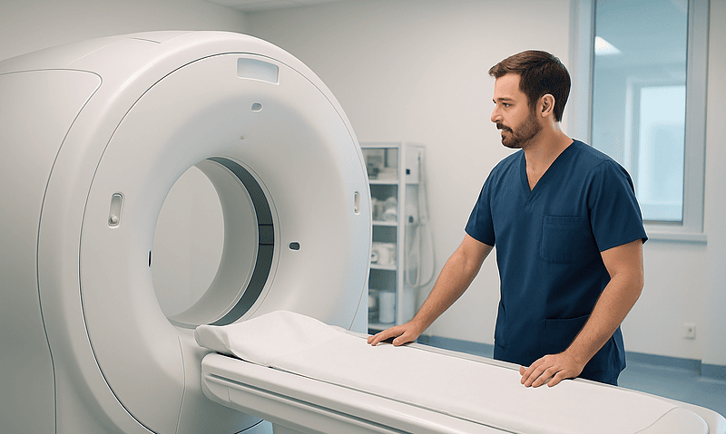
The simulation suite looks much like any CT room, with one difference you may notice: the couch top is flat carbon fiber rather than curved, so it matches the treatment machine. Overhead, wall and floor-mounted lasers throw thin red or green lines across the table. Cabinets hold the raw materials of immobilization: sheets of thermoplastic, bags of foam beads, knee wedges and headrests in graduated sizes.
Timing varies with the body region and the number of devices being made. The NHS describes the planning CT appointment as a single visit that maps the treatment area; many people are in the department for an hour or so, with the scan itself taking only minutes. Ask your own department for its typical range rather than relying on a general figure, because a head-and-neck mask with a mouthpiece takes longer to build than a simple leg mold.
Expect several checks before the scan runs. Therapists confirm your name and date of birth, review pregnancy status where relevant, and ask about metal in the body such as hip replacements, dental work or a pacemaker. Metal does not usually prevent planning, but it creates streak artifacts on CT and alters proton range, so the team needs to know about it in advance and may choose beam angles that avoid it.
After positioning, the couch slides into the scanner and you hear a soft whir. You are asked to breathe normally, or, for some chest and abdominal sites, to hold your breath in a rehearsed pattern. Once the images are checked for quality, marks are placed and photographed, and the position is written up in a set-up sheet that travels with your plan to every treatment session.
Who is asked to prepare in this way, and who is usually asked to wait
Not every proton patient needs every element described here. Immobilization is nearly universal, because reproducible position is the foundation of the whole approach. Bladder and bowel preparation, by contrast, is mostly reserved for pelvic sites: prostate, bladder, cervix, uterus, rectum and some lymph node fields. Johns Hopkins lists these among the cancers commonly treated with protons, alongside brain, head and neck, eye, spine, lung, breast and pediatric tumors.
Children form a distinct group. Proton therapy is often considered for pediatric tumors because growing tissue beyond the target can be spared, and the Mayo Clinic notes its use in children’s cancers. Small children may not manage a mask or stay still, so the pathway can include play therapy, practice visits and, in some centers, general anesthesia for both the simulation and daily treatment. Those decisions rest entirely with the pediatric oncology and anesthesia teams.
Some people are asked to wait before simulation. Recent surgery is a common reason, since swelling and healing tissue change the anatomy that will be scanned, and the team often prefers to plan once the surgical bed has settled. Active infection, uncontrolled pain that prevents lying still, or a pending decision about chemotherapy sequencing can also delay planning. Pregnancy requires an individual discussion about timing and shielding.
Others are steered toward a different technique altogether. If a tumor is very mobile, or sits behind dense metal hardware, the radiation oncologist may judge that photon-based therapy or another modality offers a more predictable plan. That is not a downgrade. It reflects the physics of range uncertainty, and the recommendation belongs to the treating team, who weigh your scans, your other treatments and your overall health.
Radiation therapy immobilization devices explained: masks, molds and cushions
An immobilization device is any custom or standard object used to hold your body in the same position at every session. The NHS notes that molds or masks may be made at the planning appointment so that you stay still and are positioned identically each time. The goal is not to restrain you but to make your Tuesday position indistinguishable from your Friday one.
Devices fall into a few families. Thermoplastic masks are mesh sheets that soften in warm water and harden as they cool, used for brain, head and neck, and sometimes upper spine treatments. Vacuum-formed bags are large cushions filled with tiny polystyrene beads; air is drawn out while you lie on them, and they set into a rigid cradle of your torso or legs. Foam molds work on a similar principle with a chemical foam that expands and hardens around you.
Simpler aids matter just as much. Knee wedges reduce lower-back arching and stabilize the pelvis. Indexed headrests, chosen from a numbered set, fix the tilt of the neck. Arm boards and wrist grips keep the shoulders out of the beam for chest and breast treatments. Each is recorded by size and position on your set-up sheet.
For protons specifically, the material of the device is part of the plan. Anything that sits in the beam path slows the particles, so physicists prefer thin, low-density devices and account for them in the calculation. You may notice therapists avoiding placing bulky items in front of the treated side; that is deliberate.
Comfort still counts. A position you cannot hold for twenty minutes without shifting is not reproducible, so speak up during the fitting if something pinches or strains. Small adjustments at this stage are easy; changes later can mean a repeat scan.
How a thermoplastic mask is made and what it feels like
The mask is the device people worry about most, and the reality is usually gentler than the imagination. You lie on the couch with your head in a fitted headrest. A therapist lifts a perforated plastic sheet from a warm-water bath, shakes off the drops and lowers it over your face. It feels like a warm, damp towel with holes in it. Over the next several minutes, staff smooth it against your cheeks, forehead and chin so it captures the contours, then clip it to a frame at the sides of the couch.
Hardening takes roughly the time it takes the material to cool, after which the mask is lifted off and labeled. Because the mesh is perforated, breathing is unrestricted, and most people can see light and movement through it. Some departments cut openings for the eyes or mouth once the mask has set, particularly if treatment will run for several weeks.
A mouthpiece, or bite block, is sometimes added for head-and-neck cancers. This is a small molded piece you rest your teeth on, holding the jaw and tongue in a fixed position and, for some plans, moving the tongue out of the beam.
Claustrophobia is common and nothing to be embarrassed about. Tell the team before the visit. Options include a practice session with an unlabeled mask, listening to music, learning slow breathing, or a discussion with your physician about short-term anxiety medicine, a decision that sits with the prescribing clinician. Cleveland Clinic and Mayo Clinic both describe daily proton sessions as lasting well under an hour, with the beam on for only a few minutes, so the mask is worn briefly each day.
Why a full bladder for radiation therapy matters with protons
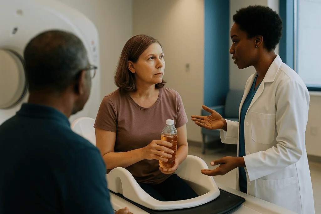
Bladder preparation puzzles many patients because it seems unrelated to the tumor. The logic is anatomical. When the bladder fills, it rises out of the pelvis and lifts loops of small bowel up and away from the prostate, cervix, uterus or rectum. A full bladder also nudges the prostate and the top of the vagina into a more consistent position. Less bowel in the treatment field generally means less bowel dose, and a stable target means the beam can be shaped more tightly.
Protons add a second reason. A bladder that is half full one day and empty the next changes the density map between skin and target. Because proton range depends on that map, a different bladder volume can shift where the beam stops by a clinically meaningful amount. Photon plans are also affected by anatomy changes, but the sharp stopping edge of protons makes them more sensitive to it.
The instruction, therefore, is not simply “drink water.” It is “arrive with the same bladder volume you had at simulation.” Your team will give you a specific routine describing when to empty your bladder and how much fluid to drink over what period. Follow their numbers, not a general figure from the internet, because the right amount depends on your bladder capacity and the treatment site.
Many departments check the bladder on the treatment machine using cone-beam CT, a low-dose scan taken in position, or a small ultrasound probe on the lower abdomen. If the bladder is too empty, you may be asked to drink and wait; if it is uncomfortably full, you may be allowed to void a little. The NHS describes this kind of daily image guidance as a routine part of modern radiotherapy.
Bowel prep, gas and the "same every day" rule
The rectum sits directly behind the prostate and beside the cervix, so its state matters as much as the bladder’s. A rectum distended by stool or gas pushes the prostate forward; an empty one lets it fall back. The difference can be several millimeters, which is a lot when a plan is designed to spare the rectal wall.
Gas deserves special mention for protons. Air is far less dense than tissue, so a pocket of gas in the beam path lets protons travel farther than the plan predicted, potentially pushing the Bragg peak past the target and into healthy tissue beyond. That is why some proton teams ask patients to avoid carbonated drinks, beans and other gas-forming foods during treatment, and why therapists may check the daily image for gas before delivering the beam. If a large pocket is present, you may be asked to walk around or pass gas and return.
Bowel routines vary. Some departments ask patients to empty the bowel each morning before treatment; others use a small enema, a liquid inserted into the rectum to prompt a bowel movement, before simulation and sometimes before each session. Whether that is right for you, and which product if any, is a decision for your radiation oncologist, who will also consider any history of hemorrhoids, inflammatory bowel disease or previous pelvic surgery.
The overarching rule is consistency. Whatever your bowel and bladder state was on the planning day is the state the plan assumes. Eating similar meals at similar times, keeping to the same fluid routine and telling staff about diarrhea or constipation all help. The NHS lists bowel changes among common radiotherapy side effects, so alert the team early if your usual pattern shifts, as the plan may need review.
Skin marks, tattoos and reference dots
Near the end of the simulation, therapists mark your skin at the points where the room lasers cross. These marks become the daily reference for lining you up before image guidance fine-tunes the position. The NHS explains that the planning team may make small, permanent ink marks on the skin, often described as tattoos, so the same area is treated each time.
Each dot is about the size of a freckle, made with a drop of ink and a fine needle. Most people describe a brief scratch. Typically two to four are placed: often one at the center of the treatment area and one on each side, so the therapists can check for rotation as well as position. Some centers now offer alternatives, such as fluorescent ink visible only under ultraviolet light or surface-guided systems that track your skin contour with cameras, reducing or removing the need for permanent marks. Availability differs by department, so ask.
Temporary marks are another option. A felt-tip cross covered by a clear waterproof dressing can survive showers for several days, but it can fade or shift, so you may be asked not to scrub the area and to return if the dressing peels away.
Do not redraw a faded mark yourself, even with the best intentions. A line a few millimeters from the original can send the day’s set-up astray. Let the therapists refresh it using your photographs and measurements.
Moisturizers are fine on the surrounding skin unless told otherwise, though many teams ask you to avoid applying anything to the marked area in the hour before treatment. Skin reactions are among the most common side effects reported by the NHS, so keep to any skincare advice you are given.
Which device and which prep for which treatment site?
Departments differ in the details, but the pairing of body region, immobilization and preparation follows a fairly predictable logic. The table below summarizes what patients commonly encounter, drawn from the general descriptions of radiotherapy planning published by the NHS, Mayo Clinic and Johns Hopkins. Your own set-up may vary, and your team’s instructions always take precedence.
| Treatment region | Typical immobilization | Typical preparation | Why it matters for protons |
|---|---|---|---|
| Brain | Thermoplastic mask, indexed headrest | None beyond removing hairpins and earrings | Skull shape is fixed; mask reproducibility is the main variable |
| Head and neck | Extended mask covering shoulders; sometimes a bite block | Dental review; consistent neck position | Dental fillings and swallowing motion alter beam path |
| Chest, lung, breast | Vacuum bag or wing board with arms overhead | Breath-hold coaching or 4D CT | Breathing moves the tumor and changes lung density in the path |
| Upper abdomen, liver, pancreas | Vacuum bag; abdominal compression in some cases | Fasting instructions; breath-hold | Stomach contents and diaphragm motion shift stopping point |
| Prostate, bladder, gynecologic | Leg and foot cradle; knee wedge | Full-bladder routine; empty rectum; gas avoidance | Bladder and rectal volume move the target and change density |
| Spine, sacrum | Vacuum bag or full-body mold | Usually none | Bone dominates the path; position reproducibility is key |
| Pediatric sites | Age-appropriate mask or mold; anesthesia in some cases | Play preparation; fasting if anesthesia planned | Sparing growing tissue depends on a still, reproducible position |
Reading down the last column, one theme repeats: anything that moves or changes density between skin and tumor has to be controlled or accounted for. Immobilization handles position; preparation handles the contents. Together they turn a single planning scan into a template that can be trusted for weeks.
What happens after your simulation: from CT to first treatment
Once you leave the department, the quiet part of the work begins. Your radiation oncologist sits with the CT images, often fused with earlier MRI or PET scans, and draws the target and the healthy structures to avoid, a process called contouring. A dosimetrist, a specialist in calculating radiation distributions, then designs beam angles and energies so that the Bragg peaks stack across the target. A medical physicist checks the plan and, for protons, runs the treatment on a phantom, a stand-in object, to confirm the machine delivers what the computer predicts.
The NHS states that radiotherapy usually starts a few days to a few weeks after the planning session, and proton planning tends to sit toward the longer end because the range calculations and quality checks are more involved. Your team will give you a start date once the plan has passed those checks; a delay of a week or two at this stage is normal and does not mean something has gone wrong.
Many centers schedule a short verification visit before the first fraction. You lie in your device, images are taken on the treatment machine, and the set-up is compared against the simulation. No beam is delivered. This is a good moment to raise anything that has changed: new pain, weight loss, a change in bowel habit or difficulty keeping the bladder routine.
Treatment itself then runs on a schedule the Mayo Clinic describes as typically five days a week for several weeks, with each visit lasting well under an hour and the beam on for only a few minutes. Daily image guidance continues to check position and, for pelvic sites, bladder and rectal filling, against the simulation reference throughout the course.
What people often get wrong about proton therapy simulation
Misunderstandings cluster around a few points, and correcting them tends to lower anxiety before the visit.
The first is that simulation is itself a treatment. It is not. The CT scanner uses a diagnostic-level X-ray dose to build a map; no therapeutic beam is involved. The NHS is explicit that planning and treatment are separate steps.
The second is that the mask is meant to be tight or that you will be unable to breathe. The mesh is perforated and molded to your face rather than clamped onto it. It should feel snug and slightly odd, not painful. If it hurts, that is a fitting problem to be fixed, not something to endure.
A third belief is that more bladder filling is always better. Overfilling can be uncomfortable enough that you move on the table, and an overstretched bladder on planning day sets a target you may not reproduce for weeks. The aim is a comfortable, repeatable volume defined by your team.
Some people assume that because proton therapy is described as precise, preparation matters less. The opposite is true. The sharp stopping edge that makes protons attractive is exactly what makes them sensitive to anatomical drift, so consistency in position, bladder and bowel matters more, not less.
Finally, patients sometimes think a faded skin mark can be touched up at home with a pen. Please leave that to the therapists, who work from your photographs and measurements.
One nuance worth stating plainly: precision in planning is a description of the physics, not a promise about outcomes. How well any course of treatment controls a tumor depends on the cancer, its stage and the whole treatment plan, and those are conversations for your oncologist.
Questions to ask your care team before and after the planning visit
A simulation visit moves quickly, and questions surface afterward. Writing a list in advance helps you use the time with your radiation oncologist and therapists well. The following prompts come up often and are worth adapting to your own situation.
- Which immobilization device will I have, and can I see or try one before the day?
- Will I need contrast for the planning CT, and is there anything in my medical history that affects that decision?
- Do I need to prepare my bladder or bowel for simulation itself, or only for daily treatment, and exactly what routine should I follow?
- How will you check my bladder and rectum on the treatment machine each day, and what happens if the filling is different?
- Are permanent tattoos used here, and are there alternatives such as surface guidance?
- How long is the expected gap between simulation and my first treatment, and who will contact me with the date?
- What changes in my body, such as weight loss, swelling or new pain, should prompt a repeat planning scan?
- If I have claustrophobia or difficulty lying flat, what support is available?
- Which of my current medicines, supplements or dietary habits could affect the preparation routine?
- Who do I call if I cannot keep to the bladder or bowel instructions on a given day?
Bring a companion if you can; a second set of ears catches instructions that slip past when you are anxious. Ask for the bladder and bowel routine in writing, and confirm the phone number for the department’s treatment desk, since practical questions often arise on the morning of the first session rather than in the consulting room. The answers, and any adjustments to your plan, rest with your treating team.
When to call your doctor
Simulation is a low-risk visit, but a few situations warrant a call to the department or your oncology team rather than waiting for the next scheduled appointment.
After contrast, seek help promptly if you develop hives, swelling of the face or lips, wheezing or difficulty breathing, or feel faint. These can indicate an allergic reaction. Call emergency services for any breathing difficulty. Milder warmth, a metallic taste or brief nausea during the injection is common and usually settles quickly.
Contact the team the same day if a skin mark or dressing is lost, if you notice redness, spreading warmth or pus at a tattoo site, or if pain prevents you from lying in your planned position. New or rapidly worsening pain, particularly back pain with numbness, weakness or changes in bladder or bowel control, needs urgent assessment regardless of where you are in the pathway, because it can signal pressure on the spinal cord.
Once treatment begins, call if you cannot pass urine, see blood in your urine or stool, develop a fever, or have diarrhea or vomiting that makes the bladder and bowel routine impossible. The NHS lists urinary and bowel changes among expected pelvic radiotherapy side effects, but a sudden change is different from a gradual one and may mean the plan needs review or that a second issue, such as infection, is present.
Significant weight loss, new swelling in the treated area, or a device that no longer fits are also reasons to speak up. Anatomy that has changed since the planning CT can shift where protons stop, and the team may choose to rescan. None of these calls is a nuisance. They are exactly what the set-up sheet, the daily images and the phone number on your appointment letter are for.
Frequently asked questions
What is a proton therapy simulation, in plain terms?
It is the planning appointment before proton treatment begins. You lie in the treatment position, a custom mask or mold may be made, and a CT scan maps your anatomy so the physics team can design beams that stop precisely inside the target. No radiation treatment is delivered at this visit, and the plan is built and checked afterward.
Is the proton therapy planning CT scan different from a diagnostic CT?
The scanner is similar, but the purpose differs. A planning CT is taken on a flat couch in your exact treatment position, often with your immobilization device in place and room lasers marking your skin. The images are converted into proton stopping-power maps rather than read for diagnosis, which is why the position and preparation matter so much.
How long does the proton therapy simulation appointment take?
Most people are in the department for an hour or so, though it varies with the site and the devices being made; the scan itself lasts only minutes. Ask your own center for its typical range. The NHS describes planning as a single visit, with treatment starting a few days to a few weeks later once the plan is checked.
What is a radiation therapy immobilization device made of?
Common materials include thermoplastic mesh that softens in warm water and hardens as it cools, vacuum bags filled with polystyrene beads that set rigid when air is removed, and expanding foam molds. Simple wedges, headrests and arm boards are also used. For protons, thin low-density materials are preferred because anything in the beam path slows the particles.
Will the mask stop me from breathing or seeing?
No. The mask is perforated, so air flows freely and you can usually see light and movement through it. It should feel snug rather than painful. If you have claustrophobia, tell the team ahead of time; practice sessions, breathing techniques and eye or mouth openings are commonly offered, and any medicine to ease anxiety is a decision for your prescribing clinician.
Why do I need a full bladder for radiation therapy to the pelvis?
A full bladder lifts small bowel out of the treatment area and holds the prostate, cervix or uterus in a more consistent position. With protons, a consistent bladder volume also keeps the tissue density between skin and tumor stable, so the beam stops where it was planned. Your team will give you a specific routine to follow each day.
What if I cannot hold my bladder on a treatment day?
Tell the therapists before you lie down. They can check the volume with imaging or ultrasound and may ask you to drink and wait, or to void a small amount if you are too full. Struggling repeatedly is worth reporting to your oncologist, who may adjust the routine or, in some cases, repeat the planning scan.
Are the skin tattoos permanent, and are there alternatives?
Traditional planning tattoos are permanent dots about the size of a freckle, placed with a drop of ink and a fine needle. Some departments use ultraviolet-visible ink or camera-based surface guidance that tracks your skin contour, reducing or removing the need for permanent marks. Availability varies, so ask your team what is used locally.
How long after proton simulation does treatment start?
The NHS notes that radiotherapy generally begins a few days to a few weeks after the planning session. Proton plans often need the longer end of that range because range calculations and physics quality checks are more involved. Your team will confirm a start date once the plan passes those checks, and a short wait is normal.
Does bowel gas really affect proton treatment?
Yes. Air is much less dense than tissue, so a gas pocket in the beam path lets protons travel farther than planned, which can move the stopping point beyond the target. Many proton teams check the daily image for gas and may ask patients to avoid carbonated drinks and gas-forming foods during a course of pelvic treatment.
References
This article is for general information only and is not a substitute for professional medical advice. Please consult a qualified doctor about your individual situation.

More from the Blog
What a Cancer Rehabilitation Program Includes: Physiotherapy, Occupational Therapy and Goals
Cancer rehabilitation is a coordinated program that helps people regain strength, movement, energy and everyday function before, during and after cancer treatment. It usually…

Life After Testicular Cancer Treatment: Fertility, Hormones and Getting Back to Sport
Life after testicular cancer treatment usually involves a period of physical healing, regular follow-up checks over several years, and adjusting to changes in fertility…
Partial or Radical Nephrectomy for Kidney Cancer: How Surgeons Decide What to Remove
Surgeons choose between partial and radical nephrectomy for kidney cancer mainly on tumor size, where the tumor sits in the kidney, how well both…

Eating Safely After a Stem Cell Transplant: The Low-Microbial Diet and When It Relaxes
After a stem cell transplant, most teams ask patients to follow a low-microbial or neutropenic diet: fully cooked foods, pasteurized dairy and juices, washed…
What Staging Tests for Head Cancer Reveal: Biopsy, Imaging and Endoscopic Examination
Head and neck cancer staging tests combine three things: a biopsy, which confirms what the cells are; an endoscopic examination, which lets the team…

Planning Your Stay in Istanbul for HIPEC: Hospital Days, Follow-Up and When to Fly Home
After cytoreductive surgery with HIPEC, most people spend roughly one to two weeks in hospital, often beginning with a day or more in intensive…