

The Brostrom Procedure: Fixing Chronic Ankle Instability, Step by Step
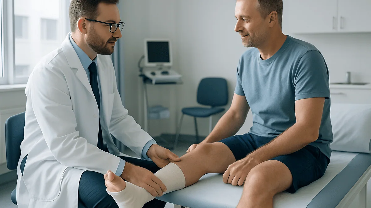
Key Takeaways
- Roughly 10 to 20 percent of acute lateral ankle sprains progress to chronic instability, according to estimates in the orthopedic literature, most sprains heal, but a meaningful minority don't.
- The Brostrom repair is typically a 60- to 90-minute outpatient operation through a 3- to 5-centimeter incision, with no bone cut in the standard version.
- Most rehab protocols allow walking in a protective boot within two to four weeks and a regular shoe around six to eight weeks after surgery.
- Long-term published series report good or excellent outcomes in roughly 85 to 95 percent of patients, with repair failure more likely in people with generalized joint laxity.
- The regional nerve block used for surgery often masks pain for the first 12 to 24 hours, so starting the pain-control plan before it wears off matters more than most patients realize.
- Balance and peroneal-strengthening rehab is what restores the reflexes that protect the repair, studies consistently show proprioceptive training reduces re-sprain risk, making the final rehab phase the least skippable.
The Brostrom procedure is an outpatient operation that tightens and reattaches the stretched outer ankle ligaments, usually the anterior talofibular and calcaneofibular ligaments, to correct chronic ankle instability after repeated sprains. Most people spend about two weeks in a splint or cast, start walking in a protective boot within two to four weeks, and return to full activity over roughly three to six months of guided rehab.
You can spot them at any pickup game: the player who tapes one ankle with the focus of a ritual, wraps it again at halftime, and still steps off a curb sideways on the way to the car. They call it a “trick ankle,” as if the joint were playing a prank. The joke wears thin around the tenth sprain.
What’s actually happening is less mysterious. A ligament on the outside of the ankle stretched during a bad sprain years ago, healed long and loose, and never regained its job as the joint’s seatbelt. Braces and tape can substitute for a while. For some people, they stop being enough.
That’s where a Swedish surgeon named Lennart Broström enters the story. The repair that carries his name, first described in 1966, remains the workhorse operation for chronic lateral ankle instability, and it’s worth understanding exactly what it involves before you’re the one signing the consent form.
What is the Brostrom procedure, exactly?
The Brostrom procedure is an anatomic repair of the ankle’s lateral ligaments, most often the anterior talofibular ligament (ATFL), sometimes joined by the calcaneofibular ligament (CFL). “Anatomic” is the key word: the surgeon works with your own tissue, shortening the stretched ligament and stitching it back down to the fibula, the outer ankle bone, where it belongs. Nothing is rerouted, and no tendon is borrowed from elsewhere.
That distinguishes it from ligament reconstruction, an older and more invasive family of operations in which a tendon graft is woven through drill holes in bone to substitute for the failed ligament. Reconstruction still has a place, typically for revision cases, very loose-jointed patients, or heavy athletes, but it sacrifices normal anatomy and can stiffen the joint. The Brostrom repair preserves the ankle’s native mechanics, which is a large part of why it became the default.
The problem it solves is chronic lateral ankle instability: the pattern of an ankle that rolls inward again and again on trivial provocation: a pebble, a stair, wet grass. Roughly one ankle sprain occurs per 10,000 people per day by common epidemiologic estimates, and while most heal well with rehabilitation, a meaningful minority do not. For that group, published surgical series over several decades report good or excellent outcomes after Brostrom repair in roughly 85 to 95 percent of patients, which is an unusually durable track record for any orthopedic operation. It is not a guarantee, no surgery is, but the evidence base is long and consistent.
Why doesn't a badly sprained ankle just heal on its own?
Ligaments are not muscles. A muscle rebuilds with use; a ligament heals like a rope splice, and if it heals in a lengthened position, it stays long. The ATFL is the weakest of the ankle’s lateral ligaments and the first to tear when the foot rolls inward, which is why it’s involved in the great majority of sprains, according to sports medicine literature summarized by sources such as Mayo Clinic and Harvard Health.
After a significant sprain, two separate problems can develop, and they matter differently:
- Mechanical instabilitythe ligament itself is physically lax. A clinician can often demonstrate this with stress tests, feeling the talus bone slide too far forward or tilt too far inward.
- Functional instabilitythe ligament may be intact, but the nerve endings inside it, which tell your brain where your foot is in space, were damaged. The ankle gives way because the reflexes fire late, not because the rope is loose.
Estimates in the orthopedic literature suggest somewhere between 10 and 20 percent of acute lateral sprains progress to chronic instability, with some studies quoting higher figures in athletes. The distinction above drives treatment: functional instability often responds beautifully to balance and strengthening work, while true mechanical laxity is the problem surgery exists to fix. Many people have some of both, which is why nearly every credible pathway runs through physical therapy first.
Do I need surgery, or can physical therapy fix chronic instability?
Start with therapy. That’s not a hedge; it’s what the evidence supports. Structured rehabilitation, peroneal muscle strengthening, balance retraining on unstable surfaces, and calf flexibility work, resolves symptoms for a substantial share of people with unstable-feeling ankles, particularly when the instability is functional rather than mechanical. Guidance from the NHS and Harvard Health both emphasize progressive rehabilitation as the foundation of sprain recovery, and surgeons generally want to see a real attempt at it, typically three to six months, before discussing an operation.
Surgery enters the conversation when a specific pattern emerges:
- The ankle keeps giving way despite months of dedicated, supervised rehab, not just a few home exercises.
- A clinical exam shows measurable mechanical laxity, often confirmed by stress testing or MRI showing a torn or attenuated ATFL.
- Instability is changing behavior: you’ve quit sports, you plan walking routes around terrain, or you’ve had repeat sprains within a single year.
There’s also a quieter argument for not waiting indefinitely once mechanical instability is established. Each giving-way episode is a small collision inside the joint, and chronic instability is associated in the literature with cartilage injury and, over decades, ankle arthritis. The causal picture isn’t perfectly settled, association is not proof, but it’s one reason surgeons don’t treat a fifteenth sprain as casually as a first. The honest framing: therapy first, always; surgery when the hardware is genuinely loose and life is shrinking around it.
Is the Brostrom procedure a major surgery?
By the usual measures, no. It’s typically an outpatient operation lasting about 60 to 90 minutes, done through a small curved incision, often 3 to 5 centimeters, over the outside of the ankle. In the standard version no bone is cut, no joint is replaced, and you go home the same day. Compared with a fracture repair or an ankle replacement, it sits firmly on the minor end of the orthopedic spectrum.
But “minor for the body” is not the same as “minor for the calendar,” and this is where people get blindsided. The repair itself is quick; protecting it while the tightened ligament heals to bone is not. You’re looking at roughly two weeks of strict protection in a splint or cast, several more weeks in a walking boot, and a strengthening program that runs months. A repaired ligament regains most of its structural integrity over about six to twelve weeks, and rushing that window is the classic way to stretch the repair back out.
So the honest answer splits in two. Anesthesia risk, incision size, blood loss, hospital time: minor. Impact on your next three months, driving, stairs, work that involves standing, the school run: significant and worth planning for. People who arrange their lives around the recovery (groceries delivered, a shower chair, a knee scooter, a desk setup that allows elevation) consistently describe an easier experience than those who assumed a small incision meant a small disruption.
What does the "Gould modification" actually add?
If your surgeon says “Brostrom-Gould,” you’re getting the modern standard rather than something exotic. In 1980, the surgeon Nathaniel Gould described a reinforcement to Broström’s original repair: after the ligament is tightened and reattached, a nearby band of strong fibrous tissue called the inferior extensor retinaculum: the natural strap that holds your toe-lifting tendons against the front of the ankle, is pulled up and stitched over the repair like a second layer of lashing.
Why bother? Three reasons show up consistently in the surgical literature:
- Load sharing. The retinaculum takes some of the strain off the healing ligament in the vulnerable early weeks, essentially splinting the repair from the inside.
- Resistance to inward roll. Because of where the retinaculum attaches, the extra layer specifically resists the inversion movement that caused all those sprains.
- Proprioceptive real estate. The tissue is rich in nerve endings, and some authors argue it contributes to restored position sense, though this benefit is harder to isolate in studies.
Biomechanical cadaver studies have generally found that the Gould augmentation adds measurable strength to the construct, and most foot-and-ankle surgeons in the United States now perform the modified version routinely, to the point that “Brostrom” in everyday usage almost always means Brostrom-Gould. It adds minutes, not risk, to the operation. If you’re comparing notes with someone who had “the Brostrom” a decade ago, odds are good you had the same procedure under slightly different shorthand.
Open, arthroscopic, or suture-tape augmented: which version will I get?
The Brostrom family has grown branches, and it helps to know the menu before your consultation.
Open repair is the classic: a small incision, direct visualization of the ligament, sutures or small bone anchors in the fibula. It has the longest track record, decades of follow-up data, and remains the benchmark against which everything else is measured.
Arthroscopic or arthroscopy-assisted repair uses a camera and instruments through keyhole portals. Its underrated advantage isn’t cosmetic: studies of chronic ankle instability consistently find that a large share of patients, commonly reported in the range of half or more, harbor additional problems inside the joint, such as cartilage lesions, scar tissue impingement, or loose bodies. A scope lets the surgeon find and address those at the same sitting. Comparative studies to date suggest arthroscopic repairs achieve outcomes broadly similar to open repair in appropriately selected patients, though the technique is more demanding and long-term data are thinner.
Suture-tape augmentation adds a strong synthetic ribbon anchored alongside the repaired ligament, acting as a seatbelt while tissue heals. Biomechanical studies show the augmented construct is stiffer at time zero, and some surgeons use that margin to accelerate rehab, earlier weight-bearing, earlier boot removal. What the evidence doesn’t yet show is a proven long-term superiority over a well-done standard repair; head-to-head trials are limited and follow-up periods short. It’s a reasonable option, especially for larger athletes or people eager to shorten protected phases, but it’s an evolving story rather than a settled one. Ask your surgeon which version they perform most, and why, for an ankle like yours.
Step by step: what actually happens in the operating room
Demystifying the day helps. Here’s the typical sequence for a Brostrom-Gould repair, acknowledging that individual surgeons vary the details.
- Anesthesia. Most patients receive general anesthesia or sedation combined with a regional nerve block: an injection that numbs the leg below the knee for many hours afterward. You will not feel the operation.
- Exam under anesthesia. With your muscles fully relaxed, the surgeon stress-tests the ankle to confirm exactly how loose the ligaments are, often more revealing than any awake exam.
- Arthroscopy, if planned. Through two small portals, the surgeon inspects the joint, smooths cartilage damage, and clears impinging scar tissue.
- The incision. A curved cut is made along the front edge of the lateral malleolus, the bony knob on the outside of your ankle, carefully avoiding nearby skin nerves.
- The repair. The stretched ATFL (and CFL when involved) is either divided and overlapped like shortening a belt, or detached and reanchored to freshly prepared bone using small suture anchors, tiny implants that stay in permanently and don’t set off airport metal detectors.
- The Gould layer. The extensor retinaculum is drawn up over the repair and sewn down, reinforcing everything beneath it.
- Closure and splint. Skin is closed, and the ankle is immobilized in a padded splint with the foot held slightly outward, protecting the repair from day one.
Start to finish, you’ll usually spend more time in the recovery room than the surgeon spends operating.
How painful is Broström surgery, honestly?
Less than most people fear, more than a few discharge pamphlets imply. Here’s the realistic arc.
The first 12 to 24 hours are often strangely comfortable, because the regional nerve block placed before surgery keeps the leg numb. This is a known trap: patients feel fine, do too much, and then the block wears off, frequently overnight, and the ankle announces itself. Surgeons typically advise starting your prescribed pain-control plan before the block fades and keeping the leg elevated above heart level almost continuously for the first several days. Elevation is not a courtesy suggestion; a dependent, swollen ankle throbs, and gravity is either your ally or your adversary all week.
Days two through five are usually the peak: a deep ache with sharper moments when the foot hangs down. Most patients describe it as very manageable with the plan their team provides, and soft-tissue repairs like this are generally reported as less painful than operations that cut bone. By the end of week one, many people are using little or no prescription medication; by weeks two to three, discomfort has typically settled into stiffness and pressure rather than true pain.
Two honest caveats. First, pain is personal, nerve sensitivity, sleep, and anxiety all modulate it, and a minority of patients have a harder ride. Second, new burning, electric, or numb sensations along the top or side of the foot deserve a call to your surgical team, since small skin nerves run close to the incision. Most such irritation fades over weeks to months, but it should be tracked, not ignored.
How long after Broström surgery can I walk?
This is the question that decides how you’ll live for two months, so it deserves a precise answer: most protocols have you walking in a protective boot somewhere between two and four weeks after surgery, with unprotected walking in a regular shoe around six to eight weeks. The exact schedule depends on your surgeon, your tissue quality, and whether the repair was augmented with suture tape, which some surgeons use to justify earlier weight-bearing.
A representative timeline, treat it as a map, not a contract:
| Timeframe | Weight-bearing status | What it looks like day to day |
|---|---|---|
| Weeks 0–2 | None, or touch-down only | Splint or cast; crutches or knee scooter; leg elevated most of the day |
| Weeks 2–4 | Progressive, in a boot | Walking boot; gradually adding weight as comfort allows; gentle motion may begin |
| Weeks 4–6 | Full, in a boot | Walking freely in the boot; physical therapy under way |
| Weeks 6–10 | Full, in a shoe | Transition to supportive shoe, often with a lace-up brace; strengthening intensifies |
| Weeks 10–16 | Unrestricted walking | Normal gait; jogging progression usually begins in this window |
Two practical notes. Weaning off the boot is gradual: a few hours a day in a shoe at first, not a single triumphant morning. And a limp that persists past the boot phase is usually a strength and confidence problem, not a repair problem; it’s exactly what the middle phase of physical therapy is designed to fix.
How long does full recovery from Broström ankle surgery take?
Plan on three to six months to feel reliably functional, and up to a year before the ankle stops occupying mental space. Those are two different finish lines, and conflating them is the most common source of recovery frustration.
The three-to-six-month milestone covers what most people mean by recovered: walking any distance, taking stairs normally, standing through a workday, returning to running and then to sport. Where you land in that range depends heavily on the demands you’re returning to: a desk worker who walks for exercise reaches their finish line far sooner than a soccer player who needs to cut and land on the ankle at full speed.
The twelve-month horizon reflects quieter biology. Swelling after ankle surgery is famously stubborn; a ring of puffiness around the joint at day’s end can persist for six months or more and is normal, not a sign of failure. Deep remodeling of the repaired ligament, collagen fibers reorganizing along lines of stress, continues for a year or longer. Many patients report that the ankle felt “good” at five months but didn’t feel forgettable, the true goal, until closer to a year.
A few realistic markers along the way: driving typically resumes once you’re out of the boot and off prescription pain medication (sooner for a left ankle with an automatic transmission, ask your surgeon); desk work is often feasible within one to two weeks with elevation; standing or labor-intensive jobs commonly require six to twelve weeks. Ask your surgical team to tailor these to your actual job rather than accepting generic ranges.
What does rehab look like, phase by phase?
The operation tightens the rope; rehabilitation reinstalls the alarm system. Skipping the second half squanders the first, because a repaired ligament with untrained reflexes is still an ankle waiting to roll. A typical protocol moves through four overlapping phases, always progressed by milestones rather than the calendar alone.
Phase one: protect (roughly weeks 0–4). The job is simply to let ligament heal to bone. Elevation, swelling control, wound care, and, critically, no inversion, the inward-rolling motion the repair was built to resist. Toe wiggling and gentle up-and-down ankle motion usually begin early to discourage stiffness.
Phase two: restore motion (weeks 4–8). A physical therapist works on dorsiflexion and plantarflexion range, calf flexibility, and gait retraining out of the boot. Inversion stretching is deliberately delayed; it’s the last motion returned, on purpose.
Phase three: rebuild strength (weeks 6–12). The stars here are the peroneal muscles running down the outside of the lower leg: the ankle’s active guy-wires against rolling. Resistance-band work, calf raises progressing from two legs to one, and early balance drills dominate. Single-leg stance on flat ground, then foam, then with eyes closed is a classic progression.
Phase four: retrain reflexes (weeks 10–16 and beyond). Proprioceptive and plyometric work, wobble boards, hopping, landing mechanics, direction changes, teaches the nervous system to protect the new ligament in the milliseconds before your conscious brain notices trouble. Research on ankle sprain prevention consistently shows balance training reduces re-sprain risk, which is exactly why this phase, the one people are most tempted to skip, matters most.
What are the risks, and how often does the repair fail?
Every credible discussion of surgery includes the downside ledger, so here is the Brostrom’s, drawn from decades of published series.
- Nerve irritation is the signature nuisance. The superficial peroneal and sural nerves, thin sensory nerves supplying skin on the top and outer foot, pass near the incision. Temporary numbness or tingling is relatively common; persistent symptoms are much less so, but they’re the complication surgeons work hardest to avoid.
- Stiffness, particularly some permanent loss of inversion range, occurs in a minority of patients. Interestingly, a slightly tight repair is often preferable to a loose one for this operation’s purpose.
- Wound problems and infection are uncommon with a small incision but rise with smoking and poorly controlled diabetes, two factors genuinely worth optimizing before surgery.
- Blood clots after lower-limb immobilization are rare but real; your team will assess your individual risk.
- Recurrent instabilitythe repair stretching out again, is the failure that matters most. Long-term studies generally report recurrence in the single digits to low double digits over many years, with higher rates in people with generalized ligamentous laxity (very flexible joints throughout the body), high body weight, or a return to high-risk sport without completing rehab.
That last group deserves candor: patients with marked generalized laxity have historically fared worse with tissue-only repair, which is precisely why surgeons may steer them toward augmented repair or reconstruction from the start. A good preoperative exam should include a laxity assessment; if yours didn’t, ask.
When can I get back to running and sports?
Running typically resumes around three to four months; cutting and pivoting sports, basketball, soccer, tennis, trail running, usually wait until four to six months. Those numbers come with an asterisk worth reading twice: the calendar is a floor, not a ticket. Return should be earned through testing, not granted by a date.
Most sports medicine teams look for objective criteria before clearing full sport:
- Calf and peroneal strength on the surgical side within roughly 90 percent of the other leg, often checked with single-leg heel raise counts.
- Symmetric performance on hop tests, single hop for distance, timed side-to-side hops, without pain or apprehension.
- Confident single-leg balance on an unstable surface, eyes open and closed.
- Sport-specific movement (cutting, landing, backpedaling) at full speed without guarding.
The psychological piece is not a footnote. After years of giving way, many athletes have built an entire movement vocabulary around protecting the ankle, landing stiffly, avoiding their plant leg, hesitating on defense. Part of late-stage rehab is unlearning that vocabulary, and athletes who report trusting the ankle tend to perform better on return than those who are merely cleared. Many surgeons also recommend a lace-up brace or taping for the first season back; the evidence for bracing as re-sprain prevention in previously injured ankles is reasonably supportive, and the cost of wearing one is close to zero.
One more honest note: published return-to-sport rates after Brostrom repair in athletes are high, commonly cited above 80 to 90 percent, but “returned” and “returned at prior level” are measured differently across studies. Ask your surgeon what their own athletes typically experience.
When should you see a doctor about an unstable ankle?
Long before surgery is on the table, certain patterns warrant a professional evaluation rather than another roll of athletic tape.
Make an appointment if any of these describe you:
- Your ankle has given way or fully sprained more than twice in the past year, especially on flat, predictable ground.
- You feel the joint shift, click, or catch, or you have deep pain and swelling that returns after activity, possible signs of cartilage or tendon involvement alongside the ligaments.
- Six or more weeks of consistent rehabilitation exercises haven’t restored your confidence on uneven surfaces.
- You’ve started avoiding activities, hiking, sports, even certain shoes, because you no longer trust the ankle.
Seek prompt care, rather than a routine appointment, after any acute injury where you cannot bear weight for more than a few steps, where there’s bony tenderness at the ankle knobs or midfoot, or where deformity, numbness, or rapidly expanding swelling appears: these are the situations in which clinicians rule out fracture, as guidance from the NHS and Mayo Clinic outlines. And after any ankle surgery, contact your surgical team without delay for fever, spreading redness or drainage at the incision, calf pain or swelling in either leg, chest pain, or shortness of breath.
The larger point is simple: a chronically unstable ankle is a diagnosable, measurable, usually fixable mechanical problem. “I just have weak ankles” is a shrug, not a diagnosis, and the people who get evaluated tend to be surprised by how specific the answers, and the options, actually are.
Frequently asked questions
Is the Brostrom procedure a major surgery?
No: it’s generally classified as a minor, outpatient soft-tissue procedure lasting about an hour, done through a small incision without cutting bone. The recovery, however, is more demanding than the operation: expect roughly two weeks of strict protection, several weeks in a walking boot, and three to six months of rehabilitation before the ankle is fully back in service. Plan your calendar for the recovery, not the incision size.
How painful is Broström surgery?
Most patients describe it as very manageable. A regional nerve block usually keeps the leg numb for the first 12 to 24 hours; pain then peaks over days two through five as a deep ache, controlled with the plan your team prescribes plus near-constant elevation. Soft-tissue repairs like this are generally reported as less painful than bone procedures, and many people need little prescription medication after the first week. Pain tolerance varies, so some have a harder stretch.
How long after Broström surgery can I walk?
Most protocols allow progressive walking in a protective boot starting two to four weeks after surgery, reaching full weight-bearing in the boot by about weeks four to six. Walking in a regular shoe typically begins around six to eight weeks, often with a lace-up brace at first. Before that, you’ll use crutches or a knee scooter. Timelines vary by surgeon and repair type, suture-tape-augmented repairs are sometimes advanced faster, so follow your specific protocol.
How long does it take to recover from Broström ankle surgery?
Plan on three to six months to feel reliably functional for daily life, running, and most work, and up to a year for the ankle to feel completely normal. Everyday walking usually returns within two months; running around three to four months; cutting sports at four to six months, milestone-permitting. Lingering end-of-day swelling for six months or more is common and expected, not a sign the repair failed.
Can a Brostrom repair fail or stretch out again?
Yes, though it’s uncommon. Long-term studies generally report recurrent instability in the single digits to low double digits of patients over many years. Risk is higher with generalized joint hypermobility, high body weight, returning to sport before completing rehab, or another major inversion injury. Surgeons often recommend augmented repair or reconstruction upfront for the highest-risk profiles, and a lace-up brace during the first season back in sport for everyone else.
Can chronic ankle instability be fixed without surgery?
Often, yes. Structured physical therapy, peroneal strengthening, balance retraining, and flexibility work, resolves symptoms for many people, especially when the problem is impaired reflexes rather than truly loose ligaments. That’s why surgeons typically require three to six months of genuine, supervised rehab before offering an operation. Surgery is reserved for ankles with demonstrable mechanical laxity that keep giving way despite that effort.
Is the Brostrom procedure done arthroscopically?
It can be. Many surgeons now perform arthroscopic or arthroscopy-assisted repairs through keyhole portals, and comparative studies to date suggest outcomes broadly similar to open repair in well-selected patients. A practical advantage is that the camera lets the surgeon find and treat cartilage damage or scar-tissue impingement, present in a large share of chronically unstable ankles, during the same operation. The open approach remains the benchmark with the longest follow-up data.
Will I have a big scar after Brostrom surgery?
No: the open version leaves a curved scar of roughly 3 to 5 centimeters along the outside of the ankle, which typically fades substantially over a year. Arthroscopic versions leave two or three portal marks under a centimeter each. Scar care basics apply: protect the incision from sun for the first year and follow your team’s guidance on massage once it’s fully healed.
When can I drive after the Brostrom procedure?
For a right ankle, most surgeons advise waiting until you’re out of the boot, off prescription pain medication, and able to brake forcefully without hesitation, commonly six to eight weeks or more. A left ankle with an automatic transmission may allow driving considerably sooner, sometimes within a couple of weeks. Always confirm with your surgeon, since driving before you can perform an emergency stop is both unsafe and a potential insurance problem.
When can I run and play sports again after Brostrom surgery?
Running typically resumes around three to four months, and cutting or pivoting sports around four to six months, but clearance should be earned through testing, not the calendar. Most teams look for strength within about 90 percent of the other leg, symmetric hop-test performance, and confident single-leg balance before full return. Published return-to-sport rates in athletes are high, commonly above 80 to 90 percent, and many surgeons recommend a brace for the first season back.
References
This article is for general information only and is not a substitute for professional medical advice. Please consult a qualified doctor about your individual situation.

More from the Blog
When an ACL Graft Fails: Re-Rupture Causes and How Revision ACL Reconstruction Differs
ACL graft failure means a reconstructed ligament has torn, stretched or loosened enough that the knee is unstable again. Causes fall into three groups:…

How Quickly Does Tenosynovitis Settle? Healing Phases From Splint to Full Use
Tenosynovitis usually settles over weeks rather than days. With early rest and a splint, mild overuse cases often ease within a few weeks, and…
Shoulder Impingement vs Rotator Cuff Tear: How Doctors Tell the Two Apart and Why It Matters
Shoulder impingement is irritation of the rotator cuff tendons and the bursa as they pass beneath the bony roof of the shoulder blade; a…
Anesthesia for Arthroscopy: General, Regional or Spinal and Why Same-Day Discharge Is Usual
Arthroscopy is usually done under general anesthesia (fully asleep), spinal anesthesia (numb from the waist down, often with sedation), or a regional nerve block…
Arthroscopic Rotator Cuff Repair Step by Step: Anchors, Sutures and Reattaching the Tendon
The arthroscopic rotator cuff repair procedure reattaches a torn shoulder tendon to bone through several small incisions using a camera and thin instruments. The…
When a Rotator Cuff Repair Fails: Re-Tear Signs and What Revision Surgery Involves
Rotator cuff repair failure means the repaired tendon has not healed to bone or has torn again, most often within the first months after…