

What a Chronic Wound Care Visit Involves: Assessment, Debridement, Dressings and Compression
Key Takeaways
- Circulation is tested before compression is applied to a leg ulcer, because compressing a leg with poor arterial supply can damage tissue; the ankle-brachial index from a Doppler test is what decides.
- Debriding dead tissue is usually painless because that tissue has no nerve supply, and light bleeding afterward signals living tissue has been reached.
- Chronic wounds heal faster when kept moist and covered; the NHS notes leg ulcer dressings are typically changed about once a week rather than daily.
- According to the NHS, most venous leg ulcers heal within three to four months with compression, and ongoing stockings after healing are advised to reduce recurrence.
- Diabetic foot ulcers are often painless because of nerve damage, which is why MedlinePlus advises daily foot checks and strict offloading of the ulcer.
- Mayo Clinic advises repositioning roughly every two hours in bed and about every fifteen minutes in a wheelchair to relieve pressure on a healing pressure injury.
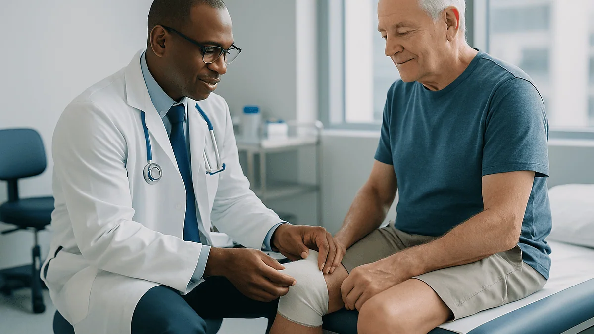
At a wound care appointment, a clinician reviews your health history, examines and measures the wound, and checks circulation and sensation in the surrounding skin. The wound is then cleaned and often debrided, meaning dead tissue is removed. A dressing is chosen to keep the wound bed moist and protected, compression bandaging is applied for venous leg ulcers when circulation allows, and you leave with a home-care plan and a follow-up date.
The referral letter says “wound clinic,” and that single word can land heavily. Maybe it is a sore above the ankle that has refused to close for two months, or a pressure injury on a parent’s heel that the home nurse has been photographing every week. You have already tried the antiseptic cream from the pharmacy, the extra pillows, the sensible shoes. Now someone is going to look at it properly, and you would like to know what that looks like before you sit down.
Knowing what happens at a wound care appointment takes most of the dread out of it. The visit is more methodical than dramatic: a careful history, a tape measure, a few simple tests, some cleaning that may briefly sting, and a bandage applied with more thought than most people expect. The real work is finding out why the wound stalled, because a wound that has stopped healing is almost always telling a story about blood flow, pressure, sugar, or nerve supply.
This explainer walks through each stage in the order it usually happens, then turns to the questions people quietly type into a search bar the night before.
What happens at a wound care appointment, step by step
Most visits follow a recognizable sequence, whether the clinic sits inside a hospital, a community health center, or a podiatry practice. The order matters, because each step answers a question the next step depends on.
It begins with talking, not touching. A nurse or clinician asks when the wound started, what caused it, what has been tried, and how it has changed. They will want your medical history in some detail: diabetes, circulation problems, prior clots, smoking, medicines that slow healing such as steroids, and anything that affects nutrition or mobility. This conversation often reveals the cause before the dressing even comes off.
Then the wound is uncovered, cleaned with saline or a mild wound cleanser, and examined. The clinician measures its length, width and depth, looks at the color and texture of the tissue inside it, checks the skin around the edge, and notes the amount and character of any fluid. A photograph is frequently taken so progress can be compared objectively at the next visit.
Next comes the hands-on part. If dead or unhealthy tissue is present, the clinician may debride it, a term explained fully below. A dressing suited to the wound’s moisture level and depth is selected and applied. For a venous leg ulcer, compression bandaging usually goes on over the dressing, provided the arteries have been checked and found adequate.
The visit ends with a plan: how often to change the dressing at home or at the clinic, what to watch for, and what the team is doing about the underlying cause. According to Johns Hopkins Medicine, addressing that cause, rather than the wound surface alone, is the part that separates a healing wound from a stalled one.
What to expect at the first wound care appointment
The first wound care appointment is the longest, and the most talkative. Expect to answer the same question several ways, because clinicians are trying to place the wound into one of a few large categories: venous (caused by weak leg veins), arterial (caused by narrowed arteries), diabetic or neuropathic (caused by nerve damage and pressure), pressure injury (caused by sustained pressure over bone), or something less common such as a surgical wound that has broken down.
Bring a list of your medicines, including over-the-counter supplements, and any photographs you have taken of the wound over time. If a district or home nurse has been visiting, ask them for their dressing records. Wear clothing that gives easy access to the wound; loose trousers or a skirt for a leg ulcer, for example, and shoes you can slip off without a struggle.
Physical examination at a first visit usually extends beyond the wound itself. For a leg wound, the clinician will feel for pulses at the ankle and foot, look for swelling, skin discoloration and varicose veins, and check the temperature of the skin. For a foot wound in someone with diabetes, they will test sensation with a fine nylon filament and inspect the footwear, since the shoe is often the culprit. MedlinePlus notes that pressure from ill-fitting shoes is a common starting point for diabetic foot ulcers.
Photographs, measurements and often a small pain assessment are recorded as a baseline. You may be asked to sign consent for debridement if it is planned. Leave time afterward: many clinics will not let you go until the dressing is secure and you understand who to call if something changes before the next visit.
How clinicians assess a wound: measuring, probing and reading the tissue
Wound assessment looks almost casual from the chair, but it is a structured examination that produces numbers as well as impressions.
Size comes first. Length and width are measured with a disposable ruler, and depth with a sterile swab gently placed in the deepest point. A wound that shrinks in area over successive visits is healing; one that holds steady or grows needs a rethink.
Then the tissue is read by color. Healthy healing tissue, called granulation tissue, is red, moist and slightly bumpy, like the surface of a raspberry. Yellow, stringy or gel-like material is slough, a mix of dead cells and protein that slows healing. Black, dry, leathery tissue is eschar, essentially a scab of dead skin that can trap infection underneath. Pale pink new skin creeping in from the edges is epithelial tissue, the finishing layer.
The clinician also probes for undermining, where the wound extends sideways under intact skin, and for tunneling, a narrow channel running deeper than the visible surface. Both are common in pressure injuries. In a foot ulcer, being able to touch bone with the probe raises concern for bone infection and usually prompts imaging.
Exudate, the fluid a wound produces, is assessed for amount, color and smell. Some moisture is normal and useful. A sudden increase, a green or cloudy color, or a persistent odor after cleaning can signal infection. The skin around the wound, the periwound, is checked for redness, warmth, and maceration, the white, soggy appearance skin takes on when it stays wet too long.
Each finding is written down. Mayo Clinic describes this kind of staging and documentation as the foundation of treating pressure ulcers, and the same logic applies to every chronic wound.
Which tests may be ordered, and why circulation comes first
A wound cannot heal without blood, so the most important test at many wound clinics measures whether enough blood is reaching the area.
For leg and foot wounds that usually means a Doppler assessment, a painless test using a handheld ultrasound probe and a blood pressure cuff. The clinician compares the pressure at the ankle with the pressure at the arm to calculate the ankle-brachial pressure index, or ABPI. The NHS explains that this test is done before compression bandaging is applied to a leg ulcer, because compressing a leg with poor arterial supply can damage tissue further. A low result redirects the whole plan toward a vascular specialist rather than toward bandages.
Blood tests are common at a first visit. A blood sugar profile helps gauge how well diabetes is controlled, since high glucose impairs the immune cells and blood vessel growth that healing depends on. A full blood count, kidney function and markers of nutrition such as albumin may be checked, because a body short on protein or iron simply lacks the raw materials to build new tissue.
Wound swabs are used more selectively than people expect. Every chronic wound carries bacteria on its surface, so a routine swab tends to grow something regardless. Clinicians reserve swabs for wounds showing genuine signs of infection, when the result will change which antimicrobial is chosen.
An X-ray, or sometimes an MRI, is requested when a deep foot ulcer might involve bone. Vein scans are used when a leg ulcer keeps recurring and vein treatment is being considered. None of these tests happens at every visit; they are ordered to answer a specific question raised by the examination.
Wound debridement: what to expect and how it feels
Debridement is the removal of dead, damaged or infected tissue from a wound so that healthy tissue can grow. It sounds alarming; in practice it is routine, and it is one of the most useful things a wound clinic does. Dead tissue is a physical barrier to new cells, a food source for bacteria, and a hiding place for infection. Clearing it also lets the clinician see the true size and depth of the wound.
Several methods exist, and they are often combined over the course of treatment.
- Sharp or surgical debridement uses a scalpel, curette or scissors to cut away non-viable tissue at the bedside. It is the fastest method. Dead tissue has no nerve supply, so removing it is usually painless, though the healthy edge can be sensitive. A topical anesthetic gel or a small local injection is sometimes used.
- Autolytic debridement uses the body’s own enzymes. A moisture-retaining dressing softens slough over days so it lifts away gradually. It is gentle and slow.
- Enzymatic debridement applies a prescription ointment containing enzymes that digest dead protein. The prescribing clinician decides whether it suits the wound.
- Mechanical debridement physically lifts debris with irrigation or specialized pads.
- Larval therapy uses sterile medical maggots that consume dead tissue. It is used in selected wounds where other methods are unsuitable.
Bleeding after sharp debridement is expected and usually light; it is a sign of live tissue. Mild soreness for the rest of the day is common. Debridement is generally avoided on dry, stable black eschar over a heel with poor circulation, because that hard cap may be protecting tissue that would not survive being opened. The decision, and the method, always rests with the treating team.
How dressings are chosen: moist wound healing explained
The instinct to let a wound “breathe” and dry out is one of the oldest ideas in home first aid, and for chronic wounds it is mostly wrong. Cells migrate across a moist wound bed far more efficiently than across a dry, crusted one, and modern dressings are engineered to hold that balance: moist, but not wet.
Choosing a dressing therefore starts with two questions. How much fluid is the wound producing? And how deep is it? A shallow, dry wound needs a dressing that adds or holds moisture, such as a hydrogel or a thin film. A wound pouring out fluid needs a dressing that absorbs it, such as a foam or an alginate, a fiber derived from seaweed that gels as it soaks. A deep cavity needs a dressing that can be packed loosely into the space so the wound heals from the bottom up rather than closing over a pocket.
Odor and infection add another layer. Dressings containing silver, iodine or honey release antimicrobial agents onto the surface, and charcoal layers absorb smell. These are tools for specific problems, not upgrades, and clinicians tend to use them for limited periods.
Two practical points matter for you at home. First, dressings are changed less often than most people assume; disturbing a wound every day cools it and tears fragile new tissue. The NHS notes that a leg ulcer dressing is typically changed about once a week, more often if it leaks. Second, if a dressing is soaked through, has slipped, or smells strongly before the scheduled change, that is a reason to call the clinic rather than a reason to improvise with whatever is in the bathroom cabinet.
Compression therapy for leg ulcers: why the bandage does the healing
For a venous leg ulcer, the dressing is almost a supporting actor. The treatment is the compression applied over it.
Here is the mechanism. Veins carry blood back up the leg against gravity, relying on one-way valves and the squeezing action of calf muscles. When those valves fail, blood pools in the lower leg, pressure inside the small veins rises, fluid and proteins leak into the tissue, and the skin becomes swollen, stained and fragile. A small knock then opens a wound that the waterlogged tissue cannot close. Compression squeezes the leg from the outside, pushing fluid back toward the heart, reducing swelling and restoring the conditions healing needs.
Compression comes in several forms: multi-layer bandage systems applied by a trained nurse, two-layer hosiery kits, and adjustable wrap devices with hook-and-loop straps. The strength is graduated, firmest at the ankle and easing toward the knee. The NHS states that most venous leg ulcers heal within three to four months when treated with compression, though some take longer, and a proportion do not close with compression alone.
Expect the bandage to feel firm and slightly strange for the first day or two. It should never cause numbness, pins and needles, blue or white toes, or pain that builds rather than settles; any of those means removing the bandage and calling the clinic promptly. Sleeping with the foot end of the bed raised and walking regularly both help the calf pump do its share.
One point often surprises people: compression does not stop when the ulcer closes. MedlinePlus advises ongoing compression stockings after healing, because the underlying vein problem remains and recurrence is otherwise common. Whether your legs can safely be compressed, and how firmly, depends on the arterial test described earlier, and that call belongs to your care team.
Who a wound clinic is usually for, and who is usually asked to wait
Wound clinics are built for wounds that have stopped following the normal script. Johns Hopkins Medicine describes a chronic wound as one that fails to progress through the usual stages of healing in a predictable time, with wounds that have not healed after roughly three months commonly falling into that category, and many services use a shorter threshold of a few weeks for people at higher risk.
Referral is typical for:
- Leg ulcers, whether venous, arterial or mixed, especially any that have lasted more than two weeks, the NHS threshold for suspecting a venous leg ulcer.
- Foot ulcers in people with diabetes, where nerve damage means the wound may be painless and dangerously easy to ignore.
- Pressure injuries at stage two or beyond, particularly over the sacrum, hips and heels.
- Surgical wounds that have opened or are draining weeks after the operation.
- Wounds in people with conditions that impair healing, such as peripheral artery disease, kidney failure, or long-term steroid use.
Who is usually asked to wait, or directed elsewhere? A fresh cut, scrape or minor burn that is only days old belongs with primary care, urgent care or simple first aid; the body is very good at closing acute wounds when they are clean and protected. A wound with spreading redness, fever and rapidly worsening pain is not a “wait for the clinic” situation either; it needs same-day assessment because infection may be moving faster than a scheduled appointment. And a suddenly cold, pale, painful foot points toward an arterial emergency, not a dressing problem.
The clinic is also not the right first stop for skin lesions that bleed, crust and never quite heal without an obvious cause; those warrant a dermatology review, since some skin cancers imitate a stubborn sore.
What is the hardest wound to heal?
People ask this hoping for a single answer. Clinicians tend to reply with a shortlist and a principle: the hardest wound to heal is the one whose cause has not been fixed.
With that caveat, three categories reliably cause the most trouble.
Arterial ulcers, and any wound on a limb with severely narrowed arteries, sit near the top. Without adequate blood flow, no dressing, enzyme or compression can deliver oxygen and immune cells to the tissue. These wounds are often small, deep, pale and painful, typically on the toes or outer ankle, and they frequently need a vascular procedure to reopen blood flow before the wound itself can respond.
Diabetic foot ulcers combine several obstacles at once: reduced sensation, so the person keeps walking on the wound; impaired circulation; high glucose that dampens immune defenses; and proximity to bone, which raises the risk of deep infection. MedlinePlus emphasizes that keeping weight off the ulcer, called offloading, is central to healing, and that is precisely the instruction hardest to follow for months on end.
Deep pressure injuries over the sacrum or heel in someone who cannot reposition themselves are the third group. Mayo Clinic notes that stage three and four pressure ulcers, which reach into fat or down to muscle and bone, can take a long time to heal and may need surgical repair. The pressure that caused them returns the moment the person lies down.
Venous leg ulcers, by contrast, are common but usually more responsive, provided compression is tolerated and worn. That is why so much of a wound visit is spent on causes rather than on the wound surface: the surface is rarely the reason it stalled.
Common chronic wound types at a glance
Recognizing which category a wound falls into explains most of what the clinic does next. The table below summarizes the typical picture for the four most common chronic wounds and the main treatment each one calls for. It is a guide to understanding your plan, not a tool for self-diagnosis; mixed pictures are common, and the clinician’s tests decide.
| Wound type | Underlying cause | Typical location and appearance | Main treatment focus |
|---|---|---|---|
| Venous leg ulcer | Failed vein valves, pooling of blood in the lower leg | Inner lower leg above the ankle; shallow, irregular edge, moist, surrounded by brown-stained, swollen skin | Graduated compression, leg elevation, walking; vein treatment to reduce recurrence |
| Arterial ulcer | Narrowed arteries limiting blood supply | Toes, foot, outer ankle; small, deep, pale or grey base, often very painful, cool skin | Restoring blood flow; compression avoided; protection and infection control |
| Diabetic foot ulcer | Nerve damage plus pressure, often with reduced circulation | Sole, toes, pressure points under bony areas; round, punched-out, thick callus at the rim, often painless | Offloading with special footwear or casts, debridement of callus, glucose control, infection surveillance |
| Pressure injury | Sustained pressure over bone, often with friction and moisture | Sacrum, hips, heels, shoulder blades; ranges from persistent redness to deep cavity | Pressure relief with repositioning and support surfaces, moisture management, nutrition, debridement |
Two threads run through every row. Blood supply is checked before anything else, because it determines what is safe. And the treatment column always includes something that happens outside the clinic, at home, every day. Mayo Clinic’s guidance on pressure ulcers, for instance, centers on repositioning and support surfaces as much as on dressings; the same is true of offloading for foot ulcers and elevation for venous disease. The visit sets the plan, but the healing largely happens between visits.
How long does a wound care appointment take, and how long does treatment usually take?
These are two different questions that often get mixed together, so it helps to separate them.
The appointment itself varies with what needs doing. A first visit is longer because it includes the full history, examination, Doppler test and often photographs and blood tests, plus time to teach you the home plan. Follow-up visits are shorter: a quick reassessment, cleaning, any debridement, and a fresh dressing or compression wrap. Multi-layer compression takes longer to apply than a simple foam dressing, and a foot ulcer that needs careful callus removal takes longer than one that does not. Clinics vary in how they schedule, so the most reliable answer is to ask when you book and to allow extra time for your first visit.
How long treatment takes depends almost entirely on wound type and on how well the underlying cause is controlled. For venous leg ulcers, the NHS gives the clearest published range: most heal within three to four months with compression, with some taking longer. Diabetic foot ulcers and deep pressure injuries have no single typical figure that mainstream sources state with confidence, because outcomes hinge on circulation, offloading and infection; Mayo Clinic notes that deeper pressure ulcers may take many months and sometimes require surgery.
Progress is judged at each visit by measurements rather than by feel. A wound that has shrunk noticeably over the first month is generally on track. One that has not changed prompts the team to look again at circulation, pressure, glucose, nutrition, and whether the dressing regime is actually being followed at home. None of these timelines is a promise; they are ranges drawn from published guidance, and your own course is set by your treating team.
What the days and weeks between visits usually look like
The rhythm of chronic wound care is weekly or twice-weekly for most people at the start, stretching out as the wound improves. Between visits, the job is mostly to leave the wound alone and to work on everything around it.
The dressing stays on unless it leaks, slips or smells. Keep it dry when washing; a waterproof cover or a plastic bag taped above the bandage works for showers, and baths are usually off the table until the wound closes. Do not lift the edge to peek. Each peek cools the wound and pulls at new tissue.
The cause gets daily attention. For venous ulcers that means walking as much as you comfortably can and raising the legs above heart level when sitting; MedlinePlus suggests elevation for periods through the day and at night. For diabetic foot ulcers it means wearing the offloading boot or shoe every time you stand, including the short walk to the bathroom at night, and checking the other foot daily since it carries the same risk. For pressure injuries it means changing position frequently; Mayo Clinic advises repositioning roughly every two hours in bed and about every fifteen minutes in a wheelchair, alongside a pressure-relieving mattress or cushion.
Nutrition quietly matters. Healing tissue is built from protein, and low intake is common in older adults. Adequate fluids and a balanced diet with enough protein support the process; your team may involve a dietitian if bloods suggest a shortfall.
Smoking constricts blood vessels and starves the wound of oxygen, and stopping is one of the few things that helps every wound type. Pain, if present, is reported rather than endured, because it can signal infection or a bandage that is too tight, and because unrelieved pain keeps people from walking and repositioning.
What people often get wrong about wound care
Wound care collects folklore the way old wounds collect callus. Some of it is harmless; some of it delays healing by months.
“Let it dry out and get air.” Acute scrapes tolerate this. Chronic wounds do not. New cells travel across a moist surface; a dry crust forces them to burrow underneath. Modern dressings are designed to hold moisture in the wound and keep excess off the surrounding skin.
“Hydrogen peroxide or antiseptic every day will keep it clean.” Strong antiseptics damage the fragile new cells you are trying to grow. Clinics clean wounds with saline or a mild cleanser, and reserve antimicrobial dressings for signs of infection.
“If it does not hurt, it is not serious.” In diabetic neuropathy the absence of pain is exactly the danger. MedlinePlus stresses daily foot checks for people with diabetes precisely because the warning system is switched off.
“Compression is just a tight bandage; any elastic wrap will do.” Therapeutic compression is graduated and applied at a measured pressure after arterial testing. A random elastic bandage can be too loose to help or too tight in the wrong place and cause harm.
“Once it closes, I am done.” The vein disease, the neuropathy, the pressure or the artery narrowing are all still there. Ongoing stockings, protective footwear, or pressure relief are what prevent the next wound.
“Something is wrong if it takes months.” For many chronic wounds, months is the expected scale. What matters is measurable progress, checked at each visit, rather than speed.
“Bleeding after debridement means damage.” Light bleeding is a sign the clinician has reached living tissue, which is the goal.
Questions to ask your care team
A wound clinic visit is dense with information, and the questions that matter most are easy to forget once the bandage is going on. Writing a few down beforehand tends to change the visit from something done to you into something done with you.
- What type of wound is this, and what do you think caused it to stop healing?
- Was my circulation tested, and what did the ankle-brachial index show?
- Are there signs of infection today, and how will we know if that changes?
- Which debridement method are you using, and what should I expect afterward?
- How often will the dressing be changed, and who changes it: the clinic, a home nurse, or me?
- Can I shower with this dressing on, and how should I protect it?
- If I am in compression, what sensations mean I should remove the bandage and call?
- What am I doing at home that is part of the treatment, and what should I stop doing?
- How will you measure progress, and when would you expect to see the wound getting smaller?
- If it is not improving in a few weeks, what is the next step or referral?
- Which of my medicines or health conditions might be slowing this down, and who manages that?
- Once it has healed, what do I need to do to stop it coming back?
You are also entitled to ask about alternatives when a particular approach is proposed, and about what happens if you choose not to have a procedure such as sharp debridement or vein treatment. Good teams welcome these questions. The answers are yours to weigh, and the final decisions about treatment, including any medicines or procedures, sit with the clinicians who have examined you.
When to call your doctor
Most chronic wounds change slowly, and most of what you notice between visits is ordinary. A few signs are not, and they should prompt a call to the clinic or your doctor the same day rather than waiting for the next scheduled appointment.
- Redness, warmth or swelling spreading outward from the wound edge, especially if it is advancing over hours rather than days.
- A sudden increase in fluid, or fluid that turns cloudy, green or foul-smelling after the dressing has been changed.
- New or rapidly worsening pain in a wound that was settled, or pain under a compression bandage that builds instead of easing.
- Fever, chills, or feeling generally unwell with no other explanation.
- Toes that turn blue, white or numb, or tingling under a bandage, which means the bandage should come off while you call.
- Bleeding that does not stop with gentle pressure, or a wound that suddenly looks deeper or larger.
- Black tissue appearing where there was none, or a foot that becomes cold and pale.
- In a diabetic foot ulcer, any of the above even without pain, since sensation may be reduced.
Seek emergency care immediately if the redness is spreading fast with high fever and confusion, if a limb becomes suddenly cold, painful and pale, or if a wound is bleeding heavily. These can indicate severe infection or an arterial blockage, both of which are time-critical.
For everything else, err toward calling. Wound teams expect these calls, and a phone description or a photograph often settles the matter quickly. Nothing in this article replaces the judgment of the clinicians who have examined your wound; their plan, and any changes to it, take precedence over general guidance.
Frequently asked questions
How long does a wound care appointment take?
It depends on what needs doing, and a first visit is always longer than a follow-up. The initial appointment includes a full history, examination, circulation testing and teaching, while later visits usually involve reassessment, cleaning, any debridement and a fresh dressing. Multi-layer compression bandaging takes longer to apply than a simple dressing. Clinics schedule differently, so ask for a time estimate when you book and allow extra time for the first visit.
What should I expect at the first wound care appointment?
Expect a lot of questions and a thorough examination that goes beyond the wound itself. The clinician will ask about the wound’s history, your medical conditions and medicines, then measure and photograph the wound, check pulses and sensation, and often perform a Doppler test on the leg. Cleaning, possible debridement and a dressing follow, and you leave with a home-care plan and a follow-up date. Bring a medicines list and any photographs.
What happens during wound debridement, and does it hurt?
Debridement removes dead or unhealthy tissue so healthy tissue can grow. Sharp debridement uses a scalpel or curette at the bedside; other methods use moisture-retaining dressings, prescribed enzyme ointments or, rarely, sterile larvae. Dead tissue has no nerve supply, so removal is usually painless, though the healthy edge may sting, and a topical anesthetic is sometimes used. Light bleeding and mild soreness afterward are expected.
How does compression therapy for leg ulcers work?
Compression squeezes the lower leg from the outside, pushing pooled blood and fluid back toward the heart, reducing swelling and restoring conditions the tissue needs to heal. It is applied as multi-layer bandages, hosiery kits or adjustable wraps, firmest at the ankle. The NHS states that most venous leg ulcers heal within three to four months with compression. It is only used after arterial circulation has been checked.
How long does wound care usually take before a chronic wound heals?
There is no single answer, because healing time depends on wound type and on how well the cause is controlled. The NHS gives the clearest published range for venous leg ulcers: most heal within three to four months with compression, some longer. Deep pressure injuries and diabetic foot ulcers vary widely, and Mayo Clinic notes deeper pressure ulcers may take many months. Progress is judged by measurements at each visit.
What is the hardest wound to heal?
Wounds on limbs with severely narrowed arteries are generally the hardest, because without blood flow no dressing can deliver oxygen and immune cells to the tissue. Diabetic foot ulcers and deep pressure injuries over the sacrum or heel are close behind, since reduced sensation, ongoing pressure and infection risk all work against healing. The common thread is that a wound stalls when its underlying cause has not been addressed.
Can I shower with a wound dressing or compression bandage on?
Usually yes, provided the dressing is kept dry with a waterproof cover or a plastic bag sealed above the bandage. Baths and soaking are generally avoided until the wound has closed. If a dressing does get soaked through, call the clinic for advice rather than replacing it yourself, because a wet dressing left in place can soften the surrounding skin and encourage infection.
Why is my wound dressing changed only once a week?
Because disturbing a wound less often helps it heal. Each dressing change cools the wound bed and can tear fragile new tissue, so modern dressings are designed to stay in place for days while managing moisture. The NHS notes that a leg ulcer dressing is typically changed about once a week, more often if it leaks. Your clinician sets the interval based on how much fluid the wound produces.
Do I need to keep wearing compression stockings after a leg ulcer heals?
Mainstream guidance says yes. MedlinePlus and the NHS both advise ongoing compression stockings after a venous leg ulcer closes, because the underlying vein problem that caused it remains and recurrence is otherwise common. Your care team will advise on the appropriate strength and how to put them on, and may discuss vein treatment as another way to reduce the chance of the ulcer returning.
What signs of wound infection should make me call the clinic?
Call the same day if redness, warmth or swelling is spreading from the wound edge, if fluid increases or turns cloudy, green or foul-smelling, if pain suddenly worsens, or if you develop fever or chills. Under a compression bandage, numbness, tingling or blue or white toes mean remove the bandage and call. Seek emergency care for fast-spreading redness with high fever, or a limb that becomes suddenly cold and pale.
References
- MedlinePlus: Wounds and Injuries
- MedlinePlus: Diabetes – foot ulcers
- MedlinePlus: Venous ulcers – self-care
- NHS: Venous leg ulcer – Treatment
This article is for general information only and is not a substitute for professional medical advice. Please consult a qualified doctor about your individual situation.

More from the Blog

Monoplace vs Multiplace Hyperbaric Chambers: What Changes for You During Oxygen Therapy
A monoplace hyperbaric chamber treats one person lying in a clear acrylic tube, usually pressurized with pure oxygen. A multiplace chamber is a walk-in…

Driving After Knee or Hip Replacement: The Honest Timelines
Most people are cleared to drive about four weeks after a right knee replacement and four to six weeks after a right hip replacement,…

Who Is Hyperbaric Oxygen Therapy For? Wounds, Radiation Injury and Other Recognized Indications
Hyperbaric oxygen therapy is used for a short, specific list of recognized conditions: decompression sickness, air or gas embolism, carbon monoxide poisoning, slow-healing diabetic…

Dissolvable Stitches: How Long They Take, Why Some Spit, and When to Check In
Dissolvable stitches usually hold a wound together for one to eight weeks, and the body fully absorbs the material within weeks to several months,…

What to Avoid After a Stent, and When You Can Fly or Drink Again
After a coronary stent, the most important things to avoid are stopping your antiplatelet medicines without your cardiologist's say-so, smoking, heavy lifting or soaking…

Arthrofibrosis: When a Knee Gets Stiff After Surgery, and What Helps
Arthrofibrosis is an excessive buildup of internal scar tissue after joint surgery or injury, most often in the knee, that causes persistent stiffness, pain,…