

What Do Follow-Up MRI Scans Look For After Brain Tumor Treatment and How Often Are They Done?
Key Takeaways
- A follow-up MRI is read as a comparison, so the postoperative baseline scan, typically obtained within 24–72 hours of surgery, is the anchor for every later image (RANO criteria, Wen et al.).
- Radiologists watch four things above all: contrast enhancement, FLAIR signal, mass effect and the surgical cavity, and they measure change in millimeters rather than describing it in adjectives.
- Enhancement marks leaky blood vessels, not tumor cells directly, which is why swelling after radiotherapy can look like growth for up to about 12 weeks (pseudoprogression).
- Guideline-based schedules commonly scan high-grade gliomas every 2–4 months early on and slower-growing or fully removed tumors every 6–12 months, with intervals lengthening after stable images.
- Brain tumors are graded 1 to 4 by the WHO system rather than staged, because primary brain tumors rarely spread outside the nervous system.
- A clear scan means no evidence of disease at current resolution, not the absence of every tumor cell, which is why surveillance continues after several stable results.
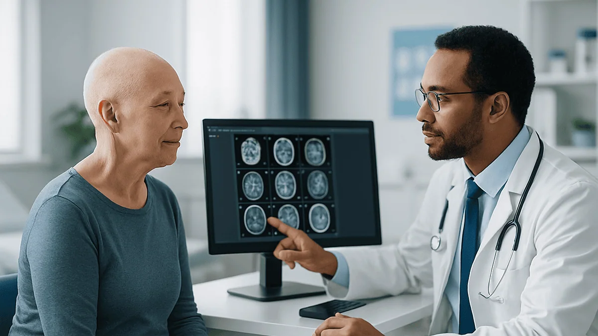
A brain tumor follow up MRI checks whether anything has changed since the last scan: new or growing contrast enhancement, expanding signal on fluid-sensitive sequences, swelling, pressure on nearby structures, or treatment effects that can mimic regrowth. Frequency depends on tumor type and grade. Guideline-based schedules often begin with scans every few months after treatment, then lengthen toward yearly imaging if findings stay stable, always set by the treating team.
The appointment letter arrives on a Tuesday, and suddenly the calendar has a new center of gravity. Everything before the scan feels like waiting; everything after it depends on a radiologist’s paragraph you have not read yet. People who have been through brain tumor treatment often describe this rhythm as scanxiety, a word that did not exist in any textbook but that every neuro-oncology nurse understands instantly.
Part of what makes a brain tumor follow up MRI so unsettling is that most people have never been told what the machine is actually hunting for. They know it is looking for the tumor. They rarely know that it is also measuring surgical scar, tracking radiation changes that can look alarming and mean little, and quietly building a timeline that only makes sense across several scans.
This explainer walks through that timeline: what each scan compares, why the first one comes so fast after surgery, how often the rest tend to follow, and which findings deserve a phone call rather than a wait.
What is a brain tumor follow up MRI actually looking for?
Strip away the jargon and a follow-up scan answers one question: has anything changed? The radiologist is not reading a single picture in isolation. They are laying the new images beside the previous ones, ideally the postoperative baseline, and searching for differences measured in millimeters.
Four kinds of change matter most. The first is enhancement, which is brightness that appears after a contrast dye is injected and marks areas where the normal barrier between blood vessels and brain tissue is leaky. Many tumors leak; healthy brain generally does not. The second is signal on fluid-sensitive sequences, usually called T2 or FLAIR, which shows swelling and infiltrating tumor cells that do not take up contrast. The third is mass effect, a plain term for tissue being pushed out of place, such as a ventricle being squeezed or the midline shifting. The fourth is the appearance of the surgical cavity itself, whose walls may thicken, shrink or fill with fluid over time.
Guideline groups have turned this into something measurable. The Response Assessment in Neuro-Oncology (RANO) working group, whose criteria are widely used in gliomas, defines progression partly as a 25 percent or greater increase in the product of two perpendicular diameters of an enhancing lesion compared with the smallest measurement recorded, along with significant growth of non-enhancing signal or clinical decline (Wen et al., PubMed). That number is not something you need to memorize, but it explains why radiologists reach for rulers rather than adjectives.
Equally important is what the scan is checking is not there. Hydrocephalus, meaning a build-up of cerebrospinal fluid, bleeding into the cavity, and infection are all complications that imaging can catch early. A stable scan is doing work too: each unchanged image extends the reassuring line on the graph.
How the scan works and what actually happens in the room
Magnetic resonance imaging uses a strong magnet and radio waves to line up and then release hydrogen atoms in your body’s water, listening to the tiny signals they give off. No ionizing radiation is involved, which is one reason MRI rather than CT is the workhorse of long-term brain surveillance (Mayo Clinic, NHS).
On the day, you change into a gown, remove anything metallic, and lie on a narrow table that slides into the tunnel of the scanner. A padded frame called a head coil rests around your skull; it is an antenna, not a restraint. The scanner is loud, producing knocking and buzzing that can reach the volume of a busy road, so you will be offered earplugs or headphones. A brain protocol for tumor follow-up typically takes between roughly 30 and 60 minutes, within the 15-to-90-minute range the NHS quotes for MRI generally.
Most tumor protocols run several sequences in turn. A T1 sequence shows anatomy in fine detail. A T2 or FLAIR sequence makes fluid and swelling bright. A diffusion sequence tracks how freely water moves, which can hint at dense tumor cells or a fresh injury. Partway through, a small cannula in your arm delivers a gadolinium-based contrast agent, and the T1 sequence is repeated so the radiologist can compare before-and-after brightness. Some centers add perfusion imaging, which estimates blood flow through tissue, or spectroscopy, which reads chemical signatures, when a finding is ambiguous.
You can usually speak to the radiographer through an intercom between sequences. Holding still matters more than anything else you do: even a few millimeters of movement blurs exactly the fine detail the comparison depends on.
Why the first MRI after brain tumor surgery comes so soon
Many people are surprised to be wheeled toward a scanner while the surgical dressing is still fresh. This early scan is not a sign that something went wrong. It is the baseline that every later image will be judged against, and its timing is deliberate.
Surgery leaves a trail of changes that all look bright on contrast-enhanced MRI: blood products, disrupted vessels, and inflamed tissue around the cavity. Within the first two to three days, that reactive enhancement has not yet fully developed, so any brightness that remains is more likely to represent tumor that could not be safely removed. The RANO working group recommends the postoperative scan be obtained within roughly 24 to 72 hours of surgery for exactly this reason; wait longer and scar and healing can masquerade as residual disease (Wen et al., PubMed).
What the baseline scan reports is the extent of resection, meaning how much of the visible tumor was removed. Surgeons describe this as gross total, near total or subtotal, and the words carry weight for planning radiotherapy or chemotherapy. The scan also confirms there is no significant bleeding, no unexpected stroke from a blocked vessel during the operation, and no early hydrocephalus.
Not everyone has surgery, of course. For people whose tumor was biopsied only, or treated with radiotherapy alone, the reference scan is usually the last one taken before treatment began. Either way, the principle is the same: the first image in the series is the anchor. If you ever change hospitals, ask that this baseline travels with you. Radiologists compare images, not descriptions, and a missing anchor makes every later reading harder.
How often is a brain tumor follow up MRI done?
There is no single schedule, and anyone who quotes one number for every patient is simplifying. The interval depends on how aggressive the tumor was, how completely it was removed, what treatment followed, and whether earlier scans have been stable. Still, guideline-based patterns exist and it helps to know their shape.
Fast-growing tumors are scanned closely. Neuro-oncology guideline schedules commonly place MRIs every two to four months in the first years after treating a high-grade glioma, because change can happen within a season. The RANO criteria also single out the first 12 weeks after chemoradiation as a period when apparent growth is often not real progression, which is why teams read scans in this window cautiously and sometimes simply repeat imaging a few weeks later (Wen et al., PubMed).
Slow-growing tumors are scanned more loosely. For low-grade gliomas, meningiomas and many benign tumors that were fully removed, intervals commonly stretch to every six to twelve months, and after several stable years some people move to annual imaging or, in selected cases, are told routine surveillance can stop. The NHS describes ongoing follow-up appointments and scans for years after brain tumor treatment without prescribing one universal rhythm, precisely because the plan is individualized (NHS).
Intervals are also not fixed for life. A new symptom, a change in medication, entry into a clinical trial or an ambiguous finding can pull a scan forward. Conversely, a run of unchanged images is often what earns a longer gap. If your schedule changes, it is reasonable to ask which of those factors prompted it. The decision rests with your treating team, but the reasoning should be shareable.
Typical surveillance rhythms by tumor type at a glance
The table below summarizes the general patterns described in neuro-oncology guideline schedules. It is a map of the territory, not a prescription. Your team may scan more or less often for reasons specific to your history, and that judgment always takes precedence over any published range.
| Situation | Early phase after treatment | Later phase if stable | What scans focus on |
|---|---|---|---|
| High-grade glioma (WHO grade 3–4) | Baseline within 24–72 hours of surgery, then roughly every 2–4 months | Intervals may lengthen after prolonged stability, decided case by case | New or growing enhancement, FLAIR expansion, pseudoprogression in first 12 weeks after chemoradiation |
| Low-grade glioma (WHO grade 1–2) | Baseline scan, then roughly every 3–6 months | Often every 6–12 months for years | Slow FLAIR growth, any new enhancement suggesting change in grade |
| Meningioma, fully removed | Early postoperative scan, then commonly at about one year | Often every 1–3 years, sometimes stopping after long stability | Regrowth along the dural attachment, new lesions |
| Meningioma or other benign tumor under observation, no surgery | Repeat scan commonly at 6–12 months | Annual or longer if unchanged | Measurable growth, new swelling or mass effect |
| After radiotherapy of any type | Reassessment scan a few months after completing treatment | Per underlying tumor type | Radiation necrosis, treatment-related swelling versus true regrowth |
Two footnotes belong beside this table. Grades are the World Health Organization’s system for describing how aggressive a brain tumor looks under the microscope and, increasingly, at the molecular level; they replace the staging language used for cancers elsewhere in the body. And the intervals quoted for gliomas trace back to guideline frameworks such as RANO and national cancer guidance rather than to any single trial, which is why they are given as ranges (Wen et al., PubMed; Mayo Clinic).
If your own schedule sits outside these ranges, that is not automatically a concern. Ask why. Reasons might include molecular features of your tumor, a clinical trial protocol, or simply a very reassuring run of previous images.
Who is scanned closely and who is usually asked to wait
Surveillance intensity tracks risk, and risk is a composite of several things your team already knows about your tumor.
People scanned most often share certain features: a higher WHO grade, incomplete removal because the tumor sat near speech, movement or vision areas, molecular markers associated with faster regrowth, or a tumor type known to recur. Recent completion of chemoradiation is another trigger for closer imaging, both to assess response and because the early post-treatment months are when true progression and its look-alikes are hardest to separate. New symptoms, whatever the tumor type, move a person into the closely watched group at least temporarily.
People asked to wait longer between scans tend to have a slow-growing tumor that was fully removed, a benign diagnosis confirmed by pathology, several consecutive stable images, or no new symptoms. Being told your next scan is a year away can feel like being dropped; more accurately, it is a statement of confidence written in calendar months.
Some people are asked to wait for a different reason: a scan taken too soon could be misleading. Immediately after radiotherapy, tissue is inflamed and enhancement can flare. The RANO group cautions that within 12 weeks of completing chemoradiation, apparent enlargement should generally not be labelled progression unless it is clearly outside the radiation field or confirmed by tissue sampling (Wen et al., PubMed). A team that schedules a scan a little later, or repeats an ambiguous one after a short gap, is often protecting you from a false alarm rather than delaying care.
Life circumstances matter too. Pregnancy, kidney disease that complicates contrast use, or certain implanted devices can shift the plan. None of these are reasons to skip surveillance; they are reasons the protocol may be adjusted, and worth raising early.
Reading your MRI report: enhancement, FLAIR and mass effect in plain words
MRI reports are written radiologist to clinician, which is why they can read like a foreign language delivered in a calm voice. A handful of phrases do most of the work.
Enhancement or enhancing component refers to tissue that brightens after contrast. Nodular enhancement means a lump-like area, which raises more concern than thin, linear enhancement along a cavity wall, a common and often benign postoperative finding. Interval increase or interval decrease simply means the change since the last comparison scan; the word interval is a cue that a comparison was made, which is what you want.
FLAIR hyperintensity or T2 signal abnormality describes brightness on fluid-sensitive sequences. It can represent swelling, radiation change, infiltrating non-enhancing tumor, or a mixture, and radiologists are usually honest that they cannot always tell which from a single scan. Vasogenic edema is swelling caused by leaky vessels, often around a lesion.
Mass effect means tissue is being displaced. Midline shift, effacement of sulci, meaning the normal grooves of the brain surface are being flattened, and ventricular compression are all descriptions of pressure. Their absence is a positive finding, and reports often state no mass effect or no midline shift for that reason.
Restricted diffusion flags areas where water moves less freely, which can indicate dense tumor, a recent stroke, or an abscess depending on context. Restricted diffusion along a fresh surgical margin is frequently a small area of surgical injury, not regrowth.
The final section, usually labelled impression, is the radiologist’s judgment. Phrases like stable appearance or no evidence of progression are as definitive as imaging gets. Phrases like indeterminate or favor treatment effect over recurrence mean the picture is ambiguous and the answer will come from time, advanced imaging, or the multidisciplinary team meeting where your case is discussed (Mayo Clinic; MedlinePlus).
Pseudoprogression on MRI and other findings that look like regrowth
The hardest conversations in neuro-oncology follow-up often begin with a scan that looks worse but may not be. Pseudoprogression is the name for an increase in enhancement and swelling after radiotherapy, usually combined with chemotherapy, that later stabilizes or shrinks without any change in treatment. It is not tumor growth; it is inflamed, injured tissue behaving like tumor on the images.
Timing is the biggest clue. Pseudoprogression most often appears within the first three months after chemoradiation, which is why the RANO criteria set a 12-week window during which enlargement inside the radiation field is not automatically called progression (Wen et al., PubMed). Teams handle this uncertainty in a few ways: repeating the MRI after several weeks, adding perfusion imaging that estimates blood flow (true tumor tends to be more vascular than dead tissue), using spectroscopy, ordering a PET scan in some settings, or, rarely, taking a tissue sample.
Radiation necrosis is a cousin that arrives later, typically months to years after radiotherapy. Irradiated tissue dies, swells and enhances, sometimes with a soap-bubble or Swiss-cheese pattern on contrast images. It can cause real symptoms and may need treatment in its own right, but it is not the tumor returning.
Pseudoresponse runs the other direction. Drugs that block new blood vessel growth, a class known as antiangiogenic agents, can tighten leaky vessels so that enhancement fades quickly while non-enhancing tumor continues to spread on FLAIR images. Radiologists reading scans of anyone on this class of medicine give the FLAIR sequence extra weight. Whether any such medicine is relevant to you is a question only your prescribing clinician can answer.
What all of these share is a lesson: a single scan is a snapshot, and context turns it into information. The report line that says cannot exclude treatment effect is not evasion. It is the honest state of the evidence at that moment.
Grades, not stages: what progression means for a brain tumor
People often ask what stage their brain tumor is, borrowing language from breast or colon cancer. The honest answer is that brain tumors are not staged in that way. Staging elsewhere describes spread to lymph nodes and distant organs; primary brain tumors very rarely travel outside the central nervous system, so the system does not fit.
Instead, the World Health Organization classification assigns a grade from 1 to 4. Grade 1 tumors grow slowly and often have clear borders. Grade 2 tumors grow slowly but infiltrate surrounding tissue. Grade 3 tumors show active cell division and other aggressive features. Grade 4 tumors are the fastest growing and most likely to recur. Modern classification also folds in molecular findings from the tissue itself, so two tumors that look similar under the microscope may be graded differently based on their genetics (Mayo Clinic; Johns Hopkins Medicine).
Progression, in follow-up language, means one of two things. Radiographic progression is measurable growth on imaging, defined in gliomas by criteria such as the RANO 25 percent rule for enhancing lesions, significant FLAIR expansion, or new lesions (Wen et al., PubMed). Clinical progression is worsening symptoms or function attributable to the tumor. A team may declare progression on either basis, and the scan and the clinic visit are read together, not separately.
Transformation is the term for a lower-grade tumor developing higher-grade features over time. This is one reason low-grade tumors are followed for many years even when they seem quiet: the scan that shows a new patch of enhancement in a previously non-enhancing tumor is exactly the kind of change surveillance exists to catch early.
Remission is a word used cautiously here. Clinicians more often say stable disease, no evidence of progression, or no visible residual tumor, because these describe what imaging can and cannot see rather than making a promise.
Will a brain tumor always show up on an MRI?
Not always, and understanding why guards against two opposite mistakes: panicking at every bright spot, and assuming a clear scan means zero tumor cells.
MRI with contrast is the most sensitive routine imaging test for brain tumors, considerably better than CT at showing soft tissue detail and small lesions (MedlinePlus; Mayo Clinic). Yet it has limits set by physics and biology. Standard clinical scanners resolve structures down to roughly a millimeter. Tumor cells are measured in micrometers. Infiltrating gliomas, in particular, send individual cells well beyond the visible edge of the abnormality, which is why surgery aims for maximal safe removal rather than claiming to remove every cell, and why radiotherapy fields include a margin around what the scan shows.
Enhancement depends on a leaky blood-brain barrier. Some tumors, especially low-grade gliomas, do not enhance at all and are visible only as FLAIR signal that can resemble swelling or old injury. Tiny recurrences may sit in a zone of postoperative scar and radiation change, where they are camouflaged. Movement during the scan, poor contrast timing or a different scanner can all reduce sensitivity from one visit to the next.
This is why the phrase no evidence of disease is chosen so carefully. It states what the images show. It does not claim biological certainty, and teams continue surveillance even after several clear scans for that reason.
The reverse also applies. Plenty of bright spots on a brain MRI are not tumor: small vessel changes common with age, old bleeds, treatment effects, or normal variation. The radiologist’s job is to weigh each finding against your history and previous images, which is why the comparison scan, not the isolated one, is the unit of information.
What the days before and after a follow-up scan usually look like
The week before a scan has a texture of its own. Sleep gets lighter. People rehearse the good result and the bad one. Practically, there is little to do: most brain MRIs need no fasting, though some centers ask you to limit food if sedation is planned. You will usually be asked about kidney function, either through a recent blood test or a questionnaire, because gadolinium contrast is cleared by the kidneys (NHS). Tell the booking team about any implants, prior surgery involving metal, or claustrophobia so adjustments can be made in advance rather than at the scanner door.
On the day, allow time for the safety screening, the cannula and the scan itself, which the NHS places within a 15-to-90-minute window depending on the protocol (NHS). Bring a list of your current medicines. If you tend to feel dizzy after lying flat, stand slowly afterward.
The result gap is often the hardest part. Radiologists typically report within days, and many neuro-oncology teams then present the images at a multidisciplinary meeting before your clinic appointment, which is why a results discussion can fall one to two weeks after the scan. Some patient portals release the report before the appointment. Whether you read it early is a personal choice; some people find the technical language frightening without context, others prefer knowing.
After a stable scan, most people return to their usual routine the same day. After an ambiguous or concerning scan, the following weeks may involve a repeat MRI, advanced imaging, or a treatment discussion. In either case, ask when the next scan is scheduled before you leave. Having the date written down turns an open-ended wait into a defined interval, which many people find easier to live alongside.
Contrast dye, implants and claustrophobia: the practical worries
Three concerns come up in almost every pre-scan conversation, and each has a straightforward answer.
Gadolinium-based contrast agents are the dyes used to highlight leaky tissue. Serious allergic reactions are uncommon, and mild effects such as a cool sensation, a metallic taste or brief nausea are more typical. People with significantly reduced kidney function are screened carefully because of a rare condition affecting skin and organs that has been linked to older contrast agents; this is why you are asked about kidney health beforehand (NHS; Mayo Clinic). Trace amounts of gadolinium can remain in the body after many scans; regulators and radiology societies continue to study this, and current evidence has not shown harm from these deposits, but it is a legitimate question to raise if you are facing years of contrast scans. Your team can explain which agent is used and whether every scan needs contrast.
Implants and metal are a safety issue rather than an inconvenience. Modern pacemakers, cochlear implants, aneurysm clips, shunts with programmable valves and some spinal stimulators may be MRI-conditional, meaning scannable under specific settings, or may need a different approach. Programmable shunt valves in particular may need checking and resetting after a scan. Always bring implant documentation; the radiographers will not guess (NHS).
Claustrophobia is real and common. Options include a wider-bore scanner where available, lying feet-first for some protocols, a mirror that shows the room, music, a hand-held call button, a companion in the room, or a mild sedative arranged with your clinician in advance. Many people find that knowing each sequence lasts only a few minutes, and that the radiographer will tell them when each one starts, makes the tunnel considerably smaller than it looks.
What people often get wrong about brain tumor surveillance scans
A first myth: a bigger bright area always means the tumor is growing. As the pseudoprogression section explained, enhancement measures leakiness, not tumor cells, and it can swell dramatically after radiotherapy while the tumor itself is responding. Radiologists build timing into their reading for exactly this reason (Wen et al., PubMed).
A second: a clear scan means the tumor is gone. Imaging shows what is large enough and leaky enough to see. Teams say no evidence of disease rather than tumor-free because the second phrase claims more than the technology can support, and surveillance continues after clear scans precisely because of that gap.
A third: a longer gap between scans means the team has stopped worrying about you. Longer intervals are earned by stable images and lower-risk pathology. They reflect evidence, not indifference. If the gap makes you anxious, say so; many teams will explain the reasoning in detail, and some will arrange an interim clinic contact.
A fourth: scans should be done at the most powerful scanner available, wherever that is. Consistency matters more than field strength for surveillance. Scans taken on the same type of scanner with the same protocol are easier to compare, and comparison is the whole point. Changing machines can create apparent differences that are purely technical.
A fifth: symptoms between scans can wait for the scheduled date. They should not. New or changing symptoms are a reason to contact the team, who may bring the scan forward. The schedule exists to catch silent change; it is not a queue you must wait in when something is clearly happening.
A sixth, quieter misunderstanding: reading the report alone is the same as getting the result. A report is one input. The result is what the team concludes after combining it with your examination, history and previous imaging, and that conversation is where questions belong.
Questions to ask your care team about your follow-up MRI
Clinic appointments are short and the anxiety is long, so a written list helps. These questions are drawn from what people commonly wish they had asked.
- What exactly are you comparing this scan with, and do you have my postoperative baseline images?
- Which findings on my scan are stable, which are new, and which are ambiguous?
- If something is ambiguous, is the plan to repeat the scan, add advanced imaging such as perfusion, or discuss it at the multidisciplinary meeting? Roughly when will I hear?
- What interval are you planning between scans, and what would make you shorten or lengthen it?
- Does every scan need contrast, and how is my kidney function being monitored?
- Which symptoms should prompt me to call before the next scheduled scan, and which number do I call?
- Will I receive the report before my appointment, and is there someone I can speak to if the wording worries me?
- Am I on any medicine that changes how my scans should be read, for example a drug that affects blood vessels or a steroid that reduces swelling?
- If I move or travel, how do I make sure my previous images go with me?
- At what point, if ever, might routine surveillance stop for a tumor like mine?
Two habits make these questions land better. Ask which staff member owns the answer, because the neurosurgeon, oncologist, radiologist and specialist nurse each hold different pieces. And write the answers down or bring someone who will; people retain less than they expect from a results conversation. None of these questions challenges your team’s authority. They invite the team to share reasoning that already exists, which is what good surveillance looks like from both sides of the desk (NHS; Cleveland Clinic).
When to call your doctor between scans
Surveillance scans are designed to catch changes you cannot feel. They are not a substitute for reporting changes you can. Contact your neuro-oncology team, specialist nurse or clinician promptly if you notice any of the following, even if your next scan is only weeks away.
- A new headache pattern, particularly headaches that wake you, are worse on lying down or in the morning, or come with nausea and vomiting.
- A first-ever seizure, or a change in the frequency, length or type of seizures you already have.
- New weakness, numbness or clumsiness in a limb or one side of the face.
- New difficulty speaking, finding words, understanding others, reading or writing.
- Vision changes such as double vision, loss of part of your visual field, or blurring that does not settle.
- Increasing drowsiness, confusion, personality change or memory problems noticed by you or those around you.
- Around a surgical wound: spreading redness, swelling, clear or cloudy fluid leaking, or fever.
- Persistent vomiting, unsteadiness or a stiff neck.
Call emergency services or go to the nearest emergency department without waiting if someone has a seizure lasting more than five minutes or repeated seizures without recovering between them, becomes hard to rouse, develops sudden severe headache described as the worst of their life, or shows sudden weakness or speech loss that could also indicate a stroke (Mayo Clinic; NHS).
Many of these symptoms have benign explanations, including medication effects, infection elsewhere, fatigue and anxiety itself. The point of calling is not to assume the worst; it is to let the team decide whether the next scan should move forward. Reaching out is part of the surveillance plan, not an interruption to it, and your team will always want to hear rather than wonder why you waited.
Frequently asked questions
How often is an MRI done after brain tumor surgery?
It depends on the tumor’s grade and how completely it was removed. A baseline MRI is typically obtained within 24–72 hours of surgery. After that, guideline-based schedules commonly repeat scans every two to four months for high-grade tumors in the early years, and every six to twelve months for low-grade or fully removed tumors, lengthening toward annual imaging if findings remain stable. Your treating team sets and adjusts the interval.
Is it possible to get remission from brain tumors?
Clinicians usually avoid the word remission for brain tumors and instead describe stable disease, no evidence of progression, or no visible residual tumor on imaging. Many benign and low-grade tumors can be treated and stay unchanged for years; some higher-grade tumors are controlled for long periods. Because MRI cannot see individual cells, teams continue surveillance even after clear scans rather than declaring the tumor gone.
What are the different stages of brain tumor progression?
Brain tumors are not staged the way most cancers are, because they rarely spread outside the nervous system. The World Health Organization instead assigns a grade from 1 to 4 based on how the cells look and their molecular features, with grade 4 the most aggressive. Progression during follow-up means measurable growth on imaging, new lesions, or worsening symptoms, and a lower-grade tumor can sometimes transform to a higher grade over time.
Will a brain tumor always show up on an MRI?
No. MRI with contrast is the most sensitive routine imaging test for brain tumors, but it resolves structures of roughly a millimeter, while tumor cells are far smaller. Some tumors, particularly low-grade gliomas, do not take up contrast and appear only as subtle FLAIR signal. Small recurrences can hide within scar and radiation change. This is why teams say no evidence of disease rather than tumor-free, and keep scanning.
What is pseudoprogression on MRI?
Pseudoprogression is an increase in enhancement and swelling after radiotherapy, often combined with chemotherapy, that mimics tumor growth but later stabilizes or shrinks without a change in treatment. It most often appears within the first 12 weeks after chemoradiation, which is why the RANO criteria discourage labelling enlargement in that window as true progression. Teams may repeat the scan, add perfusion imaging, or occasionally sample tissue to clarify.
Do you have to shave your head for brain tumor surgery?
Usually not the whole head. Most neurosurgical teams shave only a narrow strip along the planned incision, and some use techniques that require minimal or no shaving, parting the hair instead. The approach depends on the location of the tumor, the size of the incision and the surgeon’s practice. Hair typically grows back over the following months. Ask your surgical team what to expect for your specific operation.
Why do I need contrast dye for every follow-up scan?
Gadolinium contrast highlights tissue where the blood-brain barrier is leaky, which is often where active tumor sits, so it is central to comparing enhancement between scans. Not every scan strictly requires it; some low-grade or benign tumors are followed mainly on non-contrast sequences. Kidney function is checked because the dye is cleared by the kidneys. Whether a given scan needs contrast is a decision for your radiologist and treating team.
How long does a follow-up brain MRI take?
Most brain tumor protocols take roughly 30 to 60 minutes of scanning, within the 15-to-90-minute range the NHS quotes for MRI generally. Allow extra time for safety screening, changing, and placement of a small cannula for contrast. Advanced sequences such as perfusion or spectroscopy add a few minutes each. Holding still during each sequence matters more than anything else, because movement blurs the fine detail the comparison depends on.
Can I see my MRI report before the appointment?
Often yes, particularly where patient portals release results automatically. Whether to read it early is a personal choice. Reports are written for clinicians and phrases such as indeterminate or cannot exclude treatment effect can sound alarming without context. If you do read it and something worries you, contact your specialist nurse rather than waiting; many teams are happy to give a brief explanation before the full discussion.
What does no evidence of progression mean on my scan?
It means the radiologist compared your current images with previous ones and found no new lesions, no measurable growth of enhancing tumor by criteria such as RANO, and no significant expansion of FLAIR signal. It is one of the most reassuring phrases in a follow-up report. It describes what imaging shows at current resolution rather than making a biological promise, which is why scheduled surveillance continues afterward.
References
- Wen PY et al. Updated response assessment criteria for high-grade gliomas: Response Assessment in Neuro-Oncology working group (PubMed)
- NHS: MRI scan
- NHS: Brain tumours
- MedlinePlus: MRI scans
This article is for general information only and is not a substitute for professional medical advice. Please consult a qualified doctor about your individual situation.

More from the Blog

Insomnia Treatment: Why CBT-I Comes First and How a Sleep Specialist Builds Your Plan
Insomnia is treated first with cognitive behavioral therapy for insomnia (CBT-I), a structured, short-term program that retrains sleep timing, bedroom habits and anxious thinking…
How Is Neurosarcoidosis Confirmed? MRI, Lumbar Puncture, PET-CT and Biopsy Explained
Neurosarcoidosis is confirmed by combining a neurological examination, MRI of the brain or spine with contrast, analysis of spinal fluid from a lumbar puncture,…

Why Do Brain Shunts Sometimes Block or Fail? What Shunt Revision Surgery Involves
Brain shunts usually fail because the tubing becomes blocked by tissue, protein or debris, because parts disconnect or break, because infection develops, or because…

SDR Recovery Timeline: Lying Flat, First Steps and the Months of Physical Therapy That Follow
A typical SDR recovery timeline runs in stages: two to three days lying flat after surgery, roughly a week in hospital, supported standing and…
Leg Stiffness That Keeps Progressing: When a Neurologist Considers an HSP Evaluation
Hereditary spastic paraplegia (HSP) is a group of inherited conditions in which leg stiffness and weakness slowly worsen over years. A neurologist referral is…

Which Tests Shape a Brain Tumor Treatment Plan? From the First MRI to the Pathology Report
A brain tumor treatment plan is built from several tests that work in sequence: a CT scan if symptoms arrive suddenly, an MRI with…