

What Does Image Guidance Change in Endoscopic Sinus Surgery (ESS)? Navigation Explained

Key Takeaways
- Image guidance adds a real-time position display on your own CT scan to standard endoscopic sinus surgery; it changes orientation, not the incisions, instruments, anesthesia or recovery.
- Navigation is most commonly used in revision surgery, extensive polyposis, distorted anatomy, and disease near the skull base, orbit or sphenoid sinus, where visual landmarks are least reliable.
- The operation is almost always performed under general anesthesia and is usually a same-day procedure lasting roughly one to three hours, according to Cleveland Clinic.
- Post-operative discomfort is typically pressure and congestion rather than sharp pain, and nasal breathing often feels worse for the first week or two before it improves.
- Navigation is regarded as a safety adjunct, but because serious complications of sinus surgery are already rare, studies have found it difficult to prove a measurable reduction in their rate.
- Chronic rhinosinusitis is an inflammatory condition defined by 12 or more weeks of symptoms; surgery improves drainage and medicine access but ongoing medical treatment usually continues.
Image guided sinus surgery adds a real-time navigation system to standard endoscopic sinus surgery. A pre-operative CT scan is matched to the patient's head so the surgeon can see exactly where an instrument tip sits in relation to the eye socket, skull base and major vessels. It does not change the operation itself; it adds a second layer of orientation that surgeons most often use in complex, revision or anatomically distorted cases.
The consent form is on the kitchen table, and one line keeps catching the eye: “with image guidance.” It sounds reassuring and slightly alarming at the same time. Does it mean the surgeon expects trouble? Is it a different operation? Or is it, as the nurse said on the phone, “just the GPS”?
That comparison is closer than most medical metaphors. Image guided sinus surgery pairs the thin camera used in endoscopic sinus surgery with a computer that tracks the instruments and shows their position on your own CT scan, slice by slice. The sinuses sit in a crowded neighborhood. The roof of the ethmoid sinus is the floor of the brain. The paper-thin lamina papyracea separates sinus from eye socket. The sphenoid sinus wall lies a few millimeters from the optic nerve and the internal carotid artery.
So the question worth asking is not whether navigation is a gimmick. It is what, precisely, it changes for the person on the table, and what it leaves exactly as it was.
What is image guided sinus surgery, in plain language?
Endoscopic sinus surgery, usually shortened to ESS, is an operation performed entirely through the nostrils using an endoscope, a slender rigid tube with a light and a camera at the tip. There are no external cuts. The surgeon watches a monitor and uses small instruments to open blocked sinus drainage pathways, remove polyps or diseased tissue, and let the sinuses ventilate and drain again. It is most often offered for chronic rhinosinusitis, which the Mayo Clinic defines as sinus inflammation lasting 12 weeks or longer despite treatment.
Image guided sinus surgery is the same operation with one addition: a navigation system. Before surgery you have a thin-slice CT scan of the sinuses. In the operating room, that scan is loaded into a computer and “registered” to your head, meaning the computer learns how the digital scan lines up with the real you. From then on, when the surgeon touches a tracked instrument to a point inside your nose, a marker appears on the scan at that exact spot in three views at once.
Think of the endoscope as the windshield and the navigation screen as the dashboard map. The windshield is still what the surgeon drives by. The map confirms the neighborhood.
Two things follow from that description. First, navigation does not remove tissue, cauterize or make decisions; a surgeon does, using the same instruments they would use anyway. Second, the technology is only as good as the scan, the registration and the person interpreting it. A map is not a self-driving car, and no professional body describes it as one. Image guidance is best understood as an orientation aid layered onto a well-established operation.
How does a sinus surgery navigation system actually work?
The process has three stages, and none of them is exotic.
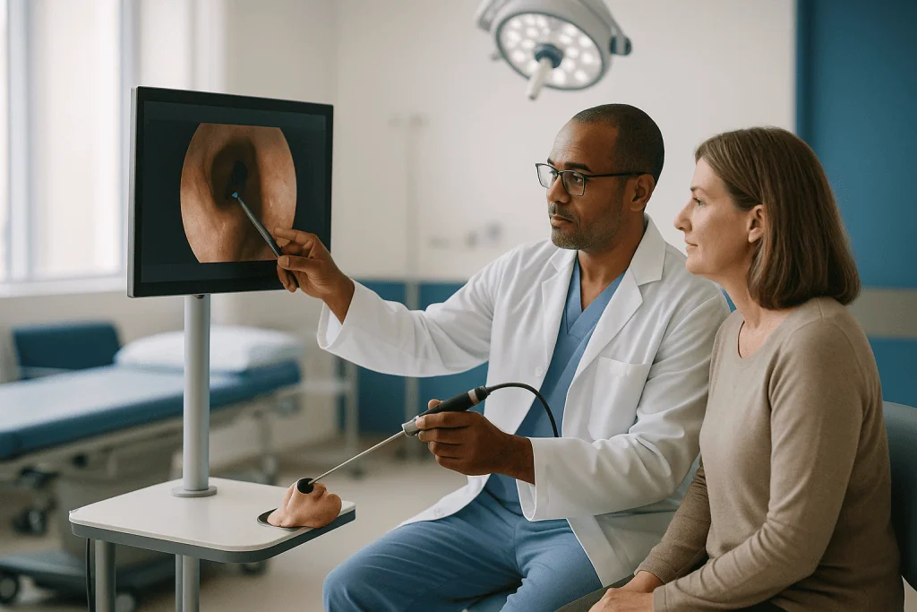
The first is imaging. A CT scan of the sinuses is taken with slices thin enough for the computer to rebuild a three-dimensional model. Some protocols use a special headset or skin markers during the scan; others rely on the bone surfaces themselves as landmarks. Your team will tell you if the scan needs to be repeated because an older one is not compatible.
The second stage is registration, which happens after you are asleep. A small tracking reference is fixed to your forehead or held by a headband. The surgeon then traces a probe over recognizable points such as the bridge of the nose or touches several bony landmarks. The computer matches those points to the scan and reports how closely they agree. Surgeons then check the match themselves by placing the probe on a structure they can see, such as the front of the middle turbinate, and confirming the screen agrees. If it does not, they re-register.
The third stage is tracking during the operation. Systems use one of two methods. Optical systems use an infrared camera that watches small reflective spheres on the instruments, which means a clear line of sight is needed. Electromagnetic systems generate a low-strength field around the head and sense tiny coils in the instrument tips, so nothing needs to be in view. Both display the tip position on axial, coronal and sagittal CT views, updated many times per second.
What the screen cannot do is update the anatomy itself. Once tissue is removed, the CT still shows it in place. This is why surgeons describe navigation as a check on position rather than a live picture of progress, and why the endoscope remains the primary view.
Why do surgeons want a map at all? The anatomy problem
The paranasal sinuses are four paired air cavities in the bones of the face: the maxillary sinuses in the cheeks, the frontal sinuses above the eyebrows, the ethmoid sinuses between the eyes, and the sphenoid sinuses deep behind the nose. Between them run some of the most consequential structures in the head.
The ethmoid roof is a plate of bone separating the sinus from the brain’s covering. The lamina papyracea, whose Latin name literally means “paper-thin layer,” is the wall between the ethmoid sinus and the orbit, the socket that holds the eye. The sphenoid sinus’s side walls often bulge over the optic nerve and the internal carotid artery, and in a minority of people the bone covering them is absent altogether. Cleveland Clinic and Johns Hopkins Medicine both list eye injury, cerebrospinal fluid leak and bleeding among the recognized, uncommon risks of sinus surgery precisely because of this geography.
Landmarks that a surgeon relies on can disappear. Polyps fill spaces and hide the usual signposts. Previous surgery removes the middle turbinate or uncinate process that normally serve as guideposts. Some people are simply built differently, with a low-hanging skull base or an ethmoid cell tucked over the frontal drainage pathway.
In a straightforward first operation, an experienced surgeon navigates by sight and by feel, guided by the CT studied beforehand. The value of a navigation system rises as the reliability of visual landmarks falls. When the picture through the endoscope is ambiguous, a second, independent source of position is exactly what a careful operator wants before advancing a millimeter further toward bone that separates sinus from eye or brain.
What does image guidance change, and what does it leave the same?
It is easier to be honest about this by separating the two lists.
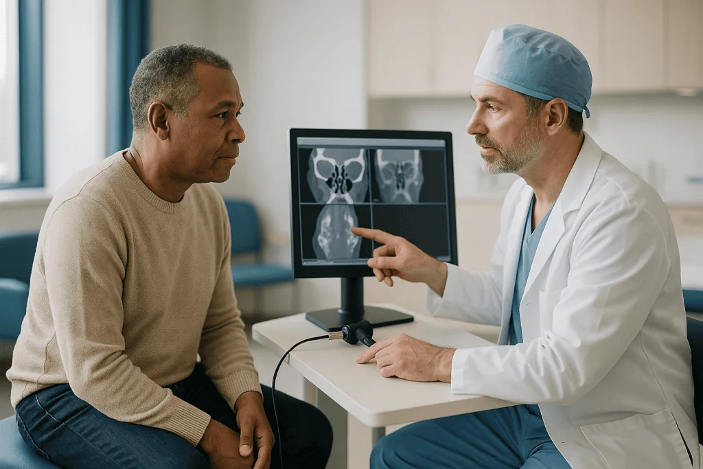
What changes is orientation. The surgeon can confirm, at any moment, where an instrument tip sits relative to the skull base, orbit and sphenoid wall. That confirmation matters most at the boundaries of the dissection: how far back to open the frontal recess, whether a remaining shell of bone is the ethmoid roof or just another cell, whether the surgeon has genuinely reached the sphenoid or a look-alike posterior ethmoid cell. Many surgeons also use navigation to be more complete, opening cells they might otherwise leave for fear of straying, because the map confirms there is room.
What also changes is planning. Because the scan is on screen throughout, the surgeon can revisit the pre-operative plan mid-procedure rather than relying on memory.
Now the list of things that do not change. The incisions, or rather the absence of them, are identical. The instruments are the same. The anesthesia is the same. The recovery is the same, because navigation adds no tissue trauma. The underlying disease is unchanged; chronic rhinosinusitis is an inflammatory condition, and Mayo Clinic notes that surgery is offered when medical treatment has not controlled it, not as a replacement for ongoing care.
One more thing that does not change, and this deserves emphasis: the complication rate is not automatically lower. Reviews of navigation in sinus surgery generally find that surgeons feel more confident and that the technology is widely accepted as a safety adjunct in complex cases, but a clear reduction in major complications has been hard to prove in controlled studies, partly because those complications are rare in the first place. Guidance reduces one source of error, disorientation. It does not remove others, such as brittle bone, bleeding that obscures the view or an unusual anatomical variant.
Who is image guided endoscopic sinus surgery usually for, and who is asked to wait?
Professional bodies in otolaryngology have published criteria describing when image guidance is considered appropriate, and the categories are consistent across them. Navigation is commonly used in:
- Revision surgery, where earlier operations have removed the usual landmarks or left scar tissue that distorts the field.
- Extensive nasal polyposis, where inflamed tissue fills the sinuses and obscures the view.
- Distorted anatomy from developmental variation, previous trauma or facial fracture.
- Disease involving the frontal, posterior ethmoid or sphenoid sinuses, the areas nearest the skull base, optic nerve and carotid artery.
- Disease reaching the skull base, orbit or optic nerve, including some benign and malignant tumors.
- Repair of cerebrospinal fluid leaks, where the exact defect must be found in a thin bony plate.
Some surgeons also use navigation routinely in primary surgery, reasoning that a tool available for every case is one the team is fluent with when it counts. Others reserve it. Neither approach is considered wrong; the decision belongs to the operating surgeon and depends on the individual scan and the surgeon’s own assessment.
Who is asked to wait? The same people who would be asked to wait for any elective sinus operation. Mayo Clinic describes surgery as an option when medical treatment has not adequately controlled chronic rhinosinusitis, so someone who has not yet completed a course of appropriate medical management is usually asked to do so first. Acute infection at the time of surgery is another common reason to postpone. People with uncontrolled medical conditions, those taking medicines that affect bleeding, and anyone whose CT does not show disease that matches their symptoms may also be advised to pause while the picture is clarified. Image guidance adds nothing to a case where surgery itself is not yet indicated.
Do they put you to sleep for endoscopic sinus surgery?
Almost always, yes. Cleveland Clinic and Johns Hopkins Medicine describe endoscopic sinus surgery as typically performed under general anesthesia, meaning you are fully asleep and unaware, with an anesthesiologist managing your breathing and vital signs. Some limited procedures can be done with local anesthetic and sedation, but when navigation is planned the case is usually complex enough that general anesthesia is the norm. The registration step also works best when the head is completely still.
The day tends to run like this. You arrive having fasted as instructed, meet the anesthesia team and confirm the plan with the surgeon. Once you are asleep, the team positions your head, fixes the tracking reference and performs registration. The nose is treated with medicines that shrink blood vessels to reduce bleeding and improve the view; these are applied topically or injected locally by the surgeon and are part of the procedure rather than something you manage.
Operating time varies with how many sinuses are addressed and whether it is a first or repeat operation. Cleveland Clinic gives a typical range of one to three hours for sinus surgery, with more extensive cases running longer. Registration and setup add some minutes at the start, which is one reason navigation was once reserved for complex cases and is now used more freely as systems have become faster.
Most people go home the same day. Johns Hopkins Medicine notes that endoscopic sinus surgery is commonly an outpatient procedure, though an overnight stay may be planned if the surgery is extensive, if a skull base repair was performed, or if you have other medical conditions the team wants to monitor. You will need someone to drive you home and ideally stay with you the first night, because anesthesia affects judgment and balance for many hours afterward.
How painful is endoscopic sinus surgery?
Less than most people expect, and different in character from what they imagine. Because there are no external incisions and no bone is cut through the face, sharp surgical pain is uncommon. What people describe instead is pressure, congestion and a dull ache across the cheeks and forehead, similar to a heavy head cold or a bad sinus flare. Cleveland Clinic and Johns Hopkins Medicine both characterize post-operative discomfort as generally mild to moderate and manageable with medicines your team prescribes or recommends.
Image guidance does not alter this. The navigation system touches nothing inside the nose except through instruments the surgeon would use anyway, so recovery discomfort is a function of how much tissue was addressed, not whether a map was on screen.
A few specifics shape the experience. Bloody drainage for the first few days is expected and is often more bothersome than pain; a gauze “mustache” dressing under the nose is common. If dissolvable or removable packing was placed, a sensation of fullness is normal, and the removal of any non-dissolving packing at the first follow-up visit is a brief, uncomfortable moment rather than a lasting one. Fatigue is nearly universal in the first week and is frequently underestimated.
Pain that escalates rather than fades, pain concentrated around one eye, or a severe headache different from your usual pattern is not typical and belongs in the red-flag category discussed later.
Your team will give specific guidance on comfort measures. Common approaches include prescribed or over-the-counter pain relievers chosen to avoid increasing bleeding risk, saline rinses once cleared to begin them, head elevation while sleeping, and cool compresses. Which of these apply to you, and in what form, is a decision for the prescribing clinician, since the right choice depends on your other medicines and health history.
How does image guided ESS compare with other sinus options?
People searching for “the best sinus surgery option” usually discover there is no single answer, because the choices address different problems. The table below summarizes what each approach involves and where it tends to fit, in neutral terms drawn from Mayo Clinic, Cleveland Clinic and Johns Hopkins Medicine descriptions.
| Approach | What it involves | Typically considered when | What it does not do |
|---|---|---|---|
| Continued medical therapy | Saline irrigation, intranasal corticosteroids, treatment of allergy or infection, and in some cases biologic medicines for polyps | First line for chronic rhinosinusitis; ongoing after any surgery | Does not remove structural blockage or extensive polyps |
| Standard endoscopic sinus surgery | Opening sinus drainage pathways and removing diseased tissue through the nostrils with an endoscope | Medical therapy has not controlled symptoms and CT shows disease | Does not stop the underlying inflammatory tendency |
| Image guided ESS | Standard ESS plus real-time navigation on the patient’s CT | Revision surgery, distorted anatomy, extensive polyps, disease near skull base or orbit, CSF leak repair | Does not change the operation, incisions, anesthesia or recovery |
| Balloon sinus dilation | A small balloon is inflated in a sinus opening to widen it, without removing tissue | Selected cases of limited disease in specific sinuses | Does not remove polyps or address ethmoid disease |
Two observations. Navigation is not a competitor to standard ESS; it is a version of it. And medical therapy is not the alternative you leave behind when you have surgery; Mayo Clinic is explicit that ongoing treatment usually continues afterward, because surgery improves drainage and access for medicines rather than switching off inflammation.
Which row applies to you depends on your scan, your symptoms, your history and your surgeon’s judgment. A specialist who explains why a given option fits your anatomy is offering exactly the conversation this table is meant to prepare you for.
Is image guided sinus surgery safe? Risks described plainly
The risks of image guided sinus surgery are, for practical purposes, the risks of endoscopic sinus surgery itself. Navigation introduces no additional incisions or tissue handling. The two small additions are the CT scan, with its modest radiation exposure, and a slightly longer setup time under anesthesia.
Cleveland Clinic and Johns Hopkins Medicine list the recognized complications of sinus surgery in broadly the same terms. Common and usually minor: bleeding, temporary changes in smell, crusting, scar bands inside the nose, and infection. Uncommon but serious: injury to the orbit or eye, including bruising, double vision or, rarely, vision loss; cerebrospinal fluid leak from a breach in the skull base, which can require repair; and heavy bleeding, in very rare cases from a major artery. Reduced sense of smell can occasionally persist. Symptoms may also return over time, since chronic rhinosinusitis is a relapsing condition.
Where does navigation sit in this picture? Surgeons and professional bodies regard it as a safety adjunct because it directly addresses the mechanism behind several of the serious complications: losing orientation near the skull base or orbit. Registration errors are the one hazard particular to the technology. If the scan and the head are misaligned by even a few millimeters, the screen can falsely reassure. This is why the confirmation step, in which the surgeon checks the probe against a structure visible through the endoscope, is treated as non-negotiable, and why the endoscopic view remains the primary guide.
Is it “safer”? The honest answer from the evidence is that navigation is widely accepted and used in complex cases, that surgeons report greater confidence and completeness, and that proving a statistically lower rate of rare complications has been difficult because those events are already uncommon. That is not a criticism of the technology. It is a description of what studies can and cannot show, and your surgeon can tell you how it applies to your case.
What do the first days and weeks after surgery usually look like?
Recovery after image guided ESS follows the same arc as any endoscopic sinus operation. Timelines below are typical ranges described by Cleveland Clinic and Johns Hopkins Medicine; your own course may be faster or slower, and your team’s instructions take precedence.
The first 24 to 48 hours. Expect bloody drainage, congestion, fatigue and a heavy-headed feeling. Sleeping with the head elevated helps. You will be told not to blow your nose, to sneeze with the mouth open, and to avoid bending, lifting and straining, all of which raise pressure in the head and can restart bleeding.
The first week. Drainage lightens from red to pink to clear. Saline rinses usually begin on your team’s schedule and become the single most useful thing you do at home, washing away clots and crusts and keeping newly opened passages from sealing. Cleveland Clinic notes most people return to work or school within about one to two weeks, sooner for desk work, later for physical jobs.
The first follow-up. Usually within the first week or two. The surgeon looks inside with an endoscope, removes any packing and clears crusts and clots, a process called debridement. It is brief and can be uncomfortable but is considered an important part of healing.
Weeks two to six. Congestion eases as swelling settles. Strenuous exercise is typically cleared in stages. The sense of smell, if it was affected, often begins returning during this window, though it can take longer.
Beyond six weeks. Johns Hopkins Medicine and Cleveland Clinic describe full healing of the sinus lining over several weeks to a few months. Medical therapy generally continues, because the surgery improved drainage and access, not the underlying inflammatory tendency. Symptoms that return later are a reason to check in, not a sign that the operation failed.
Can I breathe through my nose after endoscopic sinus surgery?
Not comfortably at first, and this surprises people who expected instant relief. The operation opens the sinuses, but the nasal lining responds to any surgery the way skin responds to a scrape: it swells. Add clots, crusts and any packing, and the nose feels more blocked in the first days than it did before. Cleveland Clinic describes congestion as expected in the early recovery period.
Breathing improves in stages rather than all at once. As swelling recedes over the first one to two weeks and the first debridement clears debris, airflow returns. Many people notice a distinct change after that first follow-up visit. Saline rinses accelerate the process by keeping the passages clear of the crusting that would otherwise narrow them.
Image guidance does not alter this timeline. What it may change, indirectly, is completeness: surgeons often report that navigation lets them open sinuses more fully in complex cases because they can confirm safe margins. Whether that translates to better long-term breathing for a given person depends on many factors, including the underlying condition, and it is not something anyone can promise in advance.
Two practical points help. First, mouth breathing at night is normal for a while and can leave the throat dry; a humidifier and water at the bedside are simple remedies. Second, a sensation of blockage that persists past the point your team expects, or that is on one side only and accompanied by pain or foul-smelling discharge, is worth reporting rather than waiting out.
Nasal breathing is also affected by things surgery does not address, such as allergy or a deviated septum that was not part of the plan. If those were discussed before the operation, your team will have set expectations accordingly; if not, the follow-up visit is the time to raise them.
What do the medicines around surgery actually do?
Several classes of medicine typically appear before and after sinus surgery. What follows describes how they work and when they tend to be used, so the conversation with your prescribing clinician makes sense. It is not a recommendation to start, stop or adjust anything; those decisions belong to the team who knows your history.
Intranasal corticosteroids are sprays or rinse additives that reduce inflammation in the nasal and sinus lining. They act locally, with limited absorption into the rest of the body, and are often continued long term because chronic rhinosinusitis is an inflammatory disease that surgery does not switch off. Mayo Clinic lists them among the mainstays of management.
Saline irrigation is not a drug, but it behaves like one after surgery. Rinsing with a salt-water solution mechanically clears crusts, clots and mucus and keeps the newly opened passages from sealing.
Oral corticosteroids are sometimes used for a short period around surgery, particularly when polyps are extensive, to shrink inflamed tissue and reduce bleeding in the operative field. Their systemic effects are why courses are kept short and are individually judged.
Antibiotics may be prescribed if there is active infection or as short-term prevention after certain procedures; they treat bacterial infection and do nothing for the inflammation itself.
Pain relievers after surgery are chosen with bleeding risk in mind. Some common over-the-counter options affect platelet function, which is why your team will be specific about what to take.
Biologic medicines, given by injection, target specific immune signals involved in nasal polyps and are used in selected people with polyp disease. They work slowly, over weeks to months, and are typically managed by a specialist. Whether any of these applies to you is a decision for your care team.
What people often get wrong about image guided sinus surgery
“Navigation means my surgeon expects trouble.” Usually it means your surgeon is preparing for a case with fewer visual landmarks than average, which describes most revision surgeries and most extensive polyp cases. Some surgeons also use it in every case simply to stay fluent with the system. Its presence on your consent form is information about the anatomy, not a verdict on you.
Also common: “The computer does part of the operation.” It does not. Navigation displays position. Every movement of every instrument is made by a person who is watching the endoscope first and the map second.
A third misunderstanding: “It makes the surgery risk-free.” Navigation addresses one mechanism of serious complication, disorientation near the skull base and orbit. It cannot prevent bleeding that obscures the view, cannot reinforce bone that is unusually thin, and cannot correct a registration that has slipped. Reviews describe it as a safety adjunct whose benefit is hardest to prove precisely because the complications it targets are already rare.
Then there is “Surgery ends the need for treatment.” Mayo Clinic is clear that chronic rhinosinusitis is a long-term inflammatory condition. Surgery improves drainage and lets medicines reach the sinuses; it does not remove the tendency to inflammation, and ongoing care is the norm.
“Recovery will be worse because it is high-tech.” The opposite is closer to true. Navigation adds no tissue trauma, so recovery is governed entirely by the extent of the surgery, not the screen in the corner.
Finally, “A newer system is automatically better.” Optical and electromagnetic systems each have trade-offs, and the surgeon’s familiarity with the system in front of them matters more than the brochure. Asking which type your team uses is reasonable; asking for a specific brand is not a question the evidence can answer.
Questions to ask your care team before image guided ESS
A good pre-operative conversation covers the disease, the operation and the recovery. The questions below are the ones patients most often wish they had asked, phrased so you can bring them as they are.
- Why is image guidance planned for my case specifically? Which part of my anatomy or history makes it useful?
- Will I need a new CT scan for the navigation system, or can the one I already have be used?
- Which sinuses will you be opening, and are any of them the frontal, posterior ethmoid or sphenoid?
- Is this a first operation or a revision, and how does that change what you expect to find?
- How do you confirm that the navigation is accurate during surgery, and what happens if it is not?
- What are the specific risks in my case, beyond the general list, given my scan?
- Will I go home the same day, and what would change that plan?
- What medicines should I stop or continue before surgery, and who decides?
- When does saline rinsing begin, and how often will you want to see me for debridement?
- What should I expect my breathing and sense of smell to do in the first two weeks, and when should I worry?
- What ongoing medical treatment do you expect after healing, and for how long?
- What symptoms should prompt a same-day call, and what number do I use out of hours?
Bring someone with you if you can. Pre-operative visits move quickly, and a second set of ears catches instructions the first misses. Write the answers down, particularly to the questions about medicines and follow-up, because those are the ones that matter most in the days when you are tired and congested and least inclined to remember.
Every one of these questions has an answer that depends on your scan, your history and your surgeon’s judgment. That is the point of asking them of the team treating you rather than of an article.
When to call your doctor after endoscopic sinus surgery
Most recovery days are dull: drainage, congestion, tiredness. A small number of symptoms are different in kind and warrant a call the same day, or emergency care if they are severe. Cleveland Clinic and Johns Hopkins Medicine describe the following as reasons to contact your surgical team promptly:
- Bleeding that is heavy, that soaks through dressings repeatedly, or that does not slow with the measures your team described.
- Any change in vision: double vision, blurring, loss of vision, or a new inability to move the eye normally.
- Swelling, bruising or increasing pain around one or both eyes.
- Clear, watery fluid dripping from the nose, especially from one side, that increases when you lean forward and tastes salty or metallic; this can signal a cerebrospinal fluid leak.
- Severe headache unlike your usual pattern, a stiff neck, or sensitivity to light, particularly with fever.
- Fever above the threshold your team specified, or chills.
- Foul-smelling discharge or worsening one-sided pain after an initial improvement.
- Confusion, unusual drowsiness or a seizure, which require emergency care rather than a routine call.
Persistent mild symptoms that simply outlast the timeline you were given, such as congestion lingering past the point your surgeon expected or a sense of smell that has not begun to return, are not emergencies but are worth mentioning at follow-up rather than waiting silently.
Image guidance changes none of this. The red flags after a navigated operation are the red flags after any sinus surgery, because the operation and its healing are the same. Your team will give you a written list and a number to call; keep both where you can find them. When in doubt, the correct decision is always to call and let the people who operated on you decide whether it is nothing, because they would far rather hear about a false alarm than miss a real one.
Frequently asked questions
What is image guided endoscopic sinus surgery?
It is standard endoscopic sinus surgery performed with a navigation system that tracks the surgeon’s instruments and shows their position on the patient’s pre-operative CT scan in real time. The surgeon still operates through the nostrils using an endoscope; the navigation screen is a second source of position information, most valuable near the skull base, eye socket and sphenoid sinus. It does not change the incisions, anesthesia or recovery.
Do they put you to sleep for endoscopic sinus surgery?
Almost always, yes. Cleveland Clinic and Johns Hopkins Medicine describe endoscopic sinus surgery as typically performed under general anesthesia, with an anesthesiologist managing breathing and vital signs throughout. When image guidance is planned, general anesthesia is especially usual because the registration step works best with the head completely still. Most people go home the same day unless the surgery was extensive or other conditions warrant monitoring.
How painful is endoscopic sinus surgery?
Most people describe pressure, congestion and a dull facial ache rather than sharp pain, because there are no external incisions and the face is not cut. Cleveland Clinic and Johns Hopkins Medicine characterize discomfort as generally mild to moderate and manageable with medicines the surgical team advises. Bloody drainage and fatigue in the first week are often more bothersome than pain. Escalating or one-sided eye pain is not typical and should be reported.
Can I breathe through my nose after endoscopic sinus surgery?
Not comfortably at first. Swelling, clots, crusts and any packing usually make the nose feel more blocked in the first days than before surgery. Airflow improves gradually as swelling settles and after the first debridement visit, typically within the first one to two weeks according to Cleveland Clinic. Saline rinses on your team’s schedule speed the process. Persistent one-sided blockage with pain or foul discharge should be reported.
Is image guided sinus surgery safe?
Its risks are essentially those of endoscopic sinus surgery itself, since navigation adds no incisions or tissue handling; the additions are a CT scan and a few minutes of setup. Recognized complications include bleeding, temporary smell changes, scarring and, uncommonly, eye injury or cerebrospinal fluid leak. Navigation is widely accepted as a safety adjunct for complex cases, though a statistically proven reduction in rare complications has been difficult to demonstrate.
Is there a single best sinus surgery option?
No. The options address different problems. Medical therapy is first line for chronic rhinosinusitis; standard endoscopic sinus surgery is considered when that has not controlled symptoms and imaging shows disease; image guidance is the same surgery with navigation for complex anatomy; balloon dilation suits selected limited disease. Which fits you depends on your scan, symptoms and history, and the decision rests with your treating surgeon.
How does a sinus surgery navigation system know where the instrument is?
Before surgery, a thin-slice CT scan is loaded into the system. After you are asleep, the surgeon registers the scan to your head by touching landmarks or tracing the skin, and the computer learns how they align. Instruments are then tracked either optically, by an infrared camera watching reflective markers, or electromagnetically, by sensing small coils in the tips. The surgeon verifies accuracy against a visible structure before relying on it.
Will I need a new CT scan for navigation sinus surgery?
Possibly. Navigation systems need a scan with slices thin enough to build a three-dimensional model, and some protocols require specific positioning or markers. An older diagnostic scan may or may not meet those requirements. Your surgical team will tell you whether your existing imaging can be used or whether a dedicated navigation scan is needed, and they weigh the small additional radiation exposure against the benefit for your case.
How long is recovery after image guided endoscopic sinus surgery?
The same as for standard endoscopic sinus surgery, since navigation adds no tissue trauma. Cleveland Clinic notes most people return to work or school within about one to two weeks, with strenuous activity cleared in stages and full healing of the sinus lining over several weeks to a few months. Follow-up visits for debridement, clearing crusts and clots, are a normal part of the process. Your team’s timeline for you takes precedence.
Does sinus surgery mean I can stop my nasal sprays and rinses?
Usually not. Mayo Clinic describes chronic rhinosinusitis as a long-term inflammatory condition, and surgery improves drainage and lets medicines reach the sinuses rather than removing the tendency to inflammation. Ongoing treatment such as saline rinses and intranasal corticosteroids is commonly continued after healing. Whether to continue, adjust or stop any medicine is a decision for your prescribing clinician based on your recovery and follow-up findings.
References
This article is for general information only and is not a substitute for professional medical advice. Please consult a qualified doctor about your individual situation.

More from the Blog

When Are Ear Tubes Recommended for a Child? How ENT Specialists Weigh Fluid and Infections
A child is usually considered for ear tubes when fluid has sat behind both eardrums for about three months or longer and is affecting…

When Does Vertigo Need an ENT Specialist Rather Than Waiting for It to Pass?
See a doctor promptly for any first episode of vertigo, and seek same-day or emergency care if spinning arrives with a severe new headache,…

When Is Earwax Build Up a Job for an ENT Specialist Rather Than Ear Drops?
Most earwax build up clears on its own or with a few days of softening ear drops, so an ENT is not the first…

After an Intratympanic Steroid Injection: Ear Fullness, Dizziness and What to Report
After an intratympanic steroid injection, most people notice temporary ear fullness, muffled hearing and brief dizziness that settle within hours to a few days,…

Living With an Implantable Hearing Aid: Swimming, Skin Care Around the Post and Daily Use
Living with an implantable hearing aid usually means a short daily routine: the external sound processor comes off before swimming, bathing or sleeping, and…

Flying and Pressure Changes After Stapedectomy Surgery: What International Patients Ask
Most surgeons ask patients to avoid flying after stapedectomy for a period of weeks, not days, because cabin pressure changes stress the healing eardrum…