

What Happens During an SBRT Session: Positioning, Imaging Checks and Beam Delivery
Key Takeaways
- SBRT is usually delivered in one to five sessions, but each session carries a far higher dose than a conventional daily fraction, so the short course is a sign of concentration, not weakness.
- Most of an SBRT appointment is positioning and imaging; the beam itself often runs for only minutes while setup and verification can take the better part of an hour.
- A cone-beam CT or X-ray is taken on the treatment couch every session so the team can shift you by millimeters before any radiation is delivered.
- External beam radiation does not make you radioactive; you can be around children and pregnant relatives the same day.
- Side effects track the treated anatomy, with fatigue common across sites and problems such as pneumonitis or pain flare tied to specific locations and time windows.
- Scar tissue after SBRT can look like residual tumor on early scans, which is why teams often wait months and sometimes use PET before judging the response.
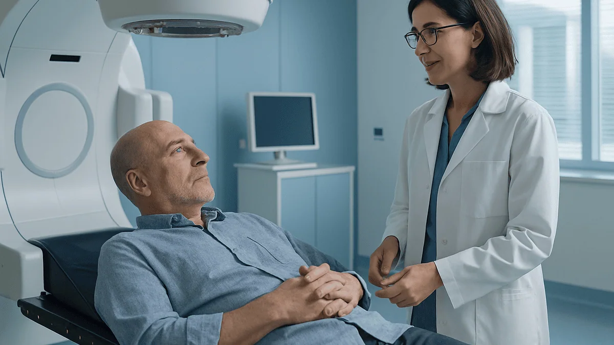
During an SBRT session, you lie in a custom mold or frame made at an earlier planning visit, the team takes a CT or X-ray scan to confirm the tumor sits exactly where the plan expects, small couch adjustments are made, and a linear accelerator then delivers focused, painless radiation beams from several angles. Most of the appointment is setup and imaging; the beam itself runs for minutes. SBRT is usually given in one to five sessions.
The waiting room has a fish tank and a stack of magazines nobody reads. A woman in a cardigan checks her phone for the third time, then asks the person next to her the question she did not ask her oncologist: “Do you actually feel anything?” He has two sessions left. “Nothing,” he says. “The hardest part is lying still.”
That exchange captures what many people want to know about what happens during SBRT treatment, and what the consent form never quite explains. The letters stand for stereotactic body radiation therapy: a way of aiming a high dose of radiation at a small target in the body with millimeter precision, usually over just a handful of visits.
What follows is the session itself, walked through in order: the mold you lie in, the scan the team studies before pressing the button, the beam you cannot see or feel, and the hours and days afterward. The details matter, because knowing why you are being asked to hold your breath or lie perfectly still turns an intimidating room into a procedure you can picture.
What happens during SBRT treatment, in plain language
Strip away the equipment and an SBRT session has three acts: positioning, imaging checks, and beam delivery. Everything else in the room exists to make those three steps as exact as possible.
Stereotactic body radiation therapy uses a machine called a linear accelerator, a device that generates high-energy X-ray beams and rotates around you on a heavy arm called a gantry. The word stereotactic simply means the target is located using three-dimensional coordinates, the way a surveyor pins a point on a map. Instead of spreading a modest dose over many weeks, SBRT concentrates a large dose into a small volume and delivers it in a short course, typically one to five sessions according to Mayo Clinic.
The catch with a large dose is that any error hurts more. If a tumor sits a centimeter from where the plan assumes, healthy tissue receives the intended dose and the tumor does not. That is why the session feels disproportionately slow before anything appears to happen. Radiation therapists, the technologists who run the machine, spend most of the appointment getting your body into the same position it held during planning, then verifying it with a scan taken on the treatment table.
The beam portion, by contrast, is brief and silent apart from a hum. Radiation is invisible, has no smell and produces no sensation at the moment of delivery. People sometimes describe feeling let down that nothing dramatic occurred. Nothing dramatic is the goal.
Behind the room, a radiation oncologist (the physician who prescribes and oversees the treatment), a medical physicist (who checks the machine and the plan) and a dosimetrist (who calculates how the beams should be shaped) have already done the invisible work. The session you attend is the last step of a process that began at the planning visit.
Before the first beam: simulation and the mold that fits only you
The most consequential SBRT appointment is one where no radiation is given. It is called simulation, and it usually happens days to a couple of weeks before treatment begins.
At simulation the team builds an immobilization device: a shell, cushion or frame shaped to your body so that you can be placed in an identical position each time. For lung or liver targets this is often a vacuum-formed bag that molds around your back and arms and then sets firm. For tumors in the spine it may be a rigid body frame. For the prostate it can be a cushion under the knees and feet with a marked position for the hands. The device is labeled with your name and stored between visits.
Lying in the device, you have a planning CT scan, sometimes combined with an MRI or PET scan from another day. If breathing moves the target, the CT may be recorded in four dimensions, capturing where the tumor sits at each phase of the breathing cycle. Small tattoo dots, tiny permanent ink marks the size of a freckle, or removable skin markers are placed so that lasers in the treatment room can line you up.
Some people also have fiducial markers placed before simulation. Fiducials are small metal seeds, typically gold, inserted into or near the tumor under imaging guidance so the treatment machine can track its position on X-ray. Whether you need them depends on the tumor site and the tracking system used; your radiation oncologist decides.
After simulation the physics team spends days shaping beams around the target on the CT images, deliberately steering dose away from the spinal cord, bowel, heart or airways nearby. The plan is checked on the machine before you ever return. That interval is not a delay in care; it is where the precision comes from.
SBRT vs conventional radiation: what changes when the dose is concentrated
People who have watched a relative attend radiation every weekday for six or seven weeks are often confused when SBRT is described as one to five visits. The tumor is not being treated less. The dose per visit is much higher, and the total course is compressed, as the National Cancer Institute describes in its overview of external beam radiation.
Compression is possible for two reasons. First, the target is small and well defined, so a steep dose gradient can be drawn around it: full dose inside, a sharp fall-off just millimeters outside. Second, the daily imaging and immobilization described above make that gradient trustworthy. Conventional radiation, by contrast, often treats a larger area including regions where microscopic disease might hide, and relies on many small daily doses to let normal tissue recover between visits.
| Feature | Conventional external beam radiation | SBRT |
|---|---|---|
| Typical number of sessions | Many, often daily for several weeks | One to five (Mayo Clinic) |
| Dose per session | Small | Large |
| Treated volume | Tumor plus surrounding margin, often wider | Small, tightly defined target |
| Imaging before each session | Periodic, sometimes daily | Every session, often more than once |
| Immobilization | Standard positioning aids | Custom mold or frame, motion management |
| Time per appointment | Usually brief | Longer setup; beam time still short |
Neither approach is universally better. A tumor pressing broadly against the bowel, or a region where the whole area needs treatment, may be safer with conventional fractions. The radiation oncologist weighs tumor size, location, what sits next to it, and your overall health. The table describes how the sessions differ, not which one you should have; that judgment belongs to your team.
Who is SBRT usually for, and who is usually asked to wait
SBRT earns its keep on targets that are small, visible on imaging, and surrounded by tissue that can tolerate a sharp dose edge. Mayo Clinic and the National Cancer Institute describe its use in lung, liver, pancreas, kidney, prostate, spine and adrenal tumors, and in a limited number of metastases, tumors that have spread from another site.
Several situations commonly bring it into the conversation. Someone with an early lung cancer who cannot safely have surgery because of heart or breathing problems may be offered SBRT as an alternative. A person whose cancer has spread to one or a few sites, sometimes called oligometastatic disease, may have those spots treated individually. Spinal metastases causing pain, or a liver tumor that cannot be resected, are other frequent uses. Some prostate cancer programs also use SBRT as a short-course option; who qualifies is a specialist decision.
The question people ask most, what size tumor SBRT can treat, has no single answer. Guidelines and center descriptions consistently refer to small tumors, but the practical limit depends less on a tape-measure figure than on what neighbors the tumor. A modest lesion wrapped around a major airway or against the stomach may be riskier than a somewhat larger one sitting in the periphery of the lung.
Who is usually asked to wait, or steered elsewhere? People whose disease is widespread, where treating one spot would not change the overall picture, are generally offered systemic therapy first. A tumor whose edges cannot be clearly seen on imaging is a poor target. Active infection, uncontrolled pain that makes lying still impossible, or a recent course of radiation to the same area may lead the team to pause. Pregnancy is a specific contraindication that must be discussed. None of these are permanent verdicts; they are reasons to plan differently.
Step one: getting positioned on the treatment couch
You will be asked to change into a gown or remove clothing over the treatment area, and to take off jewelry or anything metallic nearby. Then two or three radiation therapists walk you into a large room with a flat, narrow couch under the gantry. Your personal mold is already waiting on it.
Lying down in the mold is the first test. The therapists compare your skin marks with red or green laser lines projected from the walls and ceiling, then nudge your shoulders, hips or chin until the lines cross the marks. They may ask you to relax and let them move you rather than helping; a body that is actively adjusting itself is harder to place than one that is limp.
For chest and abdominal targets, an abdominal compression plate may be lowered gently onto your upper belly. It feels like a firm hand pressing down and limits how far your diaphragm can move, which shrinks the range the tumor travels with each breath. Head and neck immobilization uses a mesh mask made at simulation. Spine treatments may use a full-body frame with straps.
Comfort is not an afterthought. Because the session can be long, the therapists check whether your arms are numb, whether the cushion under your knees is right, and whether you need a blanket. Speak up now rather than ten minutes into the beam; changing position later means repeating the imaging.
Before leaving the room the therapists confirm your identity, the treatment site and the session number against the plan. Then the door closes. You are alone in the room, but you are not alone: cameras and an intercom keep you in constant contact, and a hand is on the pause button at all times.
Step two: the imaging checks before the beam turns on
The couch may rotate slightly, then the gantry begins to circle you. This is not treatment yet. The machine is taking a cone-beam CT, a low-dose CT scan produced by the treatment unit itself, or a pair of orthogonal X-rays from fixed angles. The purpose is to see where your tumor and organs actually are at this moment.
Outside the room, on a bank of monitors, the therapists overlay today’s image onto the planning CT. Bones are matched first; soft tissue or fiducial markers second. Differences of a few millimeters are normal and expected, because no body settles identically twice. The couch then shifts by those millimeters, in any of six directions including small tilts, to bring today’s anatomy into alignment with the plan. Cleveland Clinic describes this image guidance as a routine part of modern radiation delivery.
Some findings prompt a pause rather than a shift. A full bladder when the plan assumed an empty one, gas in the bowel next to a pancreatic target, or a lung tumor that has shrunk or moved between planning and treatment can each change the geometry. The radiation oncologist may be called to review the images before proceeding. Occasionally the session is rescheduled so the plan can be adjusted. Frustrating as that is, it is the system working as intended.
Once the shift is applied, a second image is often taken to verify it. For treatments using breath-hold or tracking, additional X-rays may be acquired during delivery itself, so the machine is continuously confirming that the target has not drifted.
From your side, this phase is quiet: some whirring, a few clicks, the couch moving in tiny increments you can barely feel. It usually takes longer than the beam that follows.
Step three: beam delivery and what you actually feel
The therapist’s voice comes through the speaker: “We’re starting treatment now. Stay still and breathe normally.” A buzz or steady hum begins. That is the only sign.
The gantry rotates around you, sometimes in a continuous arc, sometimes stopping at several fixed angles. Inside the head of the machine, a multileaf collimator, a set of dozens of thin tungsten leaves that slide in and out, shapes each beam to match the tumor’s outline from that direction. Beams from many angles converge on the target so that the tumor receives the sum of them while any single path through healthy tissue receives only a fraction.
You feel nothing from the radiation itself. Medline Plus and Mayo Clinic both describe external beam radiation as painless during delivery. There is no heat, no tingling, no light. What you do feel is the couch, the mold, the compression plate if one is used, and the effort of not shifting. Some people notice the room is cold or that their nose itches the moment they are told not to move.
If you need to cough, sneeze or move, say so. The beam stops instantly when the therapists release the button; a break is far better than treating a moved target. After repositioning, imaging is repeated and delivery resumes where it left off. Nothing is lost.
A minority of treatments use a robotic arm carrying a compact linear accelerator rather than a rotating gantry. The arm moves around you in a series of positions, pausing at each, and takes frequent X-rays to track fiducials or the tumor itself. The experience is similar: lie still, listen for instructions, and let the machine do the moving.
When the hum stops and the door opens, the session is over. Walk out; drive home if you have been cleared to; you are not radioactive.
Why you may be asked to hold your breath: managing motion in the chest and abdomen
A lung tumor near the diaphragm can travel two centimeters or more with each breath. For a treatment designed around millimeter margins, that motion is the single biggest threat to accuracy, and several strategies exist to tame it.
The simplest is the compression plate described earlier, which restricts the depth of breathing. Another is respiratory gating: a sensor on your chest or a camera tracking a marker on your abdomen tells the machine where you are in your breathing cycle, and the beam switches on only during a chosen phase, usually the pause at the end of exhaling. You breathe normally and the machine does the timing.
Breath-hold techniques ask more of you. You take a breath to a consistent depth, sometimes guided by a mouthpiece that measures the air you inhale, and hold it for a set interval while the beam runs. Many systems show you a bar or a line on a screen so you can see when you are in the right zone. The therapists coach you through it, and the hold is repeated as many times as needed. People with limited lung capacity are usually offered gating or compression instead.
Tumor tracking is the most technically involved option. Fiducial markers or the tumor’s own outline are followed on repeated X-rays, and the beam or the couch adjusts to keep pace. This is used when motion is large and irregular.
Which method you get is decided at simulation and rehearsed there, so nothing in the treatment room should be new. If holding your breath makes you anxious, tell the team early. Anxiety changes breathing patterns, and a calm, consistent rhythm is worth more to the plan than any single deep breath.
How long does SBRT take? Session length and the full course
Two different clocks are running, and people often confuse them.
The first is the course: how many visits. Mayo Clinic describes stereotactic body radiation as typically delivered in one to five treatments. Those sessions are usually spaced out rather than given on consecutive days, often every other day or a few times a week, so that normal tissue between the beams has time to begin recovering. The total course therefore usually spans one to two weeks, though the exact schedule is set by the radiation oncologist for the tumor site.
The second clock is the appointment. Here the answer surprises most people. The beam itself often runs for only a few minutes to around a quarter of an hour, depending on the number of angles and whether gating pauses it. Positioning and imaging take the rest. Mayo Clinic notes that linear-accelerator-based stereotactic treatments commonly take under an hour, while more complex setups can run longer. Plan on being in the department for an hour or more on the first day, when everything is being verified for the first time, and somewhat less on later days.
Several things stretch the time. A target that moves with breathing means gating pauses. A full-or-empty bladder protocol may require waiting. Any imaging finding that needs the doctor’s review adds minutes. None of these are signs that something is wrong.
Practical implications follow. Eat according to the instructions you were given, since some protocols ask you to fast or to drink a set amount of water. Bring something to occupy a wait. Empty your pockets of anxieties as well as keys: the slower the session feels, the more carefully the team is working.
What to expect after SBRT: the first hours and the sbrt side effects timeline
You walk out of the treatment room able to do most of what you did walking in. There is no anesthesia to shake off, no incision, and no radiation lingering in your body; external beam treatment does not make a person radioactive, as the National Cancer Institute confirms. Most people drive themselves home unless a sedative was given for anxiety or claustrophobia.
Side effects, when they come, arrive on two timelines. Early effects appear within days to a few weeks and usually fade over the following weeks. Late effects can surface months later and are less common but more durable. Which ones you might experience depends entirely on what sits inside the treated volume.
Fatigue is the most widespread early effect across all sites, described by the NHS as common with radiotherapy generally. It tends to build over the course and can persist for a few weeks afterward. Skin over the treated area may redden or feel tender, though this is milder with SBRT than with long courses because fewer entry points receive repeated dose.
Site-specific early effects follow the anatomy. Lung treatment can bring cough or mild breathlessness; treatment near the esophagus can make swallowing sore for a week or two. Abdominal targets may cause nausea or looser stools. Bone metastases sometimes produce a pain flare, a temporary increase in pain in the first days after treatment, which is expected and which your team can help manage. Prostate treatment often brings urinary frequency or urgency that peaks in the first couple of weeks.
When people ask about the hardest days after radiation, the honest answer for SBRT is often the end of the first week and the second week, when fatigue and any local irritation overlap. Your own pattern may differ. Keep the after-hours number handy and report anything that worries you.
Late effects and what the follow-up scan can and cannot show
Months after the last session, the treated tissue is still changing. Radiation triggers a slow inflammatory response that gradually gives way to scarring, and for a small minority of people that process causes symptoms.
In the lung, radiation pneumonitis, an inflammation of lung tissue in the treated region, can develop weeks to a few months after SBRT, producing cough, breathlessness or low-grade fever. It is treatable and usually resolves, but it needs to be recognized, which is one reason follow-up visits are scheduled in that window. In the liver, transient changes in blood tests are common and usually settle. Near the ribs or chest wall, aching or, rarely, a rib fracture can occur many months later. Spine treatment carries a small risk of vertebral compression fracture. Prostate treatment may leave some people with longer-term urinary or bowel changes. Cleveland Clinic and Mayo Clinic both describe late effects as depending on the treated site and the tissues within the field.
The follow-up scan brings its own puzzle. A tumor treated with SBRT does not vanish overnight; it shrinks slowly over months, and the surrounding tissue often becomes dense with scar. On a CT scan, that scar can look larger than the original tumor. Radiologists and radiation oncologists are trained to read these patterns, but the images can be genuinely ambiguous for a time, and a PET scan or a repeat interval scan may be needed before anyone can say what is happening. If your team says “we’ll look again in three months,” that reflects biology, not indecision.
Keep every follow-up appointment even when you feel well. The visit exists to catch the uncommon late effect early and to watch the treated area settle. Bring a list of any new symptoms and when they started; timing is often the clue that distinguishes a treatment effect from something unrelated.
What people often get wrong about SBRT
“It’s surgery.” The word radiosurgery, used for stereotactic treatment of the brain, has bled into how people imagine SBRT. Nothing is cut. There is no anesthetic, no incision and no operating room. The precision is surgical; the method is not.
“Fewer sessions means a weaker treatment.” The opposite is closer to the truth. Each SBRT fraction carries a far higher dose than a conventional daily fraction. The short course is possible because the dose is concentrated, not because less is given.
“I’ll be radioactive afterward.” External beam radiation passes through you and stops the instant the machine stops. The National Cancer Institute is explicit that it does not make you radioactive. You can hug children and hold a baby the same afternoon. This is different from some internal radiation treatments involving implanted sources, which have their own precautions.
“If I don’t feel anything, it didn’t work.” Radiation damage to tumor DNA is silent at the moment it happens. Absence of sensation says nothing about effectiveness.
“The scan will show the tumor gone right away.” As described above, response unfolds over months and scar tissue can mimic residual tumor on early imaging.
“Any tumor can be treated with SBRT.” It suits small, well-defined targets with a safe neighborhood. Large, diffuse or poorly visible tumors, or those wrapped around sensitive structures, may be better served by other approaches. Being told SBRT is not suitable is a statement about geometry, not about how seriously your case is being taken.
“Side effects are the same as my neighbor’s.” Effects track the treated anatomy. A person treated for a lung nodule and one treated for a spine metastasis have almost nothing in common in what they might feel afterward.
What does "success rate" really mean for SBRT?
It is one of the most searched questions and one of the hardest to answer honestly, because success has several meanings and the numbers change with every one of them.
Local control means the treated spot itself stops growing or shrinks. This is what SBRT is designed to achieve, and in trials it is measured at set intervals after treatment. Progression-free survival means the cancer has not grown anywhere, treated or untreated. Overall survival means being alive at a given time point, whatever the cause. A treatment can achieve excellent local control while the disease advances elsewhere, which is why the radiation oncologist and the medical oncologist plan together.
Published figures also depend on the cancer type, the tumor size, whether the person could have had surgery, how many spots were treated, what other therapies were given, and how long people were followed. A number quoted for early lung cancer in people unfit for surgery cannot be transferred to a liver metastasis from bowel cancer. Any single percentage floating loose on the internet has been separated from the conditions that made it true.
This article deliberately does not offer a headline number. The responsible source for your figure is the team that knows your scans, your pathology and your other health conditions. Ask them directly: what outcome is this treatment aiming for in my case, how often is that achieved for people like me, and where does that estimate come from? A good answer will name the tumor type and stage it refers to and will acknowledge uncertainty. Mayo Clinic and the National Cancer Institute both frame stereotactic radiation as a treatment whose results depend on the situation rather than a fixed rate.
What the evidence does support is the general principle: focused high-dose radiation, delivered with rigorous imaging, controls small tumors locally in a substantial proportion of appropriately chosen people. Who counts as appropriately chosen is the whole art.
Questions to ask your care team before the first session
A good consultation leaves you knowing not just that you will have SBRT but why this approach, why this many sessions and what will be watched afterward. These questions help get there.
- What exactly is the target, and what healthy structures lie closest to it? Ask to see the planning scan if that helps you understand the risks being weighed.
- Why SBRT rather than surgery, conventional radiation, ablation or systemic therapy for my situation? What would change if I chose to wait?
- How many sessions will I have, how far apart, and roughly how long should I expect to be in the department each time?
- Will I need fiducial markers, a breath-hold technique or a compression device? Can I practice at simulation?
- Are there instructions about eating, drinking, bladder or bowel preparation before each visit?
- Which side effects are most likely for my treatment site, when would they start, and how long do they usually last?
- Which symptoms should prompt a same-day call, and what number do I use after hours?
- Should any of my regular medicines or supplements be reviewed before treatment? Do not change anything on your own; ask the prescribing clinician.
- When will the first follow-up scan be, and how will you tell scar tissue from remaining tumor?
- What is the goal of this treatment in my case, and how will we know whether it has been reached?
Bring someone with you if you can, and write the answers down. People routinely forget most of what is said in a consultation, and the person beside you may remember the one detail that reassures you at two in the morning. If an answer is unclear, say so. Radiation oncology teams expect these questions and would rather explain twice than have you lie on the couch wondering.
When to call your doctor: red-flag signs after SBRT
Most people get through SBRT with fatigue, some local soreness and little else. A few symptoms, though, should not wait for the next scheduled appointment. Your department will have given you a number that works evenings and weekends; use it.
Call the same day, or seek emergency care, if you notice any of the following:
- New or worsening shortness of breath, chest pain, or coughing up blood after lung or chest-wall treatment.
- A fever, especially with cough or breathlessness, in the weeks after treatment; this can be an early sign of pneumonitis or infection.
- Sudden severe pain at a treated spine site, new weakness or numbness in the arms or legs, or new difficulty controlling bladder or bowel. These can indicate pressure on the spinal cord and need urgent assessment.
- Inability to swallow liquids, persistent vomiting, or signs of dehydration after treatment near the esophagus or abdomen.
- Black or bloody stools, blood in the urine that does not settle, or an inability to pass urine after pelvic or abdominal treatment.
- Severe or rapidly worsening abdominal pain, or yellowing of the skin or eyes after liver or pancreatic treatment.
- Any symptom that frightens you or that your instinct says is not the ordinary tiredness you were warned about.
Less urgent but still worth a call: skin that is blistering or weeping, pain that your usual approach is not controlling, nausea that stops you eating for more than a day, or any new symptom you cannot explain. The NHS radiotherapy guidance encourages patients to report side effects rather than tolerate them, since many can be eased.
Every judgment here rests with your treating team. This section is a prompt to pick up the phone, not a substitute for the person who knows your case. When in doubt, call.
Frequently asked questions
How long does SBRT take for a single session?
Plan for roughly an hour in the department, sometimes more on the first day. Mayo Clinic describes linear-accelerator-based stereotactic treatments as commonly taking under an hour. The beam itself often runs only a few minutes to around a quarter of an hour; the remainder is positioning in your mold, imaging on the couch, and the small adjustments that make the treatment accurate.
What size tumor can be treated with SBRT radiation?
There is no universal cutoff. Major sources such as Mayo Clinic and the National Cancer Institute describe SBRT for small, well-defined tumors, and the practical limit depends more on what lies next to the tumor than on a single measurement. A modest tumor wrapped around a major airway may be riskier than a somewhat larger one in the lung periphery. Your radiation oncologist judges suitability on your scans.
What are the hardest days after radiation treatment with SBRT?
For many people the toughest stretch is the end of the first week into the second, when fatigue builds and any local irritation, such as sore swallowing, cough, urinary frequency or a pain flare at a treated bone, peaks. Most early effects then fade over the following weeks. Your pattern depends on the treated site, and your team can help manage symptoms rather than leaving you to wait them out.
What is the success rate of SBRT radiation therapy?
No single figure is honest, because success means different things: local control of the treated spot, freedom from progression anywhere, or overall survival. Each varies with cancer type, tumor size, other treatments and follow-up length. Ask your radiation oncologist what outcome the treatment aims for in your case and how often that is achieved for people with your specific diagnosis and stage.
What does SBRT feel like during beam delivery?
Nothing from the radiation itself. Medline Plus and Mayo Clinic describe external beam radiation as painless during delivery; there is no heat, tingling or light. What you notice is the couch, your mold, a compression plate if one is used, and the effort of staying still while the machine hums and rotates. If you need to cough or move, say so and the beam is paused.
What is the difference between SBRT vs conventional radiation in the treatment room?
The room and machine can be identical; the process differs. SBRT uses a custom immobilization device made at simulation, takes imaging on the couch at every session, often manages breathing motion with gating or breath-hold, and delivers a much larger dose per visit over one to five sessions. Conventional radiation spreads smaller doses across many weeks and usually treats a wider area with less elaborate setup.
Will I be radioactive after an SBRT session?
No. External beam radiation passes through the body and stops the instant the machine switches off, and the National Cancer Institute states it does not make you radioactive. You can be around children, pregnant relatives and pets immediately. This differs from some internal radiation treatments that place a radioactive source inside the body and carry their own temporary precautions.
Why do I have to hold my breath during SBRT?
Because tumors in the lung and upper abdomen move with each breath, sometimes by two centimeters or more. Holding your breath at a consistent depth freezes the target so the beam can be delivered to a stationary spot. Alternatives include gating, where the machine fires only during one phase of your normal breathing, and abdominal compression. The method is chosen and rehearsed at your simulation visit.
What to expect after SBRT in the first 24 hours?
Usually very little. You can typically drive home, eat normally and resume light activity unless told otherwise. Some people feel tired or notice mild soreness over the treated area. Nausea can occur after abdominal treatment, and a temporary pain flare can follow bone treatment. Follow the specific instructions from your department and call if anything unexpected develops.
Can SBRT be repeated if the tumor comes back or a new spot appears?
Sometimes, but it depends on how much radiation the surrounding tissue has already received and how much it can safely tolerate. Treating a new spot in a different part of the body is often possible. Re-treating the same area is more complex and requires careful dose accounting by the physics team. Your radiation oncologist will weigh these factors alongside other options such as systemic therapy.
References
This article is for general information only and is not a substitute for professional medical advice. Please consult a qualified doctor about your individual situation.

More from the Blog

When Is Basal Cell Carcinoma Treated Without Surgery? Topical and Procedural Options
Basal cell carcinoma is usually removed surgically, but non-surgical treatment is considered when a tumor is superficial, low-risk and sits on the trunk or…
Living With One Kidney After Cancer Surgery: How Function Is Monitored Over Time
After a kidney is removed for cancer, the remaining kidney usually adapts to take over most of the filtering work, and most people go…

Molecular and Genetic Testing Before Targeted Therapy: How Tumor Markers Match the Drug
Molecular testing before targeted therapy examines a tumor sample for specific biomarkers, such as gene mutations, gene fusions, extra gene copies or abnormal proteins,…

CyberKnife vs Standard Radiation Therapy: Sessions, Precision and Who Suits Which
CyberKnife is a brand name for a robotic form of stereotactic radiosurgery, which delivers a small number of high-precision radiation treatments, typically one to…
Eating After Esophageal Cancer Surgery: Small Meals, Reflux Habits and a New Routine
Eating after esophageal cancer surgery usually means small, frequent meals instead of three large ones, chewing slowly, sitting upright during and after eating, and…
How a Stomach Cancer Tumor Board Decides Between Surgery First and Chemotherapy First
A stomach cancer tumor board is a scheduled meeting where surgeons, medical and radiation oncologists, radiologists, pathologists and nurse specialists review one patient's scans,…