

What the Microscope Changes in Microsurgery: Repairing Vessels and Nerves Thinner Than a Hair

Key Takeaways
- Surgical loupes typically magnify about 2.5 to 4.5 times, while operating microscopes offer an adjustable range that rarely exceeds roughly 40 times.
- Laboratory 4x, 10x, 40x and 100x objectives give 40 to 1000 times total magnification and are used for slides and cells, never for live surgery.
- Free-flap vessels commonly measure one to three millimeters, and supermicrosurgery joins vessels below about 0.8 millimeters.
- The operating microscope provides depth perception through two optical paths and shadow-free coaxial light, which loupes cannot fully match.
- Repaired nerves regrow at roughly one inch per month according to patient guidance from Johns Hopkins, and magnification cannot speed that rate.
- The first three days after a free flap are when vessel clotting is most likely, which is why flaps are checked at short intervals around the clock.
Microsurgery magnification lets a surgeon see and stitch structures roughly one millimeter across or smaller, such as facial nerve branches and the arteries that feed a transplanted tissue flap. Surgical loupes typically enlarge the field about two to five times, while an operating microscope offers a wider adjustable range and steadier optics. Magnification improves visibility and precision; it does not by itself guarantee how a vessel or nerve heals.
A woman in her forties sits in a consultation room with a mirror in her lap. One side of her face has not moved properly since a tumor was removed from her parotid gland, and the surgeon has just said the phrase that brought her here: nerve graft under the microscope. She nods, then asks the question almost everyone asks. How do you sew something you can barely see?
It is a fair question. The branches of the facial nerve that lift an eyebrow or steady a smile can be about as wide as a coarse human hair. The blood vessels that keep a transplanted flap of tissue alive are often only a few times wider than that. Nobody sutures them by squinting.
What makes this work is microsurgery magnification: a chain of optics, instruments and habits that scales the surgeon’s hands, eyes and tolerance for error down to the size of the problem. This explainer walks through what actually changes when the microscope swings over the table, what it cannot change, and what patients tend to ask afterward.
How small is thinner than a hair, really?
Numbers help here. A typical human scalp hair measures somewhere between 50 and 100 micrometers across, which is a twentieth to a tenth of a millimeter. Hold that against the structures a microsurgeon handles on an ordinary day.
The artery and vein that supply a free flap (a block of skin, fat or muscle detached from one part of the body and reconnected elsewhere) commonly run between one and three millimeters in diameter, roughly the width of a cooked spaghetti strand. Sounds manageable, until you consider that the vessel wall itself is a fraction of that, and the suture has to pass through the wall without catching the far side. Digital nerves in the fingers and the distal branches of the facial nerve are often close to a millimeter or less. Lymphatic channels, the target of some newer procedures for limb swelling, can be well under half a millimeter.
The eye alone cannot resolve detail at that scale reliably. Two points closer together than about a tenth of a millimeter blur into one at normal reading distance, and the fine layered anatomy of a vessel wall or the tiny bundles inside a nerve sit below that threshold. The microscope does not make the surgeon’s hands smaller. It makes the target large enough that a hand tremor of half a millimeter looks like what it is: a mistake in progress, visible in time to stop.
Everything else in microsurgery follows from that shift. Instruments shrink, sutures shrink, the pace slows, and the operating room quietens. The scale of the anatomy sets the rules.
How microsurgery magnification actually works in the operating room
An operating microscope is, at heart, a pair of binoculars on an arm, with a light source pointed down the same path the surgeon looks along. Three features separate it from the magnifying glass in a drawer.
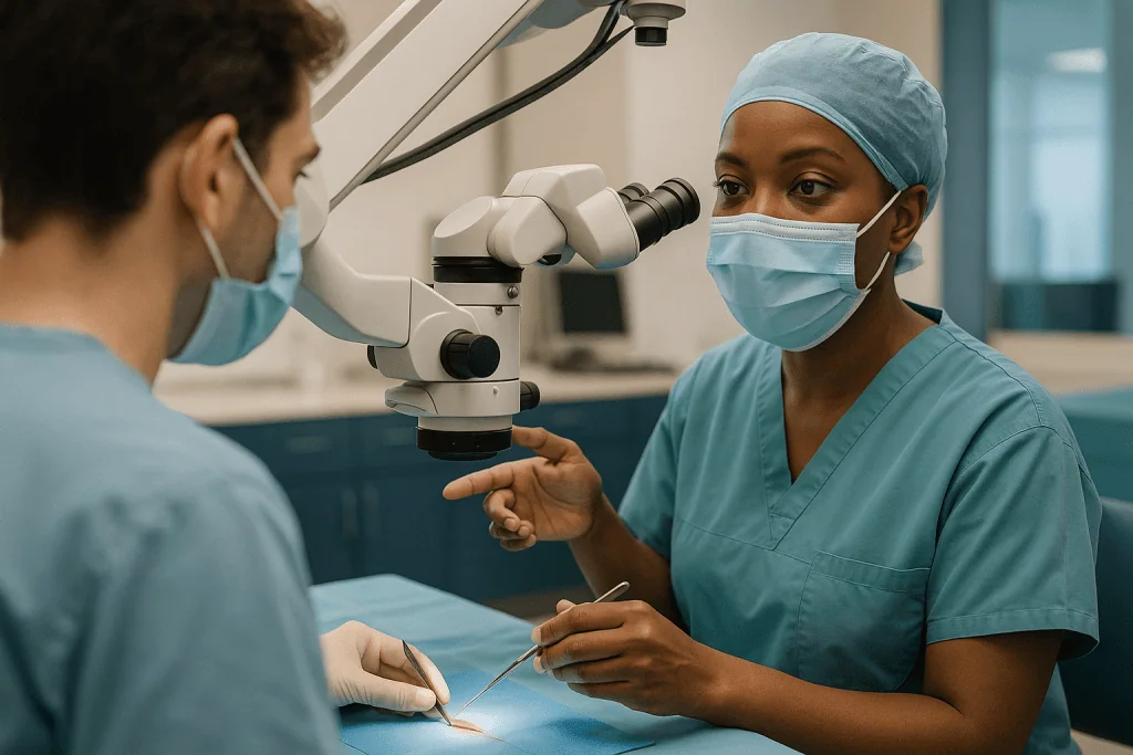
First, it is stereoscopic. Each eye receives a slightly different image through separate optical paths, so the surgeon perceives depth. Placing a needle through a vessel wall, rather than in front of it or behind it, depends entirely on that depth cue. Second, the light is coaxial, meaning it travels down the same axis as the view, so the bottom of a deep wound is illuminated without shadows from the surgeon’s own hands. Third, the working distance is long, typically twenty to forty centimeters, which leaves room for instruments underneath the lens.
Magnification on most surgical microscopes is adjustable through a zoom system rather than fixed objectives. A surgeon might dissect at low power to keep the whole field in view, then step up several times for the stitches themselves. Focus is controlled by a foot pedal or hand grip so that both hands stay on the instruments.
Many modern systems add a second eyepiece for an assistant and a camera feeding a screen, which lets the team see exactly what the surgeon sees. Some units, sometimes called exoscopes (a camera-based system that shows a magnified image on a large monitor rather than through eyepieces), replace the eyepieces entirely. The optical principle is unchanged: gather more light, enlarge the image, preserve depth, and keep the surgeon’s neck and hands free to work.
Loupes vs microscope surgery: when is each chosen?
Surgical loupes are magnifying lenses mounted on eyeglass frames. The surgeon wears them, moves with them, and sets up nothing on the table. That convenience is why loupes are the everyday tool for a great deal of hand, plastic and reconstructive work.
Their limits are physical. Common loupes enlarge the field about two and a half to four and a half times, with a fixed focal distance. Lean in and the image blurs; lean back and it blurs the other way. Higher-power loupes exist, but the field of view narrows and the weight on the nose climbs. Surgeons develop a stiff posture to hold the image still, and neck strain is a recognized occupational complaint.
An operating microscope changes the equation in three ways. Magnification can climb well beyond loupe range and can be adjusted mid-procedure without stopping. The image is stabilized by the stand, not by the surgeon’s spine. And a co-observer, whether a trainee or a second surgeon, sees the same view.
Which tool ends up in use depends on the size of the target and the surgeon’s training. Repair of a larger nerve or a vessel of two to three millimeters is frequently performed under loupes by experienced hands, and studies comparing outcomes for some digital nerve repairs have not shown a consistent advantage for one over the other. Below about one millimeter, and for lymphatic work, the microscope becomes the norm. Neither is inherently superior; they are instruments matched to a job, and the treating surgeon’s judgment about that match is part of the care.
What magnification do surgeons use? A plain comparison
People searching this question often land on laboratory microscope pages, where objectives of 4x, 10x, 40x and 100x are standard. Those numbers describe a different world. The table below sets surgical tools beside the lab bench so the two are not confused.

| Tool | Typical magnification | What it is used for |
|---|---|---|
| Unaided eye | 1x | Skin closure, larger vessels, most general surgery |
| Surgical loupes | About 2.5x to 4.5x | Tendon repair, larger nerves, vessels of a few millimeters |
| Operating microscope, low range | Roughly 4x to 10x | Dissection, identifying vessel and nerve branches |
| Operating microscope, high range | Roughly 10x to 40x | Suturing vessels near or below one millimeter, nerve fascicle matching |
| Lab microscope, 4x to 40x objectives | 40x to 400x total with a 10x eyepiece | Tissue slides, cells, pathology review |
| Lab microscope, 100x oil objective | About 1000x total | Bacteria and fine cell structure, never live surgery |
Two points deserve emphasis. Surgical magnification rarely exceeds about forty times, because higher power shrinks the field of view to a pinhole and magnifies every tremor and breath along with the anatomy. And the total magnification quoted for lab microscopes multiplies the objective by the eyepiece, which is why a 40x objective yields 400x. A surgeon working at 100x, let alone 1000x, could not see both ends of a stitch at once.
Ranges above reflect manufacturer specifications and surgical training texts rather than clinical outcome data, and individual surgeons choose settings within them case by case.
Instruments, sutures and the problem of a steady hand
Enlarge the view and everything the hands touch must follow. Microsurgical forceps taper to tips finer than a pencil point. Needle holders lock with a whisper of spring pressure so that squeezing does not jolt the tissue. Scissors are sprung, curved and small enough that the whole instrument disappears under the lens.
Sutures shrink furthest of all. The threads used for a one-millimeter vessel are thinner than the hair they are being compared with, and the attached needle is a curved sliver a few millimeters long. Under the microscope it looks substantial. Lift the eyepieces and it is nearly invisible against a drape, which is why surgical teams count and track them with particular care.
Tremor is the enemy nobody can fully abolish. Every human hand oscillates a fraction of a millimeter with each heartbeat and breath. At normal scale this is irrelevant; at high magnification it is the dominant motion on the screen. Microsurgeons manage it rather than defeat it. Forearms rest on padded supports so movement comes from the fingers alone. Stools are set so shoulders drop. Many avoid caffeine before long cases. Breathing is timed so the needle passes between breaths.
Training matters as much as equipment. Surgeons practice on synthetic tubes, then on laboratory models, long before a patient’s artery is on the table, because the skill is partly optical, partly manual, and partly a learned patience with a pace that feels unnaturally slow. A single vessel join may take the better part of an hour. That is not delay. It is the method.
Repairing a blood vessel: what happens stitch by stitch
The join between two vessels is called an anastomosis (a surgical connection that allows blood to flow from one vessel into another). Under the microscope it unfolds in a sequence that varies little between an artery in the forearm and one at the base of the neck.
The surgeon first clears the vessel ends of surrounding tissue, so that the wall itself is visible, and trims back any damaged segment. Tiny clamps hold each end still and stop bleeding. A drop of irrigation keeps the field clear, and the surgeon examines the inner lining, called the intima, because a rough or torn lining is where clots begin.
Stitches then go in one at a time, passing through the full thickness of both walls so the linings meet edge to edge. The classic technique places the first two stitches at opposite points, then fills in between, rotating the clamps to reach the back wall. A one-millimeter artery may need eight to twelve stitches. Each has to be evenly spaced and gently tensioned; too tight and the wall puckers, too loose and blood leaks or pools.
When the clamps come off, the team watches. A healthy repair fills, pulses and shows bright flow past the suture line. A vein is checked by gently emptying and refilling it. If flow is sluggish, the surgeon may redo the join. In some cases medicines that reduce clotting, such as antiplatelet or anticoagulant agents, are considered by the anesthesia and surgical teams to protect the repair; whether and how they are used is a decision made for each patient by the treating clinicians, not a fixed part of the procedure.
Repairing a nerve: why magnification helps but cannot rush regrowth
A peripheral nerve is not a single wire. It is a cable of many bundles, called fascicles (groups of nerve fibers wrapped in their own thin sheath), each carrying fibers to a particular muscle or patch of skin. When a nerve is cut, the fibers beyond the cut degenerate. Repair does not reconnect them; it lines up the tubes so that new fibers sprouting from the healthy side can grow down the right path.
Magnification earns its place here. Under the microscope a surgeon can match fascicle to fascicle, align the small surface vessels that run along a nerve as landmarks, and place stitches in the outer sheath without spearing the fibers inside. When a gap is too wide to close without tension, a graft taken from a less important sensory nerve, or a synthetic conduit, bridges the distance.
Then biology sets the timetable. Nerve fibers regenerate slowly; patient resources from Johns Hopkins describe a rate of about one inch per month, and Mayo Clinic notes that recovery after nerve injury may take months and is not always complete. A repair at the wrist may not reach the fingertips for the better part of a year. The microscope improves the odds that fibers find the right tube. It has no effect on how fast they travel.
Patients often find this the hardest part to accept. Nothing visible happens for months, tingling and odd sensations come and go, and the surgeon’s answer to “is it working” is honestly “we will know when it gets there.” That uncertainty is inherent to the tissue, not a sign that anything went wrong.
Microsurgery magnification in facial surgery: nerves, flaps and smiles
The face concentrates everything that makes microsurgery demanding. Nerve branches are small, blood vessels are small, and the margin for a poor result is measured in how a person looks when they laugh.
Facial nerve work is the clearest example. When a branch is cut during tumor removal or trauma, direct repair or grafting under the microscope aims to give regenerating fibers a route back to the muscles of expression. When the nerve cannot be repaired, surgeons may borrow signal from a neighboring nerve, such as the one that moves the tongue or the one to the chewing muscles, and route it to the paralyzed side. In longer-standing paralysis, a small muscle can be transplanted from the thigh along with its artery, vein and nerve, and connected in the cheek. Every one of those connections is a microsurgical join.
Free flaps rebuild the face after cancer surgery or major injury. A segment of bone from the leg with its blood supply can reconstruct a jaw; a patch of forearm skin can line the mouth. The flap lives or dies on the anastomosis between its vessels and those in the neck.
Facial aesthetic surgery in the narrower sense, such as face-lift procedures, does not usually involve vessel or nerve repair under a microscope. Magnification still appears in the form of loupes, used to identify and protect nerve branches during dissection. The distinction matters for expectations: a person considering cosmetic facial surgery is not typically facing microsurgery, while a person facing facial reanimation or reconstruction almost certainly is.
Supermicrosurgery vessels: what changes below one millimeter?
Supermicrosurgery is the term used for joining vessels smaller than about 0.8 millimeters, often down to 0.3 millimeters. At that scale a vessel is roughly the width of a few hairs laid side by side, and the wall is nearly transparent.
The best-known application is lymphaticovenous anastomosis, a procedure that connects blocked lymphatic channels directly to tiny veins so that fluid can drain. It is offered in some centers for lymphoedema, the chronic swelling that can follow lymph node removal. The NHS describes surgical options for lymphoedema as suitable only for selected people and notes that compression and physical therapy remain the foundation of management; the evidence base for surgery is still developing, and it is not a routine first step.
Technically, supermicrosurgery pushes every element of the craft further. Magnification runs at the upper end of the microscope’s range. Sutures are finer still. Surgeons often inject a fluorescent dye and use a special camera to find lymphatic channels that would otherwise be invisible under the skin. Fewer stitches are placed, because each one occupies a larger share of the tiny circumference.
The same techniques appear in fingertip replantation, where the vessels at the very end of a finger are joined, and in some perforator flaps, where a flap is nourished by a single small branch rather than a larger trunk. The common thread is that as targets shrink, the microscope stops being one tool among several and becomes the only way the work can be done at all.
Who is microsurgery usually for, and who is usually asked to wait?
Microsurgical repair is considered when a structure is too small to fix by other means and its function matters enough to justify a long operation. Typical situations include a severed nerve in the hand or face, an amputated finger that might be reattached, reconstruction after cancer surgery, and breast reconstruction using the patient’s own tissue.
Timing is often driven by the injury. A cut nerve is generally repaired soon, because the fibers on the far side degenerate and the muscles they supplied begin to waste with time. An amputated digit has a window of hours. Reconstruction after tumor removal is frequently planned in the same operation.
Other people are asked to wait, or are steered toward alternatives, for reasons that come back to blood flow. Mayo Clinic notes that smoking raises the risk of complications after flap surgery, and most teams ask patients to stop nicotine in all forms for a period before and after, because nicotine constricts small vessels, exactly the ones being joined. Poorly controlled diabetes, active infection, some clotting disorders and severe vascular disease can all tip the balance. Weight and general fitness enter the discussion not as judgments but because long anesthetics and slow-healing wounds carry more risk for some bodies than others.
None of this is a checklist a patient can apply alone. Surgeons, anesthetists and often other specialists weigh the whole picture, including how much the structure matters to that person’s life. A musician’s finger and a retired person’s finger are anatomically identical and may be counseled differently, and both conversations belong with the treating team.
What the following days and weeks usually look like
The first three days after a free flap are the anxious ones. A freshly joined vessel is most likely to clot in this window, and teams check the flap at short intervals around the clock. Nurses assess its color, warmth and how quickly it refills after a gentle press; many units also use a small Doppler probe, which turns blood flow into an audible whoosh, or a light-based sensor that reads oxygen levels in the tissue. Mayo Clinic describes a hospital stay of several days after flap-based breast reconstruction, and similar patterns apply to flaps elsewhere.
Position and temperature matter more than most people expect. A flap in the neck may require the head to stay neutral so the vessels are not kinked. Rooms are kept warm because cold constricts vessels. Caffeine and nicotine are typically off the menu.
Nerve repairs follow a different rhythm. The wound heals in a couple of weeks, splints or restrictions protect the join from tension for several weeks more, and then the long wait begins. Sensation or movement may take months to appear and continues to change for up to two years, according to patient resources from Johns Hopkins and Mayo Clinic. Physical or occupational therapy is commonly part of the plan, retraining the brain to interpret signals from fibers that may have found slightly different endings than before.
All of these are typical ranges drawn from general guidance, not commitments. Individual recovery depends on the site, the injury and the person, and the treating team’s instructions override anything written here.
Risks and alternatives, in neutral terms
Microsurgery shares the risks of any operation under anesthesia: bleeding, infection, reactions to medicines and clots in the legs. It adds risks specific to tiny joins.
The vessel connection can clot or leak. If an artery blocks, the flap loses its supply; if a vein blocks, blood enters but cannot leave and the flap becomes congested. Either can require an urgent return to the operating room to reopen the join. Despite the best monitoring, some flaps cannot be saved, and the team will have discussed beforehand what would happen in that case. The site the tissue was taken from, called the donor site, can develop its own problems with wound healing, numbness or weakness.
Nerve repairs carry the risk of incomplete recovery, painful sensitivity at the repair site, and misdirected regrowth, where fibers reach the wrong destination and produce unintended movement or odd sensations. Nerve grafts leave a patch of numbness where the donor nerve was taken.
Alternatives exist along a spectrum. For some nerve injuries, observation is reasonable because bruised rather than cut nerves often recover on their own; Cleveland Clinic notes that most people with Bell’s palsy, a facial nerve condition that is not surgical, improve over weeks to months without any operation. For reconstruction, options may include implants, tissue moved on its own attached blood supply without a microsurgical join, or accepting a simpler closure. For lymphoedema, compression and physiotherapy remain the standard approach. Each path has trade-offs the treating team can lay out for the individual situation.
What people often get wrong about surgical magnification
“More magnification is always better.” It is not. Beyond a point the field of view narrows, depth of focus shrinks and tremor dominates. Surgeons choose the lowest power that shows what they need, and step up only for the finest steps.
“Surgeons use 100x like a lab microscope.” A 100x objective is an oil-immersion lens used to view bacteria on a slide, giving about a thousand times total magnification with the eyepiece. No live tissue is operated on at that scale. Surgical microscopes top out around forty times.
“The microscope does the delicate part.” Optics enlarge the image; hands, training and judgment do the rest. Two surgeons at the same magnification can produce very different repairs.
“If the nerve is repaired well, feeling comes back quickly.” Regeneration is slow by nature, roughly an inch a month according to patient guidance from Johns Hopkins, and the repair only gives fibers a path. Nothing about the operation shortens the journey.
“Robots have replaced the microscope.” Robotic platforms exist for some microsurgical tasks and can filter tremor, but they remain uncommon, still rely on magnified imaging, and evidence comparing them with conventional technique is early.
“Loupes are a lesser tool.” For many repairs, comparative studies have not shown a clear outcome difference between loupes and the microscope. The right tool is the one suited to the target and the surgeon’s training, not the most impressive one in the room.
Questions to ask your care team before microsurgery
A good consultation leaves a person with fewer open loops, not more. These questions tend to surface what matters.
- Which structures will be repaired under magnification, and how small are they?
- Will the operation use loupes, an operating microscope or both, and does that choice affect what I should expect?
- If a nerve is being repaired, what is a realistic timeline for the first signs of recovery, and what might full recovery look like?
- If a flap is involved, how will it be monitored afterward, and what happens if the blood flow becomes a concern?
- What would the alternative be if this repair is not attempted or does not succeed?
- Are there habits, such as smoking or certain medicines, that I need to change before surgery, and who will guide that?
- Where will tissue or nerve be taken from, and what numbness or weakness could that leave?
- How long is the expected hospital stay, and what restrictions on movement or position should I plan for?
- What kind of therapy will follow, and when does it begin?
- Who do I contact, and how, if something worries me after discharge?
Bring a notebook or a second pair of ears. Answers about microsurgery involve timescales that stretch across months, and it is easy to lose the thread between the first appointment and the day of the operation. Any medicine question, including whether to pause or continue something already prescribed, belongs with the prescribing clinician and the surgical team, never with a magazine article.
When to call your doctor after microsurgical repair
Most recoveries are uneventful, but the tiny vessels and nerves at the heart of this surgery can signal trouble early, and early matters. Contact the surgical team without delay, using the emergency number they provided, if any of the following appear after discharge.
For a flap or replanted part: a change in color to pale white, dusky purple or blue; the area becoming cold, or swelling tightly and rapidly; new bleeding that soaks a dressing; or a Doppler signal you were shown how to check going quiet. These can indicate a problem with blood flow, and the window to correct it is short.
For any wound: spreading redness, increasing pain rather than steady improvement, pus or a foul smell, or a temperature of 38°C (100.4°F) or higher, which can point to infection.
For nerve repairs: sudden loss of movement or sensation that had started to return, or severe, escalating pain at the repair site.
Seek emergency care immediately for chest pain, sudden shortness of breath, or a painful swollen calf, which can signal a blood clot after any operation, and for any bleeding that will not stop with firm pressure.
When in doubt, call. Teams that perform microsurgery expect these calls and would far rather hear about a false alarm than learn about a real one late. Decisions about what to do next, including whether to return to the hospital, rest with the treating team, who know the specifics of the repair.
Frequently asked questions
What magnification do surgeons use during microsurgery?
Most microsurgery is performed between about 4 and 20 times magnification, with brief steps toward 40 times for the finest stitches. Loupes worn on the head give roughly 2.5 to 4.5 times. Surgeons choose the lowest power that shows the detail they need, because higher settings narrow the field and amplify hand tremor.
What is the magnification of 4x, 10x, 40x and 100x objectives?
Those figures describe laboratory microscope lenses, not surgical tools. Total magnification multiplies the objective by the eyepiece, usually 10x, giving 40x, 100x, 400x and 1000x. The 4x objective scans a slide, 10x and 40x examine tissue and cells, and the 100x oil-immersion lens resolves bacteria.
What is 100x magnification used for?
A 100x objective, combined with a standard eyepiece for about 1000 times total, is used in laboratories to view bacteria and fine structures inside cells on a prepared slide, with a drop of oil between lens and glass. It has no role in live surgery, where such power would show only a pinpoint of tissue.
What are the four types of magnification on a microscope?
In a lab microscope the four standard objectives are 4x scanning, 10x low power, 40x high power and 100x oil immersion. In the operating room, magnification arrives through four broad routes: loupes, the operating microscope, camera-based exoscopes that display on a screen, and endoscopic or robotic video systems.
How does an operating microscope in surgery differ from loupes?
An operating microscope sits on a stand, offers adjustable magnification, stereoscopic depth, shadow-free light and a shared view for assistants. Loupes are lightweight lenses on glasses with fixed, lower power that move with the surgeon. For very small vessels and nerves the microscope is standard; for larger structures either can be appropriate.
Why can nerve repairs take so long to show results?
Because a repair only realigns the pathway. New nerve fibers must then grow from the healthy side down to the muscle or skin, at roughly one inch per month according to Johns Hopkins patient guidance. A repair far from its target can take many months, and improvement may continue for up to two years.
What are supermicrosurgery vessels and when is that technique used?
Supermicrosurgery refers to joining vessels smaller than about 0.8 millimeters, sometimes down to 0.3 millimeters. It is used for fingertip reattachment, some perforator flaps and lymphaticovenous anastomosis for lymphoedema, a procedure the NHS describes as suitable only for selected people alongside compression and physiotherapy.
Is microsurgery used in cosmetic facial surgery?
Usually not in the vessel-and-nerve sense. Face-lift and similar procedures rely on loupes to identify and protect nerve branches, not on microscopic repair. Microsurgery in the face is mainly for reconstruction after cancer or trauma and for facial reanimation after nerve injury, where nerve grafts or transplanted muscle are connected under the microscope.
Why am I told to stop smoking before flap surgery?
Nicotine constricts small blood vessels, which are exactly the structures being joined. Mayo Clinic lists smoking among factors that raise complication risk after flap surgery. Teams generally ask for complete avoidance of nicotine, including vaping and patches, for a period before and after; the exact plan comes from the treating team.
Do robots replace the microscope in microsurgery?
Not at present. Robotic systems designed for microsurgery can filter tremor and scale hand movements, but they are uncommon, depend on magnified imaging themselves, and have limited comparative evidence so far. The operating microscope, and in many cases loupes, remain the standard tools in most centers.
References
This article is for general information only and is not a substitute for professional medical advice. Please consult a qualified doctor about your individual situation.

More from the Blog
Before Eye Laser Treatment: Corneal Thickness, Eye Pressure and Stopping Contact Lenses
An eye exam before laser treatment measures far more than your glasses prescription. Clinicians map the cornea's shape, measure its thickness in micrometers, check…

Can Xanthelasma Return After Surgical Removal? Recurrence and the Next Options
Yes. Xanthelasma can come back after surgical removal, and published case series report recurrence often enough that surgeons discuss it before treating. The plaques…
Is Surgical Tattoo Removal an Option for You? Size, Location and Skin Elasticity Explained
Yes, tattoos can be surgically removed, but excision suits a narrow group: small to medium tattoos on areas with loose, elastic skin, in people…

How Masseter Botox Is Injected: Locating the Chewing Muscle and What the Session Involves
In a masseter botox procedure, a clinician first asks you to clench your teeth so the masseter, the thick chewing muscle at the back…

Fibroblast Plasma Skin Tightening Results: What Changes, How Gradually and How Long It Lasts
Fibroblast plasma treatment results tend to be modest and gradual rather than dramatic. Small dark crusts usually lift within one to two weeks, pinkness…

How Long Does a Thread Lift Last? Honest Longevity and Who It Suits
A thread lift typically lasts one to three years, depending on the suture material, your age, skin quality, and how much lift was attempted.…