

When Is a Wound Considered Chronic? How a Wound Clinic Finds the Cause Behind Stalled Healing

Key Takeaways
- Published wound-care reviews define a chronic wound as one that has not progressed through normal healing in about four weeks or has not closed within roughly three months.
- Diabetic foot ulcers that shrink by less than about half their area in the first four weeks are markedly less likely to close by week twelve, which is why clinics reassess at that point.
- The four causes behind most chronic wounds are arterial insufficiency, venous insufficiency, diabetes-related nerve and vessel damage, and sustained pressure, and treatment differs for each.
- Stage 4 in pressure-injury classification means full-thickness loss with exposed bone, tendon or muscle; staging applies only to pressure wounds and never runs in reverse as a wound heals.
- An ankle-brachial index is checked before compression is applied to a leg ulcer because compressing a limb with unrecognized arterial disease can damage tissue.
- Every open chronic wound carries bacteria, so a positive surface swab alone does not indicate infection; spreading redness, warmth, new pain, foul drainage or fever do.
A wound is generally considered chronic when it has not moved through the normal stages of healing in about four weeks, or has not closed within roughly three months, according to published wound-care reviews. Wound clinics look for a stalled cause such as poor blood flow, unrelieved pressure, uncontrolled diabetes, infection or swelling, then address that cause rather than the surface alone.
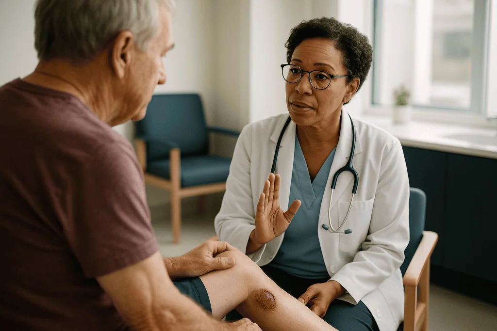
The bandage comes off the same way it did last week, and the week before. A shallow sore on the inner ankle, pink at the edges, a little wet in the middle, not larger but not smaller either. The person holding the gauze has done everything they were told: kept it clean, kept it covered, kept their fingers away from it. Six weeks in, the question changes from “how do I look after this” to “why is this still here.”
That shift is exactly what wound specialists listen for. Most cuts and scrapes close on a schedule the body has rehearsed for a lifetime. When one refuses, the skin is rarely the real problem. Something underneath, a narrowed artery, a vein that no longer drains, a pressure point nobody noticed, a blood sugar that drifts high overnight, is quietly holding the process back.
So when is a wound considered chronic, and what does a wound clinic actually do differently? The honest answer involves a calendar, a set of unglamorous measurements, and a stubborn refusal to treat the surface without first naming the cause.
When is a wound considered chronic? The 4-week and 3-month markers
Clinicians do not wait for a wound to become dramatic before calling it chronic. The most widely used working definition, summarized in a review by Frykberg and Banks in Advances in Wound Care, is a wound that has failed to progress through the normal, orderly stages of repair within about four weeks, or one that has not fully closed within roughly three months. Both markers matter, and they answer slightly different questions.
The four-week marker is about trajectory. A healthy wound should look measurably different every week: smaller, drier, with fresh pink tissue creeping in from the edges. If a nurse measures the same length and width on day 1 and day 28, the wound is not simply slow; it has stalled. The three-month marker is about outcome. Even a wound that shrinks a little each week has crossed into chronic territory if it is still open after twelve weeks.
Why does this matter to a patient rather than a textbook? Because the label changes what happens next. A stalled wound is a signal that something systemic or mechanical is interfering, and standard home care, however diligent, will not fix an artery or a pressure point. The National Health Service uses a similar logic for leg ulcers, describing them as sores that take more than two weeks to heal, a threshold low enough to prompt assessment before months are lost.
The takeaway is not that four weeks is a magic number. It is that time without visible progress is itself a clinical finding, one worth reporting rather than waiting out. A wound clinic would rather see a small, boring sore at week five than a large, complicated one at month five.
How long is a wound considered chronic before a clinic wants to see it?
People often assume a referral requires a wound that is large, deep or obviously infected. In practice, the trigger is usually the calendar combined with a risk factor. Frykberg and Banks note that most wound-care programs use the four-week point as the moment to reassess, because studies of diabetic foot ulcers have found that wounds shrinking by less than about half their area in the first four weeks are far less likely to be closed by week twelve. That percentage is a benchmark, not a promise; some wounds beat it and some do not.

Three situations shorten the wait considerably. A wound on the foot of a person with diabetes, particularly one they did not feel developing, is often assessed within days rather than weeks, because loss of protective sensation means the usual warning system is offline. A wound over a bony area in someone who spends long hours in a bed or chair is reviewed quickly because pressure damage extends under the skin faster than it shows on the surface. And any wound with spreading redness, warmth, foul drainage or a fever is not a “wait and see” situation at all.
For the rest, a reasonable rule many primary-care teams follow is this: if a wound has not clearly improved in two weeks, mention it; if it has not clearly improved in four, ask about a wound-care assessment. Clinics generally prefer earlier, smaller referrals because the search for a cause is simpler when the tissue is still relatively healthy.
Your own doctor decides the timing, and local pathways vary. What you can control is documentation: a photo taken from the same distance each week, with a ruler in frame, gives the clinic a trajectory to read instead of a memory to reconstruct.
How wounds normally heal, and where the process stalls
Healing is less a single event than a relay with four runners, and a chronic wound is a relay in which one runner never receives the baton. MedlinePlus describes the sequence plainly. First comes hemostasis, the clotting that stops bleeding within minutes. Then inflammation, in which white blood cells arrive to clear bacteria and debris; the area looks red and slightly swollen, which is normal for the first few days. Third is proliferation, when new blood vessels sprout, a protein scaffold called collagen is laid down, and skin cells migrate across the gap. Finally remodeling, in which that scaffold is reorganized and strengthened over months.
Chronic wounds are, almost by definition, stuck in the inflammation stage. Frykberg and Banks describe a wound bed flooded with enzymes that break down proteins faster than new tissue can be built, an environment in which the cells that should be constructing collagen behave as if they are exhausted. The wound is not resting; it is actively dismantling itself while trying to rebuild.
Several things can keep a wound in that loop. Tissue that receives too little oxygen cannot support the energy-hungry work of proliferation. Bacteria organized into a protective film keep the inflammatory alarm ringing. Repeated pressure or friction reopens whatever fragile new tissue forms. High glucose stiffens small vessels and blunts white-cell function, so both defense and repair run slowly.
Understanding this changes expectations. A wound clinic’s first job is not to speed up healing with a special dressing. It is to find the reason the relay stalled, remove that obstacle, and let the body’s own sequence resume. Dressings support that work; they rarely substitute for it.
What makes wounds chronic? The big four causes
Ask any wound nurse what makes wounds chronic and you will hear four answers almost every time, because the large majority of non-healing wounds fall into these categories.
Arterial insufficiency. Narrowed leg arteries, most often from peripheral artery disease, cannot deliver enough oxygen-rich blood. Wounds are typically on the toes, heel or outer foot, look pale or dry, and may hurt more when the leg is elevated. Skin heals only as well as its blood supply allows.
Venous insufficiency. Here the problem is drainage, not delivery. When valves inside leg veins weaken, blood pools, pressure rises, and fluid leaks into the tissue. The NHS describes venous leg ulcers as the most common type of leg ulcer, usually sitting just above the ankle, often surrounded by swollen, discolored skin.
Diabetes. The National Institute of Diabetes and Digestive and Kidney Diseases explains the double hit: nerve damage removes the pain that would normally make a person stop walking on a blister, and reduced circulation slows repair. A pebble in a shoe can become an ulcer nobody noticed for days.
Pressure. Sustained load over a bony prominence, the tailbone, heels, hips or elbows, compresses small vessels until the tissue starves. Mayo Clinic notes these injuries develop quickly in people with limited mobility.
Beneath these four, contributing factors stack up: infection, poor nutrition, smoking, kidney disease, certain medicines that suppress inflammation or immunity, and simple age. A wound clinic’s assessment is designed to find which combination applies to the person in front of them, because a venous ulcer treated as if it were arterial can get worse rather than better.
What is the hardest wound to heal? An honest ranking
Patients ask this hoping for reassurance that theirs is not it. The evidence-based answer is that difficulty depends less on the wound’s appearance than on how many of the big four causes are present at once and how fixable they are.
By most measures, wounds combining diabetes, poor arterial flow and infection sit at the top. Frykberg and Banks cite estimates that a foot ulcer precedes the large majority of diabetes-related lower-limb amputations, a statistic that reflects how these wounds combine absent sensation, reduced blood supply and an immune system running below par. When bone is involved, the challenge deepens, because bone infection is slow to clear and hard to confirm without imaging.
Pure arterial ulcers are also formidable, for a simple reason: no dressing can substitute for blood. Until flow is restored, through medicines, procedures or both as decided by a vascular team, the wound often cannot close. Deep pressure injuries, particularly stage 4 wounds reaching bone, are difficult because the original cause, immobility, is frequently permanent and the tissue loss is extensive.
Venous ulcers, by contrast, are common but usually more tractable. The NHS notes that most venous leg ulcers heal within three to four months when treated with appropriate compression, although some take longer and a small number do not close despite treatment. That figure describes typical experience under care, not a guarantee for any individual.
The practical point is encouraging in its own way. “Hardest” almost always means “most causes unaddressed.” Each cause that a clinic identifies and manages, whether swelling, pressure, glucose or infection, moves a wound down the difficulty scale, even if the wound itself looks the same on the day the plan begins.
What is a stage 4 wound classification, and who uses it?
The word “stage” in wound care almost always refers to pressure injuries, and the staging system measures depth of tissue damage, not how long the wound has existed. Mayo Clinic describes the progression.
Stage 1 is intact skin with a persistent area of redness, or in darker skin tones a patch that looks different in color or temperature and does not fade when pressure is relieved. Stage 2 is partial loss of the skin’s outer layers: a shallow open sore or an intact blister. Stage 3 is full-thickness skin loss in which fat is visible but bone, tendon and muscle are not. Stage 4 is full-thickness loss with exposed bone, tendon or muscle, often with dead tissue and sometimes tunneling under the surrounding skin.
Two additional categories matter. “Unstageable” means the base is obscured by dead tissue, so depth cannot be judged until it is cleared. “Deep tissue pressure injury” describes a purple or maroon area of intact skin that signals damage below the surface, which may evolve rapidly.
A few clarifications save confusion. Staging is used only for pressure-related wounds; a diabetic foot ulcer or venous ulcer is graded with different systems. Stages do not run in reverse: a healing stage 4 wound is documented as a “healing stage 4,” not reclassified as stage 2, because the tissue that fills in is scar rather than a rebuilt original. And stage does not equal prognosis. A shallow stage 2 wound in someone who cannot be repositioned may be more worrying than a deeper wound in someone whose pressure has been relieved.
Wound clinics, hospital nursing teams and home-health services all use this common language so that a wound described in one setting means the same thing in another.
What actually happens at a wound clinic visit
A first appointment usually feels more like a detective interview than a dressing change, and that is deliberate. Before anyone touches the wound, the team wants the story: when it started, what caused it if anything, what has been tried, whether it hurts and when, and what the person’s days look like in terms of walking, sitting and sleeping.
The medical history is combed for the usual suspects. Diabetes and its control, any known heart or artery disease, previous clots or varicose veins, smoking, kidney function, nutrition, and current medicines, particularly steroids, chemotherapy or immune-modulating drugs that slow repair. None of these is judged; each is a clue.
Then the examination. The wound is measured in length, width and depth, and often photographed, because a number written today becomes the baseline against which next month’s progress is judged. The base is described: healthy red granulation tissue, yellow slough, black dead tissue, or exposed structures. Edges are inspected for rolling or undermining. The surrounding skin is assessed for swelling, color change, warmth and the fragile texture that repeated wetting produces.
The team looks beyond the wound as well. Pulses in the foot are felt. Sensation is tested. Footwear may be inspected for worn insoles or seams. If the person uses a wheelchair or spends long hours in bed, cushions and mattresses come into the conversation.
Only then is the wound cleaned and, if dead tissue is present and it is safe to do so, debrided, a term meaning removal of non-viable tissue so the base can heal. A dressing is chosen to match the wound’s moisture level rather than by habit. The visit ends with a plan that names the suspected cause and the tests that will confirm it.
Tests a wound clinic uses to find the cause
Most of the tests behind a chronic-wound assessment are simple, painless and fast. Their power lies in combination: each one rules a cause in or out so treatment aims at the right target.
| Test | What it checks | What it can reveal |
|---|---|---|
| Ankle-brachial index | Ratio of blood pressure at the ankle to the arm, using a cuff and handheld probe | Narrowed leg arteries; guides whether compression is safe and whether a vascular referral is needed |
| Doppler or duplex ultrasound | Sound waves map blood flow in arteries and veins | Blockages, faulty vein valves, old clots |
| Monofilament sensation test | A thin nylon fiber pressed on points of the foot | Loss of protective sensation from nerve damage, per NIDDK guidance on diabetic foot risk |
| Blood tests | Glucose control, kidney function, protein and iron status, inflammation markers | Systemic factors slowing repair |
| Wound culture or tissue biopsy | Sample of tissue, not surface swab, sent to the laboratory | Which organisms are present when infection is suspected; unusual causes when a wound looks atypical |
| X-ray, MRI or bone scan | Imaging of underlying bone | Bone infection, foreign bodies, structural foot deformity |
| Pressure mapping | Sensor mat under a seated or lying person | Where load concentrates, to guide cushions and repositioning |
A point worth underlining: surface swabs of a chronic wound almost always grow bacteria, because every open wound carries some. A positive swab alone does not mean infection, and clinics increasingly rely on clinical signs plus tissue sampling to decide. Likewise, an ankle-brachial index is not optional decoration; applying firm compression to a leg with unrecognized arterial disease can harm tissue, which is why the NHS describes this check as part of standard venous ulcer assessment.
Not every patient needs every test. The history and examination usually narrow the field to two or three, and the ordering clinician decides which.
Who is usually referred to a wound clinic, and who is asked to wait
Referral patterns vary by health system, but the underlying logic is consistent: specialist assessment is prioritized where the cause is likely to be hidden, the consequences of delay are serious, or standard care has already been given a fair trial.
People commonly referred early include anyone with diabetes and a foot wound, however small; anyone with a wound on a leg that is swollen, discolored or has had previous ulcers; anyone with known peripheral artery disease or a wound that is pale, dry and painful at rest; people with limited mobility who have developed a pressure injury; and those with wounds related to radiation, surgery that has reopened, or conditions affecting immunity. Wounds that look unusual, with irregular edges, unexpected pigmentation or tissue that bleeds easily, are referred so that rarer causes, including skin cancers, can be considered.
Who is generally asked to continue with primary care for now? Someone with a straightforward cut, scrape or surgical incision that is steadily improving on schedule, with no risk factors, is usually managed by their own doctor or nurse. So is a person whose wound is less than two to four weeks old and still shrinking week on week. The criterion is trajectory, not size.
Waiting is not the same as neglect. A primary-care team can begin the fundamentals, addressing glucose, encouraging leg elevation, checking footwear, reviewing nutrition, and can escalate the moment progress stalls. In many systems, a photograph sent to a wound service can speed that decision.
Whoever sits in the chair, the choice of when and whether to refer belongs to the treating clinician, who weighs the wound alongside everything else about the person’s health.
When is a wound considered chronic if it keeps reopening?
A wound that closes for a fortnight and then splits again presents its own puzzle. Technically each reopening is a new wound, but clinically the pattern is treated as chronic, because the underlying cause never went away. The skin that formed was too thin, too poorly supplied or too heavily loaded to survive.
Venous ulcers are the classic example. The NHS notes that once a venous ulcer has healed, recurrence is common unless the swelling that caused it continues to be controlled, which is why long-term compression garments are frequently advised after closure. The new skin is not the failure; the untreated vein pressure is.
Diabetic foot ulcers recur for a related reason. NIDDK explains that the nerve damage and any foot deformity that produced the first ulcer remain, so the same spot experiences the same pressure with every step. Protective footwear and regular foot checks are recommended precisely because closure is only the midpoint of care.
Pressure injuries reopen when the person returns to the position or surface that caused them. A healed sacral wound in someone who still sits twelve hours a day on an unsuitable cushion is, in practical terms, a wound on pause.
Wound clinics therefore think of “healed” in two ways. Epithelialized means skin has covered the surface. Remodeled means that skin has matured and strengthened, a process MedlinePlus notes continues for months. A wound is reasonably considered chronic, or at least high risk, until both are true and the driving cause is under control. The most useful question to ask at discharge is not “is it closed” but “what will keep it closed.”
What the following weeks usually look like
Once a cause is identified, the rhythm of care becomes predictable, even if the wound’s own progress is not. Most clinics schedule reviews weekly or every two weeks at first. Each visit repeats the same measurements so change can be seen in numbers, not impressions.
The first two to four weeks focus on removing obstacles. Compression is fitted if venous swelling is the driver and arterial flow has been confirmed adequate. Offloading, meaning any device or footwear that shifts weight away from a foot ulcer, is arranged. Cushions or mattresses are changed for pressure injuries. Glucose targets are discussed with the person’s diabetes team. Dead tissue is debrided as needed. If infection is confirmed, antibiotics chosen by the prescribing clinician run their course, typically over one to a few weeks depending on depth, with bone involvement taking longer.
Around week four, the clinic asks its central question: is the wound at least meaningfully smaller? Frykberg and Banks describe the roughly 50 percent area-reduction benchmark used in diabetic foot ulcer studies. Wounds on track continue the plan. Wounds that have not moved prompt a rethink rather than more of the same: repeat vascular testing, a tissue biopsy, imaging for bone infection, or a conversation about advanced therapies.
From weeks four to twelve, improving wounds typically shrink steadily, with dressings simplified as drainage falls. The NHS timeline of three to four months for many venous ulcers under treatment gives a sense of scale; foot and pressure wounds are more variable.
Discomfort, itching and skin fragility around the wound are common along the way. What should not happen is silence. If something changes between visits, the clinic wants to know then, not at the next appointment.
Treatments a wound clinic may discuss, and what the evidence shows
Treatment is layered, and the base layer is always cause correction. Everything else supports it.
Compression for venous ulcers has the strongest track record of the mechanical treatments. The NHS describes multilayer bandaging or stockings as the mainstay of venous ulcer care, applied only after arterial flow is checked.
Offloading for diabetic foot ulcers ranges from special footwear to removable boots and, in some cases, a non-removable cast. NIDDK emphasizes that keeping weight off the ulcer is central to healing. Devices that cannot be taken off tend to work better in studies for the unglamorous reason that people actually wear them.
Debridement removes dead tissue and disrupts bacterial biofilm, a protective community of microbes clinging to the wound bed. Methods include sharp instruments, moisture-retaining dressings that soften tissue, and enzymatic or biological approaches; the team chooses based on the wound and the person’s circulation and pain.
Moisture-balanced dressings keep the bed neither soggy nor dry. Research summarized by Frykberg and Banks has not shown one dressing type to be clearly superior for healing across wounds; matching the dressing to drainage level matters more than the product.
Negative pressure wound therapy uses a sealed dressing and gentle suction to draw off fluid and encourage tissue growth. Evidence suggests benefit for certain wound types, especially after surgery or debridement, though results across chronic wounds are mixed.
Skin substitutes, growth factors and hyperbaric oxygen are used selectively. Reviews describe encouraging results for specific diabetic foot ulcers, but the overall evidence is of variable quality, and these are adjuncts, not replacements for offloading and infection control.
Vascular procedures to restore arterial flow, and surgery to close large defects, are decisions for vascular and plastic surgery teams. In every case, the option chosen, and whether to choose it, rests with the treating clinicians and the patient together.
Infection and biofilm: colonized is not the same as infected
Every chronic wound harbors bacteria, and this fact causes both over- and under-treatment. Understanding three states helps.
Contamination is bacteria present but not multiplying. Colonization is bacteria multiplying on the surface without harming tissue; this is the normal condition of an open wound and does not require antibiotics. Infection is bacteria invading tissue and provoking a host response: spreading redness, increasing warmth, new or worsening pain, swelling, thick or foul drainage, and sometimes fever. Only the last state generally warrants antimicrobial treatment, and the decision belongs to the prescribing clinician.
Biofilm sits awkwardly between colonization and infection. Frykberg and Banks describe it as a structured community of microbes encased in a self-produced slime that shields them from antibiotics and from the body’s white cells. Biofilm rarely causes fever or dramatic redness; instead it keeps the wound in low-grade inflammation, producing the pale, glossy, non-progressing base that experienced nurses recognize. It is a leading explanation for why some clean-looking wounds refuse to close.
Biofilm is difficult to sample and cannot be reliably detected by a swab, which is one reason clinics rely on clinical judgment and on physical removal. Regular debridement, followed by dressings that discourage regrowth, is the approach most commonly used. Topical antiseptics have a role in some protocols; systemic antibiotics are generally reserved for true infection because they penetrate biofilm poorly and carry resistance risks.
Bone infection, called osteomyelitis, is the most consequential form, particularly in diabetic foot wounds where a probe can reach bone. Imaging and, where appropriate, bone sampling confirm it, and treatment runs longer than for soft tissue. If a clinic tests your wound with a blunt probe, this is what they are checking.
What people often get wrong about chronic wounds
Some beliefs about wounds are harmless folklore. Others actively slow healing. A few deserve correcting.
“Let it breathe.” Air does not heal wounds; cells do, and they migrate best across a moist, covered surface. MedlinePlus advises keeping wounds covered and moist, not dried into a scab, for exactly this reason. A dry crust is a barrier the new skin has to burrow under.
“If it isn’t painful it isn’t serious.” Diabetic foot ulcers are dangerous partly because they often do not hurt. NIDDK’s guidance to check feet daily exists because absence of pain is a symptom of nerve damage, not a sign of health.
“Pus means infection; no pus means no infection.” Some infected wounds are dry; many wet wounds are simply draining serum, the clear yellowish fluid that healing tissue produces. Infection is judged by the whole picture.
“Stronger cleaning is better.” Scrubbing with hydrogen peroxide, alcohol or undiluted antiseptics damages the fragile cells trying to close the gap. Gentle rinsing with clean water or saline is what most guidance recommends unless a clinician advises otherwise.
“A special dressing will fix it.” No dressing corrects arterial disease, drains a swollen leg or lifts a heel off a mattress. Dressings are the last layer of care, not the first.
“Elevation and compression are the same as rest.” Venous wounds improve when the leg is above heart level and compressed; they worsen when the person sits for hours with the leg dependent. Rest in a chair can be the wrong kind of rest.
“Chronic means permanent.” It means stalled. Many stalled wounds progress once the cause is found, which is the whole premise of a wound clinic’s work.
Questions to ask your care team
A wound clinic visit is more useful when the person arrives with questions, because the answers shape the daily care they will do at home. Consider bringing a few of these.
- What do you think is the main reason this wound has not healed, and how confident are you?
- Which tests have you done or ordered to confirm that cause, and what would change if the results are different from what you expect?
- How are you measuring progress, and what change should I expect to see by the next visit?
- What is my role between appointments: how often should the dressing be changed, and by whom?
- Is compression safe for my legs, and has my arterial circulation been checked?
- If I have diabetes, what glucose targets is my diabetes team aiming for, and how does that interact with this wound?
- What should I do about walking, standing, sitting or lying on the area?
- Which signs would mean I should contact you before the next appointment?
- Are any of my current medicines slowing healing, and who should I discuss that with? (Never change a prescribed medicine without that conversation.)
- If the wound is not smaller in four weeks, what is the next step in your plan?
- Once it closes, what will keep it closed?
Writing the answers down, or asking permission to record them, helps. So does bringing the shoes you wear most, a list of medicines and, if you have taken them, your weekly photographs. Clinicians consistently say the most valuable thing a patient brings is an accurate account of what actually happens at home, including the parts that did not go to plan.
When to call your doctor
Most chronic wounds change slowly, and slow change is a reason for the next scheduled review, not an emergency. Certain changes should not wait.
Contact your doctor or wound clinic the same day if you notice redness spreading outward from the wound edge, especially if it advances over hours; increasing warmth or swelling around the wound; new or rapidly worsening pain, or pain in a wound that was previously painless; thick, cloudy or foul-smelling drainage, or a sudden increase in the amount; a wound that becomes deeper, larger or develops black or gray tissue; or bleeding that does not stop with gentle pressure after a few minutes.
Seek urgent care or emergency services if a wound is accompanied by fever, chills, shaking, confusion, a racing heart or feeling faint, since these can signal infection spreading into the bloodstream. Do the same if a foot or lower leg becomes suddenly cold, pale, blue or numb, which can indicate a blocked artery, or if a limb swells rapidly and painfully, which can indicate a blood clot. Red streaks tracking up the limb toward the body, or a crackling feel under the skin, also warrant immediate assessment.
For people with diabetes, NIDDK advises reporting any new blister, cut, sore or color change on the foot promptly, even without other symptoms, because sensation loss removes the usual early warning.
A guiding rule is worth keeping in mind: the fact that a wound has been present for months does not make a sudden change less important. If something looks or feels different from last week, that is reason enough to call. The clinical team would rather hear from you and reassure you than learn of a change at the next visit.
Frequently asked questions
How long is a wound considered chronic?
A wound is commonly considered chronic once it has failed to progress through normal healing in about four weeks, or has not closed in roughly three months, according to published wound-care reviews. The four-week point is when most clinics reassess, because a wound that has not measurably shrunk by then usually has an underlying cause, such as poor circulation, pressure or infection, that needs to be identified rather than waited out.
What makes wounds chronic in the first place?
Most chronic wounds stall because something interferes with blood supply, drainage or protection of the tissue. The main drivers are narrowed arteries, failing leg veins, diabetes-related nerve and vessel damage, and sustained pressure over bone. Infection, bacterial biofilm, poor nutrition, smoking and certain medicines that dampen immunity or inflammation can add to the delay. The wound bed becomes stuck in prolonged inflammation, breaking tissue down faster than it can rebuild.
What is a stage 4 wound classification?
Stage 4 is the deepest category in the pressure-injury staging system: full-thickness tissue loss with exposed bone, tendon or muscle, often with dead tissue and tunneling beneath surrounding skin. The scale runs from stage 1 (persistent redness on intact skin) through stage 2 (shallow open sore or blister) and stage 3 (fat visible, no bone or muscle). Staging applies only to pressure wounds; diabetic and venous ulcers use different grading systems.
What is the hardest wound to heal?
Wounds combining several unaddressed causes are hardest, and the classic example is a diabetic foot ulcer with poor arterial flow and infection, particularly when bone is involved. Pure arterial ulcers are also difficult because no dressing can substitute for restored blood supply. Deep pressure injuries in people with permanent immobility are challenging for similar reasons. Difficulty falls with each cause that is identified and managed, which is the central aim of wound-clinic care.
Why does a wound clinic check blood pressure in my ankle?
The ankle-brachial index compares blood pressure at the ankle with pressure in the arm to detect narrowed leg arteries. It matters for two reasons: arterial disease is a common hidden cause of stalled healing, and compression bandaging, the main treatment for venous ulcers, can harm tissue if applied to a leg with poor arterial flow. The test is quick, painless and uses a cuff and a handheld probe.
Does a positive wound swab mean my wound is infected?
Not on its own. Every open chronic wound carries bacteria on its surface, a state called colonization, so a swab will almost always grow something. Infection is diagnosed from the clinical picture: spreading redness, increasing warmth or swelling, new or worsening pain, thick or foul drainage, or fever. Clinicians increasingly use tissue samples rather than surface swabs when infection is genuinely suspected, and antibiotics are reserved for true infection.
Should I let a slow-healing wound dry out and scab over?
No. Skin cells migrate across a wound most efficiently when the surface is moist and covered, and a dry scab is a barrier they must burrow beneath. MedlinePlus advises keeping wounds clean, covered and moist. Harsh antiseptics such as hydrogen peroxide or alcohol damage the fragile new cells. Gentle rinsing with clean water or saline is generally recommended unless your clinician advises a specific product.
How long does a venous leg ulcer usually take to heal with treatment?
The NHS notes that most venous leg ulcers heal within three to four months when treated, principally with compression bandaging or stockings after arterial flow has been confirmed adequate. Some take longer and a small number do not close despite treatment. Once healed, recurrence is common unless swelling continues to be controlled, which is why long-term compression garments are frequently advised. Your treating team sets the plan for your circumstances.
What does a wound clinic do at the first visit?
The first visit is largely investigative. The team takes a detailed history, reviews risk factors such as diabetes, circulation, smoking and medicines, measures and photographs the wound, examines the base, edges and surrounding skin, checks foot pulses and sensation, and often inspects footwear or seating. Simple tests like the ankle-brachial index may be done on the spot. The wound is then cleaned, debrided if appropriate, dressed, and a plan naming the suspected cause is agreed.
Can a chronic wound be a sign of something more serious?
Occasionally. Wounds with irregular edges, unusual pigmentation, tissue that bleeds easily or an atypical location may be biopsied so that rarer causes, including certain skin cancers and inflammatory conditions, can be considered. More commonly, a chronic wound points to unrecognized peripheral artery disease, venous disease or poorly controlled diabetes, all of which carry their own health implications. This is one reason clinics investigate causes rather than treating only the surface.
References
- MedlinePlus: How wounds heal
- NHS: Venous leg ulcer
- NIH NIDDK: Diabetes and foot problems
- Frykberg RG, Banks J. Challenges in the treatment of chronic wounds. Advances in Wound Care (PubMed Central)
This article is for general information only and is not a substitute for professional medical advice. Please consult a qualified doctor about your individual situation.

More from the Blog

How Is a Central Venous Port Implanted? Sedation, Imaging Guidance and the Small Chest Incision
A chemo port is placed in a short procedure, usually under local anesthetic with light sedation rather than general anesthesia. Using ultrasound, the clinician…

Dropless Cataract Surgery Explained: How It Works, Who It Suits and Recovery
A dropless cataract treatment is cataract surgery in which the surgeon places a small amount of anti-inflammatory and antibiotic medication inside the eye at…

Life with a Pacemaker: Being Alone After Surgery, Flying, MRI, Lifting Your Arm
Many people can be alone after pacemaker surgery once they are home, but most hospitals ask that someone stay with you for the first…

What Happens at a Clinical Nutrition Therapy Consultation? From Food Diary to a Written Plan
A clinical nutrition consultation is a structured appointment with a registered dietitian who reviews your medical history, current medicines, recent blood results and what…

Are Cataracts Hereditary, and How Long Until They Affect Sight?
Cataracts can be hereditary, but genes are rarely the whole story. Rare congenital cataracts often run in families and affect sight from birth or…

Nonunion Fractures: When Bones Refuse to Heal and What Can Be Done
A nonunion fracture is a broken bone that has stopped healing, typically showing no progress on X-rays six to nine months after the injury.…