

When Is an Impacted Tooth Monitored, Exposed or Removed? How Dentists Decide

Key Takeaways
- UK national guidance and the Cochrane review agree that symptom-free, disease-free impacted wisdom teeth do not need removal simply because they are impacted.
- The NHS lists decay, gum disease, pericoronitis, cellulitis, abscess and cysts as the problems that turn a monitored impacted tooth into one recommended for removal.
- Impacted upper canines are the second most commonly stuck teeth and are often surgically exposed and guided into place with braces rather than extracted.
- Dry socket pain typically begins one to three days after extraction and is the most common complication, according to Mayo Clinic.
- Swelling after wisdom tooth removal is usually worst for the first three days and jaw stiffness should settle within seven to ten days, per the NHS.
- Nerve numbness of the lip, chin or tongue after lower wisdom tooth removal is usually temporary, though the NHS notes it can rarely be permanent.
An impacted tooth is usually monitored when it is free of pain and disease and sits where it is unlikely to harm neighboring teeth. Removal is recommended when it causes repeated gum infection, decay, gum disease, damage to the adjacent tooth or a cyst. An impacted canine is often surgically exposed and guided into place with braces instead. Dentists decide using an examination, X-rays and your age, together with you.
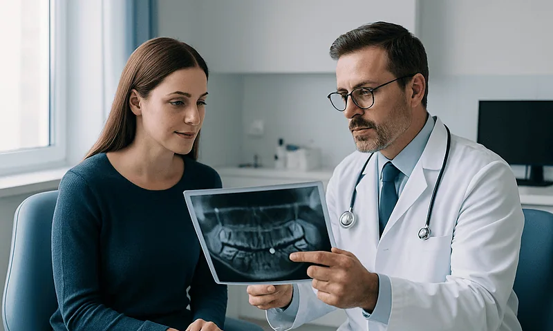
The screen above the dental chair shows a wide, curved X-ray of a whole jaw, and tucked behind the last molar on the lower left there is a tooth lying on its side like a passenger asleep on a train. ‘That one is impacted,’ the dentist says, and a 19-year-old who came in for a routine check now has a question she did not bring: do I leave it, or do I have it out?
Whether to monitor or remove an impacted tooth is one of the most common decisions in dentistry, and one of the most misunderstood. Plenty of people assume the answer is always surgery. Plenty of others assume that if nothing hurts, nothing needs to happen. Both are wrong often enough to matter.
The honest answer depends on where the tooth is, what it is touching, whether disease has already started and, for some teeth, whether it can be coaxed into position rather than taken out. This explainer walks through how clinicians weigh those factors, what the research actually supports and what the weeks after surgery tend to look like.
What does ‘impacted tooth’ actually mean, and why do wisdom teeth get stuck?
A tooth is called impacted when it has failed to come fully into the mouth by the time it normally would, because something stands in its way: bone, gum, another tooth or simply a shortage of room. Impaction is not a disease. It is a position.
Wisdom teeth, the third molars at the very back of each side, are the usual culprits because they arrive last, typically between the ages of 17 and 25, when the jaw has finished growing and the space it has is the space there is (Mayo Clinic). The upper canines, the pointed teeth beside the front incisors, come second on the list, and they matter for a different reason: they sit at the corner of the smile and help guide the way the jaws slide against each other (MedlinePlus).
Dentists describe impactions by angle and depth. A tooth may tilt forward into the molar ahead of it (mesioangular), tilt backward (distoangular), lie flat (horizontal) or stand upright yet buried (vertical). It may have broken partway through the gum, sit fully under soft tissue or remain encased in bone. Each combination changes two things at once: how likely the tooth is to cause trouble, and how involved any surgery would be.
That distinction is the foundation of everything that follows. A tooth fully buried in bone, pointing nowhere in particular and bothering no one is a very different proposition from one poking halfway through the gum with a flap of tissue over it that traps food. Both are impacted. Only one has an open door for bacteria. The word on your X-ray report describes where the tooth is. It does not, on its own, say what should happen next.
Impacted tooth: monitor or remove? The three paths dentists choose between
Strip away the jargon and there are three roads: watch it, free it, or take it out. Most impacted teeth travel one of the first two before anyone reaches for a surgical instrument.

Monitoring means periodic examination and X-rays at an interval your dentist sets, with clear instructions about what should bring you back sooner. Exposure means a small operation to uncover a buried tooth, usually a canine, so an orthodontist can attach a bracket and guide it into the arch. Removal means extraction, which for an impacted tooth often involves lifting the gum, removing a little bone and sometimes dividing the tooth into pieces so each part can come out through a smaller opening.
| Path | Usually chosen when | What it involves | What comes after |
|---|---|---|---|
| Monitor | No symptoms, no decay, no gum pocketing, no cyst, no threat to the neighboring tooth | Examination and X-rays at set intervals | Lifelong review; plan changes if disease appears |
| Expose and align | A canine or premolar is buried but the root is healthy and there is, or can be made, space in the arch | Gum (and sometimes bone) uncovered; bracket and chain bonded; orthodontic traction | Braces for a period set by the orthodontist; then retention |
| Remove | Repeated infection, decay, gum disease, damage to the adjacent tooth, a cyst, or the tooth blocks planned treatment | Local anesthetic with or without sedation; gum lifted, bone removed, tooth sectioned if needed | Healing over roughly two weeks; review if problems |
| Partial removal (coronectomy) | A lower wisdom tooth needs to come out but its roots hug the nerve | Crown removed; roots deliberately left in bone | X-ray follow-up to check the retained roots |
UK national guidance takes the position that impacted wisdom teeth free of disease should not be removed simply because they are impacted, and the NHS continues to describe removal as something offered when a tooth causes problems rather than as a preventive routine (NHS). That stance reflects evidence-based humility: trials have never shown a clear benefit from removing healthy, symptom-free wisdom teeth, as the next sections explain.
How dentists decide: what the examination and X-ray actually show
The decision begins before any image appears. Your dentist will ask whether the area has ever been sore, swollen or tasted unpleasant, how often, and whether anything made it worse. Then comes the mouth itself: is the tooth visible at all, is the gum around it inflamed, does a probe slip into a deep pocket between the impacted tooth and its neighbor, and is the neighbor showing early decay on the surface that faces backward where a brush cannot reach?
Next, the X-ray. A panoramic radiograph, the wide image that shows both jaws in a single sweep, reveals the angle of the tooth, how deep it sits, whether its roots have finished forming and how close the lower wisdom teeth lie to the inferior alveolar nerve canal, the channel carrying the nerve that gives feeling to the lower lip and chin. When that relationship looks intimate or unclear, some clinicians request a cone beam CT, a three-dimensional dental scan, to see whether the root wraps around the canal or merely overlaps it in two dimensions.
The picture is then set against the person. Age matters because roots complete over time and bone becomes denser, which tends to make later surgery more involved (Mayo Clinic). General health matters: medicines that thin the blood or affect bone turnover, conditions that slow healing, and smoking all enter the conversation. So does practicality, such as whether the tooth sits in the path of planned braces or under the area where a denture will rest.
What the dentist is really weighing is a probability against a risk. On one side, how likely is this tooth to cause disease in the coming years, and how much damage could it do before anyone noticed? On the other, what are the risks of operating now, and would operating later be harder? There is no formula that spits out an answer. There is a trained judgement, and it should be explained to you in plain words.
Who is usually asked to wait, and who is usually offered removal
Waiting is the most common recommendation for a reason. If an impacted tooth is fully covered by bone or healthy gum, has never caused symptoms, shows no decay or cyst on the X-ray and is not pressing into the root of the tooth beside it, MedlinePlus notes that no treatment may be needed at all. That describes a large share of impacted wisdom teeth found incidentally on routine X-rays.

Waiting is also commonly advised when the balance of risk tips the other way. A deeply buried lower wisdom tooth whose roots curl around the nerve canal in an older adult is an example: the tooth may never cause trouble, while surgery carries a real chance of numbness. Clinicians will also defer non-urgent dental surgery during pregnancy, during active treatment for another serious illness or while a medicine that affects clotting or bone healing is being adjusted, always in discussion with the prescribing team.
Removal moves up the list when disease has already arrived or is predictable. The NHS lists the usual triggers: tooth decay in the impacted tooth or its neighbor, gum disease around it, pericoronitis (infection of the gum flap over a partly erupted tooth), cellulitis (infection spreading into the soft tissues of the face), an abscess, and cysts or benign growths forming around the buried crown (NHS). A tooth that blocks orthodontic movement, sits in a planned jaw-surgery cut or lies under a future denture is also often removed, not because it is diseased but because it is in the way.
Age is a factor without being a deadline. Younger people tend to have incomplete roots and softer bone, which is why some surgeons prefer to operate earlier when removal is clearly going to be needed (Mayo Clinic). That is a practical observation about surgical difficulty, not evidence that early removal is better for everyone, and it does not make removal urgent for a tooth that is causing no problems.
Does an impacted tooth need to be removed? The problems that change the answer
The default answer is no. The honest answer is: not until, or unless, one of a short list of problems appears, and some of those problems can begin without pain.
Decay is the quiet one. A wisdom tooth tilted into the molar in front of it creates a cramped space where food packs and toothbrush bristles cannot reach. Acid produced by bacteria eats into the back of the second molar, a tooth you would very much like to keep, and often does so without any sensation until the cavity is deep. When decay in that location cannot be reliably restored, removing the impacted tooth protects its neighbor.
Gum disease behaves similarly. A pocket forms between the two teeth, bone slowly recedes from the second molar’s root, and the damage is measured in millimeters lost rather than pain felt. The Cochrane review of asymptomatic, disease-free impacted wisdom teeth found insufficient evidence to support or refute routine removal, but it did note low-certainty evidence that keeping such teeth may be associated with more gum disease on the adjacent molar over the long term (NIH PubMed). That is precisely why ‘monitor’ means monitor, not forget.
Cysts form when the sac that surrounded the developing crown fills with fluid and expands, hollowing out bone. They are uncommon, usually painless and found on X-ray, and they generally lead to removal of the tooth along with the cyst lining (Mayo Clinic).
Then there is infection. A single episode of pericoronitis is often managed by cleaning the area, and the tooth may settle. Repeated episodes, or a severe one that spreads, shift the balance firmly toward extraction, because each flare carries a chance of becoming the kind of facial infection that needs urgent care.
Notice what is missing from this list: ‘because it is impacted.’ Position alone is not a diagnosis, and the research has never shown a benefit from treating it as one.
What are the symptoms of an infected wisdom tooth? Understanding pericoronitis
When a wisdom tooth has partly broken through, a flap of gum called the operculum often drapes over the back of the crown. Food slips underneath; a toothbrush cannot follow. Bacteria multiply in the warm, sheltered pocket, and the flap becomes inflamed. That is pericoronitis, the most frequent reason an impacted wisdom tooth makes itself known.
People who have had it describe a dull ache at the very back of the jaw that sharpens when the upper tooth bites down on the swollen gum. The area may feel puffy to the tongue, taste unpleasant or smell bad, and opening wide can become uncomfortable (MedlinePlus). Some notice tenderness under the jaw where lymph nodes sit. These sensations are the body’s inflammatory response and not, by themselves, a sign that the infection is spreading.
Spreading infection looks and feels different, and it is the reason this section exists rather than a tidy checklist. When swelling moves from the gum into the cheek, under the jaw or toward the throat, when a fever develops, when swallowing becomes difficult or the jaw will barely open, the infection has moved into the soft tissues of the face. The NHS names this cellulitis, and it needs same-day care (NHS).
Treatment for an ordinary flare starts with the mechanics: the dentist irrigates under the flap, removes trapped debris, and may smooth or adjust the opposing tooth that is biting on the gum. Antibiotics, which work by slowing bacterial growth so the body can clear the infection, are reserved for cases with spreading signs or general unwellness, a decision that rests with the clinician who examines you. Over-the-counter pain relief and warm salt-water rinses are commonly suggested for comfort.
Once the acute episode settles, the real conversation begins: was this a one-off, or is the tooth going to keep doing this? A recurrent pattern is one of the clearest reasons a watched tooth becomes a removed one.
Impacted canine exposure: when the plan is to bring the tooth in, not take it out
Not every impacted tooth is a wisdom tooth, and not every impacted tooth is unwanted. The upper canine is the clearest example. It is the second most commonly impacted tooth (MedlinePlus), it has the longest root in the mouth, and losing it leaves a gap at the corner of the smile that is difficult to fill well. So when a canine fails to appear, the first question is usually not whether to remove it but whether it can be rescued.
Canines get stuck for reasons that differ from wisdom teeth. They travel a long path from high in the jaw, and if the baby canine is slow to fall out, the arch is crowded or the small incisor beside it is missing or peg-shaped, the adult canine can drift toward the palate or lodge against the roots of the front teeth. That last scenario is why orthodontists like to know a canine’s position early: a tooth pressing on an incisor root can slowly dissolve it, a process called root resorption, and the damage is permanent.
Exposure is the surgical half of the rescue. In an open exposure, the surgeon removes the gum and a thin layer of bone over the crown and leaves it uncovered to erupt or be bracketed. In a closed exposure, a bracket with a short gold chain is bonded to the crown during surgery and the gum is stitched back, with the chain emerging through it for the orthodontist to attach to the braces. Which technique suits a given tooth depends on its depth and position.
The orthodontic half is slow by design. Gentle force applied through the chain moves the tooth through bone a fraction at a time, and your orthodontist will give a range for your tooth rather than a universal figure. Removal of the canine, with the gap closed by moving other teeth or restored later, remains an option when the tooth is badly placed, its root is bent or resorbed, or the person prefers a shorter course of treatment.
What actually happens when an impacted tooth is removed
Most impacted tooth removals happen in a dental chair, awake, with the area numbed. Local anesthetic blocks the nerve signals from the tooth and surrounding gum, so you feel pressure and movement but not pain. Sedation, given through a vein or breathed in, may be offered for anxiety or for a longer procedure, and a general anesthetic in hospital is reserved for more complex cases or particular medical needs (NHS).
Once the area is numb, the surgeon makes a small incision in the gum and lifts it to expose the tooth and bone. If bone covers the crown, a thin layer is removed with a fine drill cooled by water. Many impacted teeth are then divided into two or more pieces, because a tooth lying sideways cannot be lifted out whole without removing far more bone than necessary. Each section is eased out with instruments called elevators that work like gentle levers, and the socket is rinsed to clear debris.
You will hear and feel this. Drilling vibrates through the jaw; sectioning produces a cracking sensation that people often find more startling than uncomfortable. Telling the surgeon beforehand that you would like a running commentary, or that you would rather not know, is a reasonable request either way.
Timing varies more than people expect. The NHS describes simple removals taking a few minutes and more complicated ones taking longer than twenty minutes (NHS). Afterward, the gum is usually closed with stitches that dissolve on their own, gauze is placed for you to bite on to control bleeding, and you are given written instructions before you leave the same day.
Coronectomy, the deliberate removal of a lower wisdom tooth’s crown while its roots stay in the bone, follows the same opening steps but stops before the roots are touched. It is used when those roots sit against the nerve and the surgeon judges that leaving them is the safer trade.
What tooth is hardest to remove, and when does a general dentist refer to a surgeon?
Ask any oral surgeon and the answer comes quickly: a lower wisdom tooth lying horizontally, buried deep in dense bone, with roots that curve in different directions and hug the nerve canal. Each feature adds difficulty on its own. Together they explain why some extractions take a few minutes and others take far longer.
Lower teeth are generally harder than upper ones because the lower jaw bone is thicker and less forgiving. Depth matters because more bone must be removed to reach the crown. Angle matters because a tilted tooth must be sectioned rather than lifted. Root shape matters most of all: a tooth with a single tapered root slides out, while one with splayed or hooked roots must be divided and each root freed separately. Age adds density to the bone and completes the roots, which is part of why clinicians note that removal tends to be more straightforward in younger adults (Mayo Clinic). Upper wisdom teeth bring their own challenge, sitting close to the maxillary sinus, the air space above the upper back teeth.
So who should do it? Many general dentists remove impacted teeth, including broken ones, routinely. A tooth fractured at the gumline is a common everyday extraction, and a general dentist can usually remove it; the question is whether the remaining root is accessible, intact and away from structures that raise the stakes. Referral to an oral surgeon is typical when the X-ray shows the root in close contact with the nerve canal, when the tooth is deeply buried or horizontal, when a root has snapped below bone level, when sedation or a general anesthetic is needed, or when medical conditions or medicines make bleeding or healing a particular concern.
Referral is not a verdict on anyone’s skill. It is matching the complexity of a tooth to the setting best equipped for it, and a dentist who refers is doing exactly what good practice asks.
Risks, alternatives and the honest numbers behind impacted tooth removal
Surgery to remove an impacted tooth is common and generally safe, and it is still surgery. Being told that plainly is part of making a real decision rather than a reflexive one.
Dry socket is the complication people are most likely to meet. After extraction, a blood clot forms in the socket and protects the bone and nerve endings beneath while new tissue grows. If that clot dissolves or dislodges early, the bone is exposed and pain typically begins one to three days after the procedure, often with a bad taste and an ache that radiates toward the ear (Mayo Clinic). Mayo describes it as the most common complication after tooth extraction, particularly of lower wisdom teeth. It is treated with cleaning and a soothing dressing, and it heals; it is miserable rather than dangerous.
Nerve injury is less common and more consequential. The lower wisdom tooth sits near the nerve that supplies the lower lip and chin, and a second nerve serving the tongue runs along the inner gum. Bruising or stretching either can leave numbness or tingling. The NHS states this is usually temporary, while in rare cases it can be permanent (NHS). Your surgeon should tell you where your tooth sits in relation to these nerves, because the risk is not the same for every tooth.
Other recognized risks include infection, prolonged bleeding, jaw stiffness, an opening into the sinus after upper tooth removal and, very rarely, fracture of the jaw during surgery on a deeply buried tooth.
Alternatives exist. Monitoring remains an option for disease-free teeth. Coronectomy avoids the nerve at the cost of leaving roots that need X-ray follow-up. Operculectomy, trimming the gum flap over a partly erupted tooth, is sometimes tried for recurrent mild pericoronitis, though the flap can regrow.
One honest note: reliable complication percentages differ by tooth position, age and surgical technique, and the general-audience sources behind this article do not publish a single figure that applies to everyone. Ask for the numbers that fit your tooth.
Impacted tooth extraction recovery: what the first two weeks usually look like
The first evening is about the clot. You bite on gauze until oozing settles, keep your head raised, skip hot drinks and avoid anything that creates suction in the mouth, such as straws, spitting forcefully or smoking, because suction can pull the clot loose (Mayo Clinic). The numbness wears off over the hours that follow, and the ache that replaces it is usually managed with over-the-counter pain relief taken as your dentist advised.
Days one to three are typically the peak. The NHS describes swelling and discomfort inside and outside the mouth as usually worst for the first three days, though it can last up to two weeks (NHS). Cheeks feel tight, chewing on that side is off the menu and soft, cool foods are your friends. From the day after surgery, gentle rinsing with warm salt water after meals helps keep the socket clean without disturbing it (NHS).
Through the first week, the jaw often feels stiff and sore, something the NHS says should wear off within seven to ten days (NHS). Bruising on the cheek, when it happens, may take up to two weeks to fade (NHS). Dissolvable stitches loosen and fall away on their own during this period; finding one on your tongue is normal.
Most people return to desk work or school within a few days, with heavier exercise resumed as comfort allows and as your dentist suggests. The socket itself fills with new tissue over the following weeks, and bone remodels underneath for months afterward, which is why a gum that looks healed can still feel slightly different for a while.
Recovery after exposure of a canine is usually gentler, with soreness around a smaller wound and the added business of a chain and braces. Coronectomy recovery resembles a standard extraction, plus scheduled X-rays to confirm the retained roots are behaving.
What should not happen is pain that improves and then returns worse, which brings us to the warning signs below.
What people often get wrong about impacted teeth
‘Every impacted wisdom tooth has to come out eventually.’ It does not. The NHS positions removal as treatment for problems, not as a routine for position, and the Cochrane review found no clear evidence that removing symptom-free, disease-free wisdom teeth benefits people (NHS; NIH PubMed). Many such teeth are watched for a lifetime and never trouble their owner.
‘If it doesn’t hurt, it’s fine.’ Also not reliably true. Decay on the back of the second molar, bone loss between the teeth and cysts around a buried crown can all progress silently (Mayo Clinic). This is why monitoring includes X-rays at intervals and not just a glance in the mirror.
‘Wisdom teeth push the front teeth crooked.’ The idea is intuitive and the evidence for it is weak. Lower front teeth tend to crowd gradually in adulthood whether or not wisdom teeth are present, and removing wisdom teeth has not been shown to prevent it. Crowding is a reason to see an orthodontist, not by itself a reason to extract molars at the back.
‘If you’re over 30, it’s too late.’ Surgery may be somewhat more involved in older adults because bone is denser and roots are complete (Mayo Clinic), but it is performed at every age when disease makes it necessary. The threshold for removing a quiet tooth simply rises as the surgical trade-off changes.
‘Only an oral surgeon can remove an impacted or broken tooth.’ Many general dentists remove impacted and fractured teeth as part of ordinary practice, referring when the X-ray or medical history calls for it.
‘Removal is just maintenance, like a filling.’ It is a surgical procedure with recognized risks, including dry socket and, rarely, lasting nerve numbness (NHS; Mayo Clinic). Those risks are acceptable when a tooth is causing or threatening disease. They are harder to justify for a tooth that is doing nothing at all.
Questions to ask your care team before deciding on an impacted tooth
A good consultation leaves you able to explain the plan to someone else. These questions help get you there, and none of them is awkward to ask.
- Which teeth are impacted, and how is each one positioned: angle, depth and whether it is partly through the gum?
- Is there any sign of disease right now, such as decay on the neighboring tooth, a gum pocket, inflammation or a cyst on the X-ray?
- If you are recommending monitoring, how often should I be reviewed and re-X-rayed, and what changes should bring me back sooner?
- If you are recommending removal, which specific problem is driving that, and what is likely to happen if I wait?
- How close is the tooth to the nerve canal or the sinus, and does that change the risks for me compared with an average case?
- Would a coronectomy, exposure or another alternative be reasonable for this tooth, and why or why not?
- Will you perform the procedure here, or is a referral advisable, and what makes the difference?
- What anesthetic or sedation options are suitable for me, given my health and any medicines I take?
- What does recovery typically look like for a tooth in this position, and what should I plan around work or study?
- Which signs after surgery mean I should call the practice, and which mean I should seek urgent care?
Write the answers down or ask for them in writing. If any medicine you take affects bleeding, bone healing or immunity, make sure the dentist knows and, where needed, speaks with the clinician who prescribes it; never adjust a medicine on your own ahead of dental surgery.
Finally, ask yourself what matters most to you. Some people would rather have one planned procedure than years of check-ups; others prefer to keep a quiet tooth and accept monitoring. Both are legitimate positions when the evidence allows a choice, and the decision should be made with your treating team, not for you.
When to call your doctor or dentist about an impacted tooth
Most impacted teeth are reviewed at routine appointments, and most recoveries after surgery follow the pattern described above. The signs that warrant a call, or in some cases an emergency visit, are reasonably clear.
Before any treatment, contact your dentist promptly if the gum around a partly erupted tooth becomes swollen and painful, if you notice a persistent bad taste or smell from the area, if the tooth beside it becomes sensitive, or if your jaw becomes difficult to open. These are the typical features of pericoronitis or early decay and are best assessed within days rather than weeks (MedlinePlus).
Seek same-day urgent care, through an emergency dental service or hospital emergency department, if swelling spreads into the face, under the jaw or toward the eye, if you develop a fever with facial swelling, if swallowing becomes difficult or painful, if you have any difficulty breathing, or if you cannot open your mouth more than a finger’s width. The NHS describes these spreading infections as cellulitis, and they can progress quickly (NHS). Difficulty breathing or swallowing is an emergency: call emergency services.
After surgery, call the practice if bleeding does not slow with firm pressure on gauze for a sustained period, if pain eases over the first day or two and then returns more intensely with a bad taste, which is the classic pattern of dry socket (Mayo Clinic), if swelling or pain is still worsening after the third day, if pus appears or if you develop a fever. Numbness of the lip, chin or tongue that persists after the anesthetic should have worn off is worth reporting straight away, even though it is usually temporary (NHS), so that it can be documented and followed.
When in doubt, call. Dental teams would much rather hear about a false alarm than see a spreading infection or a dry socket a week later than they needed to.
Frequently asked questions
Is it necessary to remove an impacted tooth?
No, not unless it is causing or threatening a problem. An impacted tooth that is free of decay, gum disease, infection and cysts, and is not damaging its neighbor, can usually be monitored with periodic X-rays. Removal is recommended when disease appears, when infection keeps recurring, or when the tooth blocks planned orthodontic or other treatment. Your dentist weighs the tooth’s position, your age and your health before advising either way.
Does an impacted tooth need to be removed if it does not hurt?
Pain is not the deciding factor. Decay on the neighboring molar, bone loss between the teeth and cysts around a buried crown can all develop without any discomfort, which is why monitoring includes X-rays rather than relying on symptoms. If those checks stay clear, a painless impacted tooth can be left in place. If they show damage beginning, removal may be advised even though nothing hurts.
What are the symptoms of an infected wisdom tooth?
An infected gum flap over a partly erupted wisdom tooth, called pericoronitis, usually causes aching at the back of the jaw, a swollen tender gum, a bad taste or smell and sometimes difficulty opening wide. Swelling that spreads into the cheek or neck, fever, or trouble swallowing or breathing means the infection is spreading and needs same-day care. Only a dental examination can confirm the cause.
What are impacted wisdom tooth symptoms, and are they always obvious?
They are often not obvious at all. Many impacted wisdom teeth are found on routine X-rays in people who feel nothing. When symptoms do occur they tend to involve the gum rather than the tooth: soreness, swelling or an unpleasant taste where food traps under a gum flap, occasionally with jaw stiffness. Silent problems such as decay on the adjacent molar are picked up at check-ups, not by sensation.
What tooth is hardest to remove?
A deeply buried lower wisdom tooth lying horizontally, with curved or splayed roots close to the nerve canal, is generally the most demanding extraction. Lower jaw bone is denser than upper, depth means more bone must be removed, and complex roots require the tooth to be divided and taken out in pieces. Dense bone and fully formed roots in older adults add to the difficulty.
Can a regular dentist pull a broken tooth?
Yes, in most cases. Removing a tooth broken at the gumline is a routine procedure for general dentists. Referral to an oral surgeon is typical when the remaining root lies close to the nerve or sinus, has fractured well below bone level, is deeply impacted, or when sedation, general anesthesia or medical complexity calls for a hospital or specialist setting.
What is impacted canine exposure and why is it done instead of removal?
Exposure is a small operation to uncover a buried canine so an orthodontist can bond a bracket and chain and guide the tooth into the arch with braces. It is preferred because the canine has the longest root in the mouth, anchors the corner of the smile and is difficult to replace well. Removal is considered when the tooth is badly positioned, its root is damaged, or treatment time is a priority.
How long does impacted tooth extraction recovery take?
The NHS describes swelling and discomfort as usually worst for the first three days and lasting up to two weeks, with jaw stiffness settling within seven to ten days and any bruising fading over about two weeks. Most people return to normal routines within a few days. Recovery is not a promise, and a tooth that was deeply buried may take longer than one that was partly erupted.
Can an impacted wisdom tooth come through on its own?
Sometimes, if the obstruction is a thin layer of gum and there is enough space. A tooth partly through the gum may finish erupting over time, especially in younger adults. A tooth lying horizontally against the molar in front, or buried in bone with no room ahead of it, will not straighten itself. Your dentist can usually tell from the X-ray which situation applies.
What happens if you never remove an impacted wisdom tooth?
For many people, nothing. A disease-free impacted tooth can stay quietly in place for life, which is why guidance supports monitoring. The risks of leaving it are decay or gum disease on the neighboring molar, repeated gum infection and, uncommonly, a cyst, all of which can be caught at regular reviews. If any of those develop, removal can be arranged at that point.
References
This article is for general information only and is not a substitute for professional medical advice. Please consult a qualified doctor about your individual situation.

More from the Blog

Recovering From Laser Dentistry: Gum Tenderness, Hot Foods and the First Day
After laser gum treatment, mild tenderness, slight swelling and sensitivity to temperature are common for the first day or two, and gums usually settle…

Metal Braces vs Clear Aligners: Bite Control, Compliance and Daily Upkeep Compared
Metal braces and clear aligners both straighten teeth with gentle, sustained pressure, and for mild to moderate crowding they reach broadly similar results. Fixed…

Oral and Maxillofacial Surgery for International Patients: Stay Length and Follow-Up
Oral surgery abroad follow up is the planned series of checks after a procedure, and it starts before you travel home. Surgeons usually ask…

What Halitosis Treatment Involves: Cleaning, Gum Care, Tongue Care and Referral When Needed
A halitosis treatment plan usually starts at the dentist: a professional cleaning, treatment of any gum disease, daily tongue cleaning and practical steps to…
How an Impacted Canine Is Surgically Exposed and Guided Into Place
Impacted canine exposure surgery is a short oral surgery in which a surgeon lifts or removes the gum (and sometimes a thin layer of…

How Long to Stay in Istanbul After Orthognathic Surgery Before You Travel Home
Most surgical teams ask patients who have had orthognathic (corrective jaw) surgery far from home to stay near the hospital through at least the…