

Which Scans Come Before Deformity Correction? Standing X-Rays, CT and Gait Analysis

Key Takeaways
- A standing long leg X-ray measures alignment under body weight, which a lying-down film cannot, and it is almost always the first study ordered before deformity correction.
- Rotation, the twist along a bone's length, is invisible on a flat X-ray and is usually measured with a few thin CT slices at the hip, knee and ankle.
- Shape distortion in radiography means a turned foot can make a bone look more or less bent than it is, which is why the kneecap, not the foot, is used to position the leg.
- MRI shows growth plates and cartilage without radiation, but magnetic field non-uniformity and metal-related distortion mean it is not used as the ruler for whole-limb alignment.
- Harvard Health lists a chest X-ray at about 0.1 mSv and a chest CT at about 7 mSv, against roughly 3 mSv of natural background radiation each year, which is why CT is added only for a specific question.
- Gait analysis is a targeted study for people whose walking pattern, not just their bone shape, is in question, and it is often repeated after recovery to document change.
Before a limb deformity correction, surgeons usually order full-length standing X-rays of both legs to measure alignment under body weight, and add a CT scan when rotation, limb length or a complex three-dimensional bend must be mapped precisely. Instrumented gait analysis is added for selected patients, often children with neuromuscular conditions, to show how the deformity behaves in motion. Your treating team decides which studies apply to you.
The hardest part of the appointment, one father told his son afterward, was standing still. The boy had to plant both feet on a marked plate, kneecaps pointing straight ahead, hands off the rail, while a tall cassette slid down behind him and the technologist counted out loud. Three seconds. Then a second view from the side. The whole thing took less time than finding parking.
That plain, slightly awkward picture is the single most important study in deformity correction imaging. It looks unremarkable, yet it carries the numbers a surgeon will use to decide where a bone should be cut, how far it should turn and whether a growth plate can do part of the work instead of a saw.
What follows is a plain-language tour of the scans that typically come first, why each one is chosen, what it can and cannot show, and the questions worth bringing to the consultation.
What deformity correction imaging actually has to answer
A bowed shin or an inward-turning knee looks like one problem. To a surgeon it is at least four separate questions, and each needs a different measurement.
The first is angulation: is the bone bent, and by how many degrees? The second is the location of that bend, because a correction placed at the wrong level straightens the bone on paper while leaving the joint above or below it tilted. The third is rotation, meaning whether the bone is twisted along its own length like a wrung towel. The fourth is length, since one leg often ends up shorter than the other, and the plan has to account for that difference.
Deformity correction imaging is the set of studies chosen to answer those four questions with numbers rather than impressions. A weight-bearing X-ray handles angulation, location and, with care, length. Rotation hides from a flat picture, so it usually needs a CT scan. Motion, the way the leg actually behaves when it carries a person across a room, is not captured by any still image, which is where gait analysis enters for selected patients.
There is a fifth question that sits underneath the others: is the deformity in the bone itself, or in the joint, the soft tissue or the way the person walks? An X-ray can show a knee that looks perfectly aligned while the patient stands, and the same knee collapsing inward with every step. Imaging is chosen to separate those possibilities, because the treatment for each is different. MedlinePlus describes X-rays as the most common way to view bone structure, while noting they show soft tissue poorly, a limitation that shapes every choice described below.
Why a standing long leg X-ray is the workhorse
Most people have had an X-ray lying on a table. For alignment work that position is nearly useless. Lying down removes body weight, lets the knee drift into whatever angle is comfortable, and hides the true relationship between hip, knee and ankle. The study surgeons want is a standing long leg X-ray, a single image that runs from the pelvis to the floor with both legs on it at once.
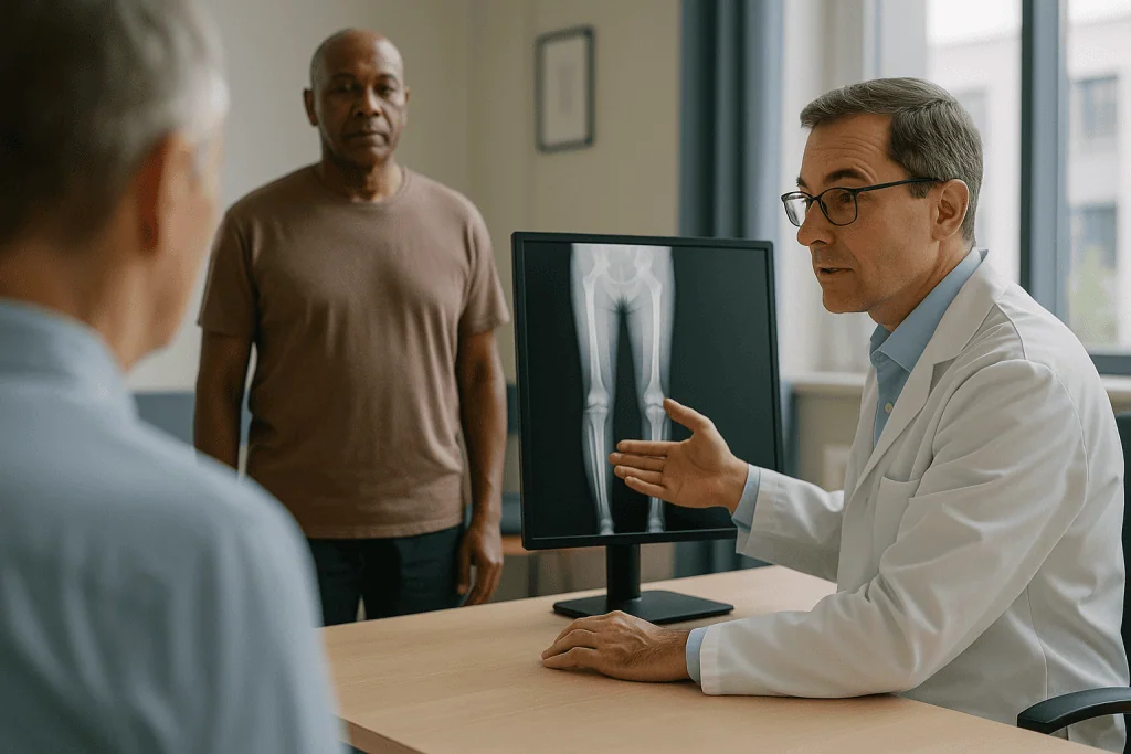
Standing matters for a plain mechanical reason. Bone alignment is a load question. Gravity pushes down through the center of the hip, and the leg is built so that line should pass close to the middle of the knee and then through the ankle. Only a weight-bearing image shows where that line actually falls. Ligaments that are loose will let the knee open on one side under load, and cartilage that has worn thin will let a joint space narrow; both effects vanish on a table.
The technologist’s fussing over foot position is not ceremony. The kneecaps are turned to face the beam directly so that the knee is imaged in its true front-to-back plane. A small block may be placed under the shorter foot to level the pelvis, and the height of that block is recorded, because it is itself a measurement of leg length difference. A ruler or a ball of known size is often included in the field so the image can be scaled.
Some centers use a low-dose slot-scanning system that captures front and side views of the whole body while a person stands in a booth. The principle is the same: full length, both legs, under load. Mayo Clinic notes that plain X-ray studies are quick and typically involve no special preparation, which is one reason this image is repeated freely during planning and follow-up.
What the surgeon measures: axis, angles and where the bend lives
Once the image is on the screen, the reading is less about looking and more about drawing. Three ideas carry most of the weight.
The mechanical axis is a straight line drawn from the center of the hip to the center of the ankle. In a well-aligned leg that line passes through or very near the center of the knee. If it falls toward the inside of the knee, the leg is in varus, the bow-legged pattern; toward the outside, valgus, the knock-kneed pattern. The distance by which it misses the center is recorded in millimeters and is one of the most quoted numbers in the whole process.
Joint orientation angles come next. Lines are drawn along the surface of the hip, the top and bottom of the knee and the top of the ankle, and each is compared with the long axis of its bone. Each of these angles has a normal range, so a surgeon can tell whether the thigh bone, the shin bone or both are responsible for the overall bend.
The third idea is the center of rotation of angulation, the exact point along the bone where the two straight segments meet. Correcting a bone at this point straightens it without creating a new step-off; correcting it anywhere else fixes the angle but shifts the segments sideways, a compromise that sometimes has to be accepted around a joint but is avoided where possible.
These drawings answer a question patients often ask: why is the cut planned there and not where the leg looks most bent? The visible curve and the mathematical apex are frequently different places. Software now does much of the drawing, but the underlying geometry is the same one that has guided this surgery for decades, and a good consultation will walk you through the lines on your own image.
What is shape distortion in radiography, and why positioning matters
Shape distortion in radiography is the change in an object’s apparent outline that happens when it is not parallel to the film or detector. An X-ray beam spreads out from a point, so anything tilted relative to the detector is stretched or squashed in the image, the same way a shadow lengthens when a lamp is moved low.
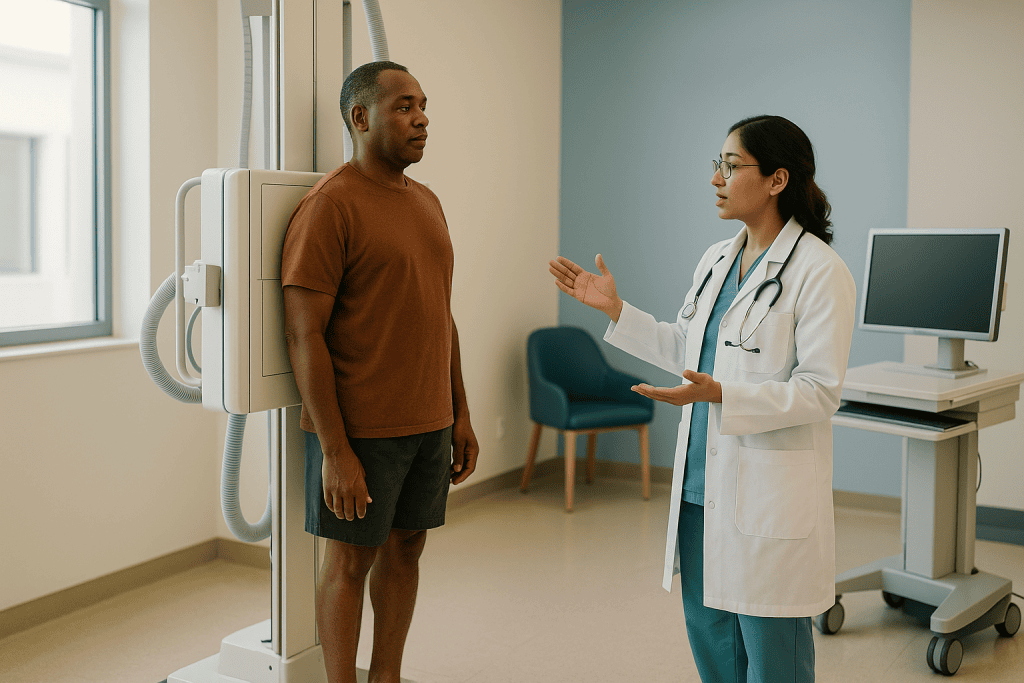
For deformity planning this is not a technicality. A shin bone that is rotated outward by even a modest amount will look more bowed or less bowed than it is, because part of a rotational twist projects onto the front-to-back image as false angulation. Two X-rays of the same leg taken with the foot pointing in different directions can disagree by several degrees on the apparent deformity. That is why the kneecap, not the foot, is used as the reference: the knee is the joint being measured, and the foot may itself be turned by a twist in the bone below.
Magnification is the related problem. Structures farther from the detector appear larger. On a full-length image, the pelvis and the ankle are at different distances from the plate, and a limb that stands slightly forward will measure longer than its partner. The scaling marker mentioned earlier exists to correct this, and the technologist’s insistence on both heels in the same plane exists to prevent it.
A practical consequence follows for patients. If you notice that your foot was turned, or you shifted weight to the more comfortable leg, tell the technologist before you leave. Repeating a three-second exposure is a small matter. Planning an operation on a distorted image is not. MedlinePlus lists correct positioning as part of every X-ray examination for exactly this reason: the picture is only as reliable as the way the body was placed in front of it.
When a CT scanogram for limb length or a 3D CT gets added
Rotation is the measurement a flat picture simply cannot deliver. A thigh bone that is twisted along its length can look straight from the front and straight from the side, yet leave the knee pointing inward when the hip is square. Computed tomography, or CT, solves this by taking cross-sectional slices at chosen levels and comparing the orientation of one end of the bone with the other.
A rotational profile CT does exactly that. Thin slices are taken through the hip, the knee and the ankle. Software measures the angle of the femoral neck against the back of the knee, and the angle of the knee against the ankle joint. The result is a number for femoral torsion and a number for tibial torsion, each compared with a normal range. This is the study most likely to be ordered when the complaint is turned-in or turned-out feet rather than a visible bow.
A CT scanogram for limb length is a different use of the same machine. A single low-dose projection image is taken of both legs, and length is measured directly between joint centers. Because the beam geometry is controlled, the magnification problem described earlier largely disappears. This is often preferred over a standing X-ray for length when a joint is contracted and cannot be straightened for the standing film.
Three-dimensional reconstruction is the third role. For a bone that is bent in two planes at once, or deformed after a badly healed fracture, the surgeon may reconstruct the whole bone as a model on screen, or print it, and rehearse the cut. Guides that fit the patient’s own bone can be made from the same data.
The NHS describes a CT scan as usually taking around 10 to 20 minutes, and notes that it is painless and generally does not require special preparation for bone studies. The trade-off is radiation, discussed below, which is why CT is added for a specific reason rather than ordered routinely.
MRI, geometric distortion and where MRI fits before correction
Magnetic resonance imaging, or MRI, builds pictures from the way hydrogen atoms in the body respond to a strong magnet and radio waves, with no ionizing radiation involved. It shows cartilage, ligaments, growth plates and bone marrow in a detail X-rays cannot approach. That makes it valuable in specific situations before a deformity correction, and largely unnecessary in others.
The clearest use is the growth plate. In a child, a deformity that is worsening may be driven by a plate that has partly closed after an injury or infection. MRI can map which part of the plate is still active and which has fused into a bony bridge, and that map changes the plan: a small bridge might be removed, a large one might mean the plate is closed deliberately and length is addressed another way. MRI also shows the state of the cartilage in a knee that has been loaded unevenly for years, which affects whether realignment alone is likely to be considered.
People sometimes ask what causes geometric distortions in MRI images. Two sources matter. The main magnetic field is never perfectly uniform, and the gradient fields used to encode position are not perfectly linear, so objects near the edges of the imaging volume can be stretched or shifted. Metal, including previous plates or screws, disturbs the local field and produces dark voids and warped edges around the implant. These effects are why MRI is generally not used for millimeter-level alignment measurement across a whole limb; the standing X-ray and CT remain the rulers, and MRI is the tissue map that sits alongside them.
The NHS notes that MRI is safe for most people but is not suitable for everyone with certain implants or devices, and that the machine is noisy and requires lying still for the length of the study. Your team will weigh whether the tissue information justifies the effort in your case.
Gait analysis before surgery: what happens in the lab
A standing X-ray captures a leg at rest. Walking is a different event, and for some patients the two disagree in ways that decide the operation.
Instrumented gait analysis is a laboratory study of how a person walks. Small reflective markers are stuck to the skin over bony landmarks: the pelvis, the sides of the knees, the ankles, the feet. A ring of infrared cameras records the position of every marker many times a second while the person walks a straight path. Plates set into the floor measure the force of each footstep, and small pads on the skin can record which muscles switch on and when. From that data a computer rebuilds the movement of each joint through the walking cycle and plots it against typical patterns.
The session is unhurried but tiring. Markers take time to place accurately, and several walking passes are needed. Children usually wear shorts and a fitted top; a parent can stay in the room. Nothing is injected and nothing hurts, though the muscle pads can feel odd when peeled off.
Gait analysis before surgery earns its place when the deformity is not purely in the bone. In cerebral palsy, for instance, an inward-turning gait may come from twisted bone, from tight muscles, from a hip that rolls in, or from all three, and the bone twist measured on CT may be only part of the story. Gait data helps a team decide which of several possible procedures, done in one session or staged, addresses what actually happens at each step. It is also used afterward to document what changed.
For a straightforward bow in a child with otherwise normal walking, gait analysis is rarely needed. The standing film and, where required, a CT carry the plan. Whether you are referred to a gait lab is a decision your treating team makes on the pattern they see, not a step every patient passes through.
Who is usually imaged fully, and who is usually asked to wait
Not everyone with a visible bend is sent for the full battery, and the reason is growth.
Toddlers are commonly bow-legged, and many preschoolers are knock-kneed. Both patterns are part of ordinary development, and both usually straighten on their own as the child grows. A first visit for a young child with symmetrical legs and no pain often ends with an examination, perhaps a single standing X-ray, and a plan to look again as the child grows. Repeated imaging in that setting adds radiation without adding information. Being asked to wait and return is, in this group, the evidence-based outcome, not a brush-off.
The picture changes when a deformity is one-sided, is getting worse rather than better, appears after an injury or infection, is linked to a known condition of bone or nerve, or comes with pain, limping or a leg length difference that is easy to see. Those children and adults are the ones who typically move to a full standing long leg X-ray, and from there to CT or MRI if rotation, length or growth plate status needs mapping.
Adults form a second group. Long-standing malalignment can overload one side of a knee, and some adults are assessed for a realignment osteotomy, a procedure that cuts and reshapes bone to shift load onto healthier cartilage. Here the standing X-ray is essential and MRI of the knee is often added to judge cartilage.
People who are asked to wait before any imaging also include those with a recent fracture still healing, where measurements would change month to month, and those whose main problem is a foot or spine issue that needs sorting out first because it alters standing posture. A short waiting period lets the team image once, well, rather than repeatedly. As always, the decision about which studies apply to you and when sits with your treating team.
How the deformity correction imaging studies compare
Set side by side, the studies stop looking like a menu and start looking like a division of labor. Each one is good at one or two things and poor at the rest, which is why a plan often uses two or three of them rather than one.
| Study | Best at | Cannot do well | Ionizing radiation | What it feels like |
|---|---|---|---|---|
| Standing long leg X-ray | Angulation, location of the bend, alignment under body weight | Rotation, soft tissue, cartilage | Yes, low | Standing still for seconds, two views |
| CT rotational profile | Twist in femur and tibia, measured in degrees | Weight-bearing alignment, cartilage | Yes, higher than X-ray | Lying still on a moving table |
| CT scanogram | Leg length difference with minimal magnification error | Angulation detail, soft tissue | Yes, low | Lying still, brief |
| 3D CT reconstruction | Complex multi-plane bends, surgical rehearsal, custom guides | Standing alignment, joint surface health | Yes, higher | Same as CT |
| MRI | Growth plates, cartilage, ligaments, marrow | Whole-limb alignment measurement | None | Lying still in a noisy tube for a longer session |
| Gait analysis | How joints and muscles behave during walking | Bone geometry, any structural measurement | None | Markers on skin, walking repeatedly |
A few patterns stand out. Everything that uses X-rays is a structural ruler; everything that does not is a tissue or motion map. Radiation rises as detail rises, from a plain film through a scanogram to a full CT, which is why Harvard Health frames every imaging choice as a balance between the information gained and the exposure accepted. Time and comfort also vary: the standing film is fast and undemanding, while MRI and gait analysis ask for patience.
What the table cannot show is sequence. In practice the standing X-ray nearly always comes first, and the others are added only when it raises a question it cannot answer on its own.
How much radiation is involved, honestly
Parents ask this more than any other question, and it deserves numbers rather than reassurance.
Medical radiation is measured in millisieverts, written mSv, a unit that accounts for the type of radiation and the tissue it reaches. Harvard Health gives useful anchors: a single chest X-ray delivers roughly 0.1 mSv, a CT of the chest roughly 7 mSv, and ordinary background radiation from the ground, air and food adds up to about 3 mSv over a year for the average person. A plain film of a limb sits at the low end of that scale, in the same order of magnitude as a chest X-ray. A full-length standing image is larger and includes the pelvis, so it sits higher than a single knee view, but it remains a low-dose study.
CT is a different tier. MedlinePlus states plainly that CT uses more radiation than regular X-rays, and that the risk from any one scan is small but adds up with repeated scans. For a rotational profile, radiologists limit exposure by taking only a few thin slices at the hip, knee and ankle rather than scanning the whole leg, and by adjusting the settings to the person’s size. The National Institute of Biomedical Imaging and Bioengineering notes that modern CT protocols are tailored to reduce dose, particularly in children, whose tissues are more sensitive and who have more years ahead in which any effect could appear.
Two principles follow. The first is that no scan is ordered simply because it is available; each should answer a question that changes the plan. The second is that the studies with no ionizing radiation, MRI and gait analysis, are not automatically better, because they cannot replace the ruler. A child who has three standing X-rays over the course of a correction is accepting a small, known exposure in exchange for a bone being cut in the right place. If the trade-off worries you, ask your team which studies are essential, whether a low-dose protocol is used, and how images from elsewhere can be reused rather than repeated.
What the days and weeks after imaging usually look like
Imaging is not a single appointment followed by a verdict. It is the front end of a planning loop that continues past the operation itself.
The first step after the scans is the read. A radiologist reports the images, and the surgical team separately draws the alignment lines described earlier, often in dedicated planning software. If the standing film and the clinical examination disagree, or if the deformity is in more than one plane, this is the point where a CT or MRI request is added. Expect a second imaging visit in that case, and do not read it as bad news; it usually means the team wants a number it does not yet have.
The planning consultation follows. You should see your own images with the lines on them, hear which bone is responsible and where the apex lies, and be told the intended correction in degrees and millimeters. For a child with growth remaining, the team may propose guided growth, a small plate placed across one side of a growth plate so the bone straightens as it grows, rather than a cut. That choice depends on an estimate of remaining growth, which sometimes prompts one more X-ray, of the hand, to read skeletal age.
After surgery, imaging continues. Plain X-rays check the position of any hardware and, over the following weeks, the healing of the cut. When a frame or lengthening device is used, repeated films track the gap opening at the interval the team sets, and a standing long leg image is taken again once weight-bearing is allowed, because only then can the result be compared with the original.
Gait analysis, where it was used before, is often repeated after recovery to document what changed. The full loop, from first standing film to final standing film, is measured in months rather than weeks; your team will give you their expected sequence, and it will be specific to your bone, your age and the method chosen.
What people often get wrong about deformity correction imaging
The most common misunderstanding is that the leg that looks most bent is the leg that needs the most work. The eye is drawn to the curve at the shin, yet the standing film often shows the thigh bone carrying half or more of the angle, or the joint line tilted in a way that no shin correction can fix. The picture, not the appearance, locates the problem.
A second myth is that a lying-down X-ray from an urgent care visit is good enough to start from. It can show a fracture or a bone lesion. It cannot show alignment under load, and measurements taken from it can be wrong enough to change the plan. Bringing old images is useful for comparison; it does not usually replace the standing study.
Third, many people assume CT is the default upgrade, the scan you get if you want the best look. In alignment work it is a specialist tool for rotation and complex three-dimensional bends, chosen for a reason, and its extra radiation is the reason it is not ordered for everyone.
Fourth, MRI is often imagined as the most accurate ruler because it is the most detailed picture. Detail of tissue and accuracy of length are different things. Because of the geometric distortions inherent to magnetic fields and the effect of metal, whole-limb measurements are not usually taken from MRI.
Fifth, gait analysis is sometimes expected as a routine step. It is a targeted study for people whose walking pattern, not just their bone shape, is in question.
Finally, some assume that a normal-looking scan means nothing is wrong. Pain and limping have causes an X-ray cannot see, and a reassuring film is an answer to one question, not to all of them. If symptoms persist after imaging that looks normal, that is a reason to go back to the team, not a reason to stop asking.
Questions to ask your care team
A planning consultation moves quickly, and the images on the screen are unfamiliar. A short written list keeps the conversation anchored on what you actually need to know.
- Which bone, or which part of which bone, is responsible for the deformity, and can you show me the line on my own X-ray?
- Is the standing X-ray enough for planning in my case, or will a CT or MRI be needed, and what specific question would it answer?
- Does my deformity include rotation, and if so how was it measured?
- Is there a leg length difference, how large is it, and how will the plan account for it?
- For a child: how much growth is expected to remain, how was that estimated, and does that open the option of guided growth rather than a cut?
- Is gait analysis being considered, and what would it add to the decision?
- What radiation dose does each proposed study involve, is a low-dose protocol used, and can any earlier images be reused rather than repeated?
- What is the alternative to operating now, including waiting and reimaging, and what would you expect to see if we waited?
- How often will X-rays be taken during recovery, and what will they be checking for?
- What does the imaging show about the joint surfaces, and does that change what you expect from correction?
Two habits make these questions more useful. Ask for the numbers in writing: the axis deviation, the angles, the torsion values and the length difference. They become the baseline against which every later image is judged, and they travel with you if you change teams or move. And ask what the team would want to see on the final standing film to consider the correction complete. That single question shifts the discussion from the operation to the outcome the imaging is meant to serve, and it makes the follow-up X-rays feel like a shared checklist rather than a mystery.
When to call your doctor
Most of this article concerns planning, but imaging also plays a part in recognizing when something has changed and needs attention sooner than the next scheduled visit.
Before any surgery, contact your care team promptly if a child’s limb deformity is worsening visibly over a short period, if a bend becomes clearly one-sided when it was symmetrical, if a new limp appears, if there is pain at night or pain that wakes the child, or if a leg length difference becomes obvious enough to cause tripping. Swelling, warmth or redness over a bone, fever alongside limb pain, or a child refusing to bear weight are reasons to seek care the same day, because infection and other urgent conditions can present this way and are diagnosed with examination and imaging rather than by waiting.
After a correction, whether by plate, nail or external frame, call the team without delay for increasing rather than easing pain, pain that is not controlled by the plan you were given, numbness or tingling in the foot, toes that are pale, blue or cold, a foot that is hard to move or that drops, redness or discharge around any pin or incision, a fever, or a sudden change in the shape or angle of the limb. Calf pain, calf swelling or unexpected breathlessness are red flags for a blood clot and need emergency assessment.
Loose, bent or broken hardware sometimes announces itself as a new click, a sudden give, or a loss of the correction you had gained. Any of those warrants an early X-ray rather than a wait for the routine one.
None of these signs is a diagnosis. Each is a reason to be seen and, in most cases, imaged, so that the team can see what is happening beneath the skin and decide what, if anything, needs to change. Keep the contact numbers you were given somewhere easy to find, and use them.
Frequently asked questions
Why do I need a standing long leg X-ray instead of a normal knee X-ray?
Because alignment is a load question and a knee-only film cannot show where the weight line from the hip to the ankle actually falls. The standing long leg X-ray captures both legs from pelvis to floor under body weight, letting the surgeon draw the mechanical axis, measure joint angles and locate the apex of the bend. A knee film taken lying down shows the joint but not the alignment that surgery aims to change.
What is a CT scanogram for limb length, and how is it different from a regular CT?
A CT scanogram is a single low-dose projection image of both legs taken on a CT scanner, used to measure leg length between joint centers with very little magnification error. A regular CT takes many cross-sectional slices and delivers more radiation. For alignment work the scanogram measures length, while a separate low-slice rotational CT measures the twist in the femur and tibia; your team chooses one, both or neither based on the question.
Is gait analysis before surgery necessary for everyone?
No. Gait analysis is used selectively, most often for people with neuromuscular conditions such as cerebral palsy, where walking problems come from a mix of bone shape, muscle tightness and joint control. For a straightforward bow or knock-knee in someone who otherwise walks normally, the standing X-ray and, if needed, CT usually carry the plan. Whether you are referred to a gait laboratory is a decision your treating team makes.
What is shape distortion in radiography?
Shape distortion is the change in an object’s apparent outline when it is not parallel to the detector, because an X-ray beam spreads from a point and stretches or squashes tilted structures, like a shadow lengthening under a low lamp. In alignment imaging, a rotated leg can appear more or less bent than it is, so technologists position the kneecap to face the beam directly and include a scaling marker to correct magnification.
What causes geometric distortions in MRI images?
Two main sources: the main magnetic field is never perfectly uniform and the gradient fields used to encode position are not perfectly linear, so structures near the edges of the imaging volume can be shifted or stretched; and metal implants disturb the local field, producing dark voids and warped edges. These effects are why MRI is valued for growth plates and cartilage but not used for millimeter-level whole-limb measurements.
What are the 7 types of imaging, and which matter for limb alignment?
Lists vary, but the seven most often named are plain X-ray, fluoroscopy, CT, MRI, ultrasound, nuclear medicine scans and PET. For deformity planning, plain standing X-rays and CT do the structural measuring, MRI maps growth plates and cartilage when needed, and fluoroscopy is used in the operating room to check cuts and hardware. Ultrasound, nuclear medicine and PET have little role in routine alignment work.
What are the five imaging techniques most often described for bone?
The five commonly grouped together are X-ray, CT, MRI, ultrasound and nuclear bone scan. X-ray and CT rely on how tissues absorb radiation and show bone geometry best; MRI uses magnetic fields and shows soft tissue and marrow; ultrasound uses sound waves and suits infant hips and soft tissue; a bone scan uses a tracer to show metabolic activity. Before deformity correction the first three do nearly all the work.
How much radiation does a child receive from these scans?
Less than many parents fear, though it is not zero. Harvard Health places a chest X-ray at about 0.1 mSv and a chest CT at about 7 mSv, against roughly 3 mSv of background radiation per year. A limb film sits at the low end; a rotational CT is limited to a few thin slices to keep exposure down. MedlinePlus notes CT uses more radiation than plain X-rays, which is why it is ordered only for a specific question.
Can old X-rays from another hospital be used instead of new ones?
Sometimes, and it is always worth bringing them. Earlier images are valuable for showing whether a deformity is changing over time, and a recent standing long leg film of good quality may not need repeating. A lying-down film, a knee-only view or an image without a scaling marker usually cannot replace the planning study. Your team will tell you which images they can reuse and which must be taken again.
Will I have more scans after the correction surgery?
Yes, plain X-rays are the routine way to check hardware position and bone healing after a correction, and a final standing long leg X-ray is taken once full weight-bearing is allowed so the result can be compared with the original. When a lengthening device or frame is used, films are repeated at the interval your team sets to track the gap. Gait analysis, where used before, is often repeated after recovery.
References
- MedlinePlus: X-Rays
- MedlinePlus: CT Scans
- NHS: CT scan
- NIH National Institute of Biomedical Imaging and Bioengineering: Computed Tomography (CT)
This article is for general information only and is not a substitute for professional medical advice. Please consult a qualified doctor about your individual situation.

More from the Blog

How Foot Surgery Is Performed: Bunion Correction, Fusion and Tendon Repair Step by Step
Foot surgery is usually performed as a day procedure under a nerve block, spinal or general anesthetic. The surgeon makes one or more small…
Hallux Rigidus Surgery Recovery: Surgical Boots, Weight-Bearing Rules and Back to Shoes
Hallux rigidus surgery recovery time depends on the operation. After a cheilectomy, which trims bone spurs, most people wear a stiff-soled boot for about…

Anatomic vs Reverse Shoulder Prosthesis: How the Rotator Cuff Decides the Design
An anatomic shoulder prosthesis reproduces the natural ball-and-socket and relies on an intact rotator cuff to center and move the joint. A reverse shoulder…

Complex Joint Reconstruction After a Failed Implant: How Surgeons Rebuild Bone and Joint
Failed joint replacement reconstruction, also called revision arthroplasty, is surgery to remove a worn, loose, infected or unstable artificial joint and rebuild the bone…

Ponseti Method vs Surgery for Clubfoot: Why Casting Usually Comes First
For most babies born with clubfoot, the Ponseti method of gentle weekly casting, a small Achilles tendon release and years of nighttime bracing is…

Driving, Typing and Lifting After Carpal Tunnel Surgery: A Realistic Return-to-Work Guide
Most people drive again after carpal tunnel surgery once the dressing is off, the hand can grip and turn the wheel without sharp pain,…