Deformity Correction
Deformity correction realigns bones or joints affected by congenital, developmental, traumatic, or degenerative conditions to improve function, posture, and mobility. Treatment may involve osteotomy, fixation, or staged orthopedic reconstruction.
Quick answer
Deformity correction is orthopaedic treatment that realigns bones, joints or limbs that have grown, healed or worn into an abnormal shape, angle, rotation or length. The surgeon usually cuts and repositions the bone, an osteotomy, then holds it with internal plates, screws or rods, or an external frame. Correction can be immediate in one operation or gradual over weeks, and bone healing takes months.
What Deformity Correction Is
Deformity correction is a group of orthopaedic procedures that realign bones, joints or limbs that have developed an abnormal shape, angle, rotation or length. It is used when a deformity causes pain, limits how you walk, stand or use your hands, damages the joints around it, or is clearly progressing. The aim is not simply to make an X-ray look straighter. The aim is a limb that carries weight along a safer mechanical axis, moves more efficiently, and puts less abnormal pressure on the joints above and below the affected bone. Some deformities are present from birth. Others develop during childhood growth, after a fracture, following infection, or through arthritis and wear over decades.
The core technique in most corrections is an osteotomy: the surgeon cuts the bone in a controlled, planned way and repositions it into better alignment. The realigned bone is then held stable while it heals, either with internal fixation such as plates, screws or rods placed beneath the skin, or with external fixation, where pins or wires connect the bone to a frame outside the body. Some corrections are completed in a single operation. Others are made gradually over days or weeks using an adjustable frame or lengthening system, which allows nerves, blood vessels, skin and muscle to adapt slowly to the new position. The choice depends on the type of deformity, the quality of the bone, the condition of the soft tissues, and your overall health.
Deformity correction also covers limb lengthening, treatment of fractures that healed in a poor position (malunion), reconstruction after infection or tumour surgery, joint-preserving osteotomies around the knee, hip, ankle and foot, and staged reconstruction when a deformity is severe. In children and adolescents, surgeons can sometimes use guided growth techniques, which steer the way a bone grows over time rather than cutting and repositioning it immediately. In adults, correction may be combined with procedures that address arthritis, tendon imbalance, joint instability, or the results of previous surgery that did not achieve its goal.
Every deformity has a three-dimensional pattern, so modern planning goes well beyond a single front-view X-ray. Surgeons assess angular alignment, rotation, limb length, the orientation of the joints, the weight-bearing axis of the whole limb, muscle balance, and how the affected limb relates to the rest of the body. This level of planning is what makes a correction stable, functional and appropriate for your age, diagnosis and goals, rather than a cosmetic adjustment that fails under load.
What does “deformity” mean in medical terms?
In medical terms, a deformity is a structural abnormality in the shape, alignment, length or rotation of a body part, most often a bone, joint or limb. The word is descriptive, not a judgement: it simply tells a clinician that the part differs from typical anatomy in a way that can be measured. Doctors usually classify a deformity by its origin. A congenital deformity is present at birth, such as clubfoot. A developmental deformity appears as a child grows, often because a growth plate works unevenly. An acquired deformity develops later, after trauma, infection, arthritis or metabolic bone disease. The classification matters because it points towards the cause, and the cause determines the treatment.
What is an example of a deformity?
Common examples of a deformity include bow legs, knock knees, a leg that is shorter than the other, a foot that turns inward, a spine that curves sideways, and fingers that bend into abnormal positions after arthritis. A fracture that heals at an angle produces a deformity too, even though the bone itself is solid. In everyday language, people use words such as malformation, malalignment or misshapenness as rough synonyms, though clinicians reserve “malformation” for structures that formed abnormally before birth. If you have been searching for help with a deformed body part, you are almost certainly describing one of these measurable patterns, an angle, a rotation, a length difference or a joint position, and each pattern has a recognised assessment pathway.
What is a step off deformity?
A step off deformity is a palpable or visible step where two parts of a bone or joint surface no longer sit flush with each other. It is most often described after a fracture: if the broken fragments heal slightly displaced, you may feel a ridge or shelf along the bone, and on X-ray the cortex shows a step rather than a smooth line. A step off inside a joint is more significant than one along the shaft of a bone, because an uneven joint surface concentrates pressure on the cartilage. That is one reason surgeons assess healed fractures carefully before deciding whether a corrective procedure is worthwhile.
When Bone or Joint Alignment Affects Daily Life
Living with a bone or joint deformity influences much more than appearance. It can change how you walk, how long you can stand, how your shoes wear, how you climb stairs, and how confidently you use your hands. Many people adapt for months or years, shifting weight to the other leg, shortening their stride, avoiding certain activities, before they seek an opinion. Adaptation has a cost: muscles and tendons tighten or weaken around the abnormal position, and the joints that compensate begin to complain. Pain in the back, hip, knee, ankle or foot is often the symptom that finally prompts assessment, even when the deformity itself has been visible for a long time.
It is natural to approach this field with questions. Is surgery truly necessary? How complex would the reconstruction be? How long is recovery, and how much independence will you have during it? Parents ask how treatment interacts with growth. Adults ask about time away from work and whether treatment can be staged. These are exactly the right questions, and honest answers depend on precise diagnosis, which is why the evaluation described below matters as much as the operation itself.
Who May Need Deformity Correction
You may be a candidate for deformity correction when abnormal alignment causes pain, functional limitation, progressive worsening, recurrent injury, or measurable risk to the joints and soft tissues around it. Some people notice the problem themselves: visible bowing, a knock-knee stance, a shortened limb, a twisted leg, an abnormal foot position, or an obvious difference between the two sides of the body. Others are referred after imaging shows that a deformity is contributing to arthritis, poor fracture healing, or uneven loading across a joint.
Typical symptoms include limping, difficulty walking longer distances, fatigue when standing, hip or knee pain, ankle or foot pain, back discomfort caused by imbalance, reduced range of motion, shoe-wear problems, calluses or skin irritation, instability, frequent falls, and difficulty with stairs. In the upper limb, a deformity can affect grip strength, reach, elbow motion, wrist position, and fine motor tasks such as writing or fastening buttons. In children, parents may notice an unusual walking pattern, a leg length difference, delayed motor milestones, or a shape difference that becomes more obvious as the child grows.
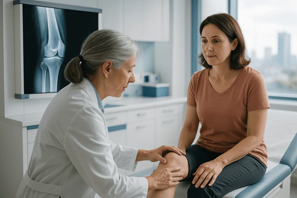
Diagnosis begins with a detailed physical examination. The orthopaedic specialist observes your posture, gait, joint range of motion, muscle strength, nerve function and limb length, and checks the position of the joints above and below the deformity, because a problem in the tibia, for example, changes how both the knee and the ankle work. Weight-bearing imaging is often essential: alignment under body weight can differ meaningfully from alignment measured lying down. Full-length standing X-rays show the mechanical axis of the whole limb, while targeted X-rays define the affected bone or joint. Depending on the case, CT is used to analyse rotation or complex bone shape, and MRI helps assess cartilage, ligaments, growth plates, bone quality and soft tissue structures.
Some patients arrive at deformity correction after previous treatment elsewhere. They may have a fracture that healed in a poor position, failed hardware, a persistent nonunion, bone loss after infection, or a deformity that recurred after childhood surgery. Others are diagnosed during evaluation for knee or hip arthritis, where an underlying alignment problem is quietly accelerating joint wear. A careful diagnostic pathway establishes three things: whether reconstruction is the right treatment at all, whether nonsurgical care is a reasonable alternative, and whether correction should be done in one stage or several.
Conditions Treated With Deformity Correction
A bone deformity can be congenital, developmental, traumatic, neuromuscular, metabolic, infectious or degenerative in origin, and the treatment strategy follows the cause. A child with a growth-related angular deformity needs a different plan from an adult with a malunited tibia, and both differ from a patient whose knee arthritis is being driven by severe bow-leg alignment.
Common indications include bow legs, knock knees, rotational deformities, limb length discrepancy, clubfoot or residual foot deformity after childhood treatment, alignment problems related to hip dysplasia, deformities after fractures, bone loss after trauma, and deformities caused by infection or growth plate injury. Patients with Blount disease, rickets or other metabolic bone disease, skeletal dysplasia, cerebral palsy, deformity after poliomyelitis, or congenital limb differences may also be evaluated, many childhood-onset conditions overlap with the broader field of congenital correction. In selected adults, a realignment osteotomy around the knee or ankle can redistribute forces across a worn joint and complement or postpone other procedures.
Post-traumatic deformity is a category of its own. When a fracture heals in an abnormal position, you may experience pain, restricted motion, difficulty bearing weight, or progressive wear in nearby joints. A corrective osteotomy can restore alignment and improve the mechanics of walking or lifting. In selected cases, deformity correction is part of treating a nonunion, a bone that has not healed, or reconstructing a bone defect.
Why does a bone bend out of shape?
A bone bent out of its normal line is usually the result of one of three processes: uneven growth, imperfect healing, or softening. In a growing child, if one side of a growth plate works faster than the other, after injury, infection or disease, the bone gradually angles away from the slower side. In any age group, a fracture that unites at an angle leaves a permanent bend unless it is corrected. And in metabolic conditions such as rickets or other disorders that soften bone, normal body weight itself slowly bows the weakened bone. Identifying which process is at work is essential, because correcting the shape without treating the underlying cause invites recurrence.
Can deformational plagiocephaly correct itself?
Mild deformational plagiocephaly, the flattened head shape some babies develop from lying in one position, often improves without surgery. The infant skull is still moulding, so measures such as varying the baby’s head position, supervised tummy time, and treating any neck tightness that keeps the head turned one way frequently allow the shape to round out as the child grows and spends more time upright. Some persistent or more pronounced cases are managed with helmet therapy, and any unusual head shape should be examined to rule out craniosynostosis, a different condition in which skull sutures fuse early and which is managed surgically by specialised teams. Plagiocephaly is not treated with the osteotomy techniques described on this page; deformities of the facial and cranial skeleton belong to a separate field, covered under facial correction.
Not every deformity requires surgery. Mild, stable deformities that cause no symptoms may simply be monitored with periodic examination and imaging. Bracing, physical therapy, activity modification, shoe lifts, orthotics, pain management, or treatment of an underlying metabolic condition may be appropriate instead. Surgery is considered when the expected functional benefit clearly outweighs the risks, and when your health, bone condition and capacity for rehabilitation support a reconstructive pathway.
Deformity Correction in Specific Joints and Regions
Because searches about deformity often concern one specific joint, it is worth answering the most common region-by-region questions directly.
Can a knee replacement correct a valgus deformity?
Yes: a knee replacement can correct a valgus (knock-knee) deformity in patients whose arthritis justifies replacement, because the surgeon’s bone cuts and soft tissue balancing re-establish the limb’s mechanical axis as part of the operation. Moderate valgus alignment is routinely corrected this way. More severe or long-standing valgus deformity is technically more demanding: the ligaments on the inner and outer sides of the knee have stretched and contracted unevenly, and the surgeon may need implants with additional built-in stability, or occasionally a staged plan that addresses bone alignment separately. In younger patients whose cartilage is still reasonably preserved, an osteotomy that realigns the bone while keeping the natural joint may be considered before replacement. Which route fits you depends on age, cartilage condition, ligament quality and the severity of the deformity.
Can swan neck deformity be corrected?
Swan neck deformity, where a finger hyperextends at the middle joint and bends down at the fingertip joint, can be corrected in many cases, and how to correct swan neck deformity depends on whether the finger is still flexible. A flexible deformity is often managed first with specially designed splints or ring orthoses that block the hyperextension while allowing the finger to bend, together with hand therapy. When the deformity is fixed, or when splinting does not restore useful function, surgical options include rebalancing the soft tissues around the middle joint, reconstructing the structures that normally prevent hyperextension, and, in advanced joints damaged by arthritis, fusing or replacing the middle joint. Because swan neck deformity frequently reflects an underlying condition such as rheumatoid arthritis or an old injury, lasting correction also requires that the underlying cause is properly managed by the treating team.
Can a wrist replacement correct hand deformity?
A wrist replacement addresses the wrist joint itself, pain, stiffness and alignment at the wrist, but it does not correct deformities of the fingers or thumb. Finger deformities such as swan neck, boutonnière or drift of the fingers towards the little-finger side have their own causes in the small joints and tendons of the hand, and they require their own procedures. In inflammatory arthritis, where wrist and finger problems commonly coexist, surgeons plan the sequence deliberately: a stable, well-aligned wrist provides a better foundation, and finger procedures are staged around it. So a wrist replacement can be one component of correcting a deformed hand, but it is never the whole answer.
What about deformity of the spine?
Spine correction is its own surgical discipline, covering curvature conditions such as scoliosis and kyphosis rather than limb alignment. The principles are recognisably similar, three-dimensional analysis of the deformity, controlled correction, stable fixation while bone heals, but the anatomy, instrumentation, risks and rehabilitation are entirely different, and the surgery is performed by dedicated spinal teams. A limb deformity and a spinal curve can also influence each other: a leg length difference tilts the pelvis and can produce a compensatory spinal curve, which is one reason whole-body assessment matters. If your concern is spinal curvature, deformity of the vertebrae or alignment of the trunk, the relevant treatment field is spine surgery.
How Deformity Correction Is Performed
Evaluation and Planning Before Treatment
Deformity correction begins with precise planning. During consultation, the orthopaedic team reviews your medical history, previous surgeries, injury records, imaging, current medicines and, importantly, your functional goals, because a plan for a manual worker differs from a plan for a sedentary office role. Existing X-rays, CT scans, MRI studies, operative reports and laboratory results are reviewed where available, which helps determine whether further tests are needed and whether the case is suitable for treatment at this stage.
Imaging is usually performed in weight-bearing positions so the limb is assessed under load. Full-length alignment radiographs identify where the mechanical axis passes through the hip, knee and ankle. CT clarifies rotational deformity and complex bone shape. MRI is requested when cartilage, ligaments, bone marrow, growth plates or soft tissues need assessment. In selected cases, gait analysis, vascular studies, nerve testing or an infection workup is added.
The surgical plan then defines the correction goal, the exact level for the osteotomy, the amount and direction of correction, the fixation method, and whether bone grafting or soft tissue balancing will be needed. Digital planning tools and three-dimensional assessment help the team simulate the correction before making it. Complex reconstructions are discussed among orthopaedic subspecialists, rehabilitation physicians, radiologists, anaesthesiologists and other relevant specialists before a plan is confirmed.
Preparation for Surgery
Before surgery you have a medical evaluation to confirm that anaesthesia and recovery can proceed safely. This may include blood tests, heart and lung assessment where indicated, and review of chronic conditions such as diabetes or thyroid disease. Your regular medicines are reviewed as part of this process; any adjustment before surgery is a decision that belongs to your treating doctor. Smoking cessation is strongly encouraged, because smoking demonstrably impairs bone healing and raises complication risk. Nutritional status, vitamin D level, bone density and infection markers may also be checked, particularly in revision or staged cases.
You are told in advance what to expect: the likely hospital stay, weight-bearing restrictions, wound care, pain control, the rehabilitation programme, and the assistive devices, crutches, a walker, sometimes a wheelchair, you will use in the early phase. The follow-up schedule is also discussed before the operation: which images will be needed at each stage of healing, and how progress will be reviewed between appointments.
The Procedure Itself
The operation is performed under general or regional anaesthesia, depending on the procedure and your condition. In broad terms, a corrective operation follows this sequence:
- The surgeon exposes the planned bone level through carefully positioned incisions.
- If an osteotomy is needed, the bone is cut in a controlled manner exactly as planned.
- The bone is realigned, either fully during the operation, or set up for gradual correction afterwards.
- Fixation is applied: internal plates, screws, nails or other implants beneath the skin, or an external frame connected through pins or wires. In some cases both are combined.
- Bone graft or bone substitute is added if there is a gap, bone loss or poor healing biology.
- Soft tissues are addressed where required, tight tendons released or lengthened, contracted ligaments balanced, muscle imbalance corrected, because soft tissue determines whether the new alignment actually functions.
- Position and implant placement are confirmed with intraoperative imaging before closure.
External fixation is often chosen when gradual correction, limb lengthening, infection management or complex multiplanar adjustment is needed, because the frame allows small, controlled changes after surgery. If the deformity involves a joint with significant arthritis, the plan may include a joint-preserving osteotomy, staged reconstruction, or, in selected adults, joint replacement as part of the broader strategy. The duration of surgery varies widely: a single-bone osteotomy takes far less time than a multi-level reconstruction involving old implants, bone loss or gradual correction. Afterwards you are monitored in recovery, then transferred to your room, where pain control, circulation checks, nerve assessment, wound monitoring and early mobilisation are the first priorities.
Technology Used During Deformity Correction
Technology supports deformity correction at every stage, from diagnosis through surgery to follow-up. Full-length standing radiographs show overall limb alignment; CT-based evaluation reveals rotational problems that standard X-rays can miss; MRI protects joint health by identifying cartilage or soft tissue issues before surgery. Digital planning systems let surgeons measure angles, simulate osteotomies and plan fixation, and in complex cases three-dimensional modelling or patient-specific planning clarifies the anatomy before a single incision is made. During surgery, fluoroscopic imaging confirms bone position and implant placement in real time. For gradual correction, adjustable fixation systems make small daily changes possible, which puts far less stress on nerves, vessels and skin than forcing a large correction at once.
Be clear-eyed about what technology is: one part of care. Its value depends entirely on the judgement behind it, surgical experience, rehabilitation planning and ongoing monitoring. The right tool is selected for the specific deformity, not applied identically to every patient.
Recovery Process After Surgery
Recovery depends on the procedure, the bone involved, the fixation method, your age, and whether correction was immediate or gradual. Some patients begin gentle motion and partial weight bearing within days; others must protect the limb from load until early healing is visible on imaging. Physical therapy concentrates on maintaining joint motion, controlling swelling, protecting the reconstruction and gradually rebuilding strength.
If you have an external frame, you and your caregivers are taught pin-site care, how to make frame adjustments if your protocol requires them, and which changes should prompt a medical review. With internal fixation, the emphasis is on wound care, swelling control, safe movement and scheduled follow-up imaging. Bone healing typically takes several months, and complex reconstructions need a longer period of supervised rehabilitation. Return to work, school, sport and travel is individualised and guided by your treating surgeon, not by a fixed calendar.
Why Acting Early Matters
Most deformities progress. A child with uneven growth plates develops increasing angulation as growth continues. An adult with malalignment loads one side of the knee, ankle or hip excessively, accelerating cartilage wear. A limb length discrepancy tilts the pelvis, alters gait and generates back pain and fatigue. Over time, muscles and tendons adapt to the abnormal position, tightening here, weakening there, in ways that make later correction more complex than earlier correction would have been.
Delaying evaluation does not automatically mean surgery becomes necessary, but it can narrow the options. In children, early diagnosis may allow guided growth procedures that are considerably less invasive than major reconstruction. In post-traumatic cases, earlier treatment of malalignment, nonunion or infection improves the environment for bone healing and may protect the neighbouring joints. In adults with degenerative overload, correction before severe arthritis develops can preserve joint function in selected cases.
Delay also multiplies compensatory problems. A deformity in the tibia affects both the knee and the ankle. Hip malalignment influences the spine. A foot deformity changes the entire walking pattern. By the time pain is constant, the original deformity is often only one link in a longer chain of mechanical problems. Timely assessment establishes whether observation, rehabilitation, bracing or surgery is the appropriate next step, and keeps the less invasive options on the table.
Benefits of Deformity Correction
The potential benefits are best understood in terms of function, joint protection, comfort and long-term mobility, not appearance alone.
| Benefit | What It Means for You |
|---|---|
| Improved alignment | Realigning the bone or joint restores a more efficient mechanical axis, so the limb carries weight in a safer, more balanced way. |
| Better mobility and gait | With the limb position and muscle mechanics improved, many patients walk with less limping, fatigue and compensation. |
| Reduced pain from abnormal loading | Correction may decrease excessive pressure on overloaded joints, soft tissues or previous fracture sites. |
| Joint preservation in selected cases | Redistributing forces across the knee, ankle, hip or foot may slow the mechanical stress that contributes to joint deterioration. |
| Improved limb symmetry | Correcting length, angle or rotation can improve standing posture, shoe wear, balance and overall body mechanics. |
| Greater independence in daily activities | With rehabilitation, patients may find it easier to climb stairs, stand for longer, return to work and take part in appropriate physical activity. |
Recovery Timeline After Deformity Correction
Recovery is individual, but most patients move through recognisable stages. Treat this as orientation, not a schedule: your surgeon sets the actual pace based on imaging and examination.
| Time Period | What Patients Can Expect |
|---|---|
| Day 1 | Monitoring of pain, circulation, nerve function, wounds and fixation. Physical therapy may begin with breathing exercises, safe transfers and protected movement. |
| First Week | Swelling and discomfort are managed with medication, elevation and guided activity. You learn mobility precautions, wound care and your weight-bearing limits. |
| First Month | Follow-up imaging checks early bone position. Therapy focuses on joint motion, muscle activation and safe daily function. Patients on gradual protocols continue frame adjustments. |
| Months 2 to 3 | Bone healing is assessed. Weight bearing advances if the surgeon confirms sufficient healing. Strengthening and gait training become more active. |
| Months 4 to 6 | Many patients increase walking endurance and daily activity. Complex reconstructions, limb lengthening and revision surgery may need a longer protected phase. |
| Longer Term | Continued strengthening, balance training and follow-up imaging protect the correction. Return to sport or high-impact activity depends on healing, stability and the surgeon’s assessment. |
Factors That Influence Outcomes
A good result depends on accurate diagnosis, careful planning, stable fixation, bone healing, soft tissue balance and consistent rehabilitation, in that order of sequence, and all of them necessary. Severity and complexity matter: a single-level angular deformity in healthy bone is more straightforward than a multiplanar deformity involving rotation, shortening, joint stiffness, previous infection or multiple prior surgeries.
Age and growth potential cut both ways. In children, remaining growth is an asset when guided growth is possible, but it can also drive recurrence if the underlying growth problem continues after correction. In adults, bone density, arthritis, muscle strength and general health shape both the surgical plan and the recovery pace. Chronic conditions such as diabetes, vascular disease, autoimmune disease, kidney disease and poor nutrition can affect wound healing and bone repair. Body weight influences the load every correction must carry, understanding your body mass index is a reasonable part of preparing for lower-limb surgery.
Bone biology is central. Smoking, uncontrolled diabetes, low vitamin D, chronic infection and certain medical treatments can delay or complicate healing; where bone grafting is planned or a nonunion is present, the team evaluates and optimises these factors before surgery. The wider subject of maintaining strong, well-mineralised bone is covered under bone health. Patients who follow weight-bearing instructions, attend therapy, protect their wounds and keep follow-up appointments give the reconstruction a measurably better environment in which to heal.
The condition of the joints above and below the deformity also shapes what surgery can achieve. If a knee, ankle, hip or foot joint already has severe arthritis or stiffness, correcting the bone alignment improves the mechanics but may not fully resolve pain arising from the damaged joint itself. This is why preoperative counselling matters: you should understand the intended goals, the recovery demands, the realistic limitations, and the possibility of staged procedures before you decide.
As with any surgery, deformity correction carries risks. These include infection, bleeding, blood clots, nerve or blood vessel injury, delayed union, nonunion, recurrence of the deformity, stiffness, irritation from hardware, pin-site problems with external fixation, and the need for additional surgery. Experienced planning reduces these risks; nothing eliminates them. A surgeon who discusses risk openly is doing their job properly.
How Deformity Care Is Organised at Acibadem
Deformity correction requires more than technical surgery. It requires precise diagnosis, coordinated planning, reliable perioperative care, rehabilitation guidance, and clear communication at every stage. At Acibadem, evaluation is based on detailed imaging, clinical examination and functional assessment, and complex cases are reviewed through a multidisciplinary approach. Depending on the patient, that review may involve orthopaedic trauma surgeons, paediatric orthopaedic surgeons, limb reconstruction specialists, spine or foot and ankle specialists, radiologists, anaesthesiologists, rehabilitation physicians, physiotherapists, infectious disease specialists, pain management teams and internal medicine physicians. This structure matters most when the deformity relates to previous trauma, congenital disease, neuromuscular conditions, infection or multiple prior operations.
The diagnostic and planning tools are chosen to fit the clinical problem rather than applied uniformly: weight-bearing radiographs for mechanical alignment, CT for rotational and complex bony anatomy, MRI for joint and soft tissue assessment, intraoperative imaging for accuracy, and fixation systems suited to either immediate or gradual correction. Treatment planning is equally individual. Some patients are best served by observation and therapy rather than surgery. Others need a focused osteotomy, gradual correction, limb lengthening or staged reconstruction. The recommendation weighs symptoms, imaging findings, age, occupation, activity goals, family circumstances and the realistic capacity to complete rehabilitation.
Second opinions are a normal part of this field. When a proposed operation is complex, when previous procedures have not achieved the intended result, or when different surgeons have recommended different approaches, a structured review of existing imaging and operative reports helps clarify the diagnosis, the realistic options and the questions worth asking before committing to treatment.
Making a Decision About Deformity Correction
Deformity correction is a significant decision, and it deserves a deliberate one. The strongest position to decide from is a precise diagnosis and an honest conversation about goals, risks, recovery demands and alternatives, including the alternative of doing nothing yet and monitoring. Whether the concern is a child’s progressive limb deformity, an adult’s post-traumatic malalignment, a limb length discrepancy or a complex reconstruction after previous surgery, careful evaluation defines the safest and most effective path. Recent imaging, previous operative notes and a clear account of your symptoms are the raw material of that evaluation; the more complete the picture, the more specific and reliable the recommendation an orthopaedic team can give.
Preparation
- Patients usually undergo orthopedic examination, X-rays, CT or MRI scans, and gait or alignment assessment when needed. Blood tests and anesthesia evaluation are completed before surgery. Blood thinners, smoking, and certain medications may need to be stopped as advised by the surgical team.
Aftercare
- Pain control, wound care, and infection prevention are monitored closely after surgery. Weight-bearing may be limited, and crutches, braces, or external fixation care may be required. Physical therapy supports safe mobility, strength, and gradual return to daily activities.
Frequently Asked Questions
What affects the cost of deformity correction?
The main factors are the deformity type and severity, the surgical technique, implant or external fixator choice, imaging needs, hospital stay, rehabilitation, and follow-up plan. A personalized quote can be prepared after a specialist reviews your medical records and imaging.
How can I get a personalized quote from Acibadem?
You can request a free consultation by sharing your diagnosis, recent X-rays or scans, previous surgery records if available, and a summary of symptoms and mobility goals. The orthopedic team can then advise on likely treatment options and package inclusions.
Are travel and accommodation included in the treatment cost?
Inclusions vary by package. International patient services may help with interpreter support, appointment coordination, transfers, and accommodation guidance, but each item should be confirmed before travel.
Why do implants or external fixators change the total cost?
Different devices have different technical requirements, planning needs, and follow-up demands. The choice depends on the deformity pattern, bone quality, stability needs, and the surgeon’s treatment plan.
Will rehabilitation affect the final cost?
Yes. Deformity correction often requires physiotherapy, gait training, wound care, and follow-up imaging. The intensity and duration of rehabilitation depend on the procedure and recovery progress.
Is this information a medical or financial recommendation?
No. This is general educational information. A specialist assessment and a formal hospital quotation are needed to understand suitability, expected pathway, and estimated inclusions for your case.
Medically reviewed by the Acıbadem International Medical Board September 13, 2026
See our medical review board →
Update history
- PublishedJune 8, 2026
- Medical review approvedSeptember 13, 2026
- Last content updateSeptember 12, 2026
Trusted care for international patients
From the Acibadem Blog
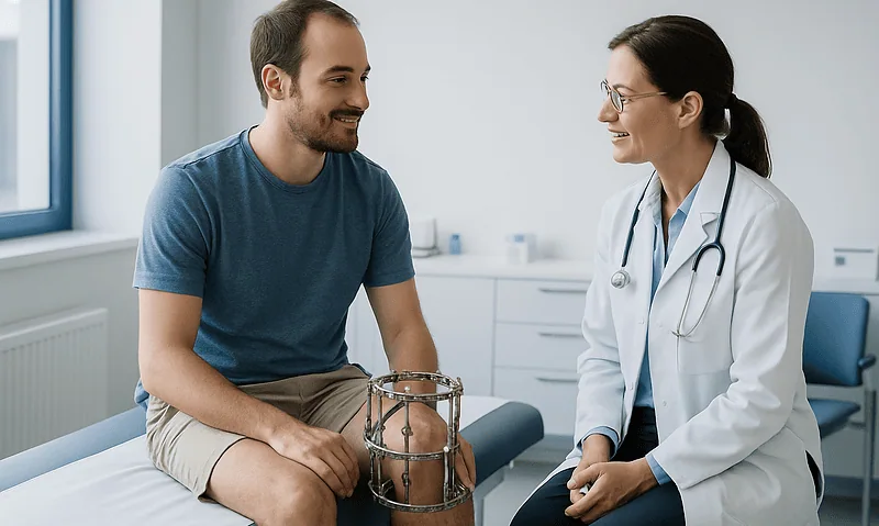
Living With an External Fixator During Deformity Correction: Pin Care and Daily Routine
Living with an external fixator during deformity correction means learning a steady daily routine: cleaning the skin around each pin as your surgical team…