

Why CT Angiography Comes Before TAVI: Planning Valve Size and the Access Route

Key Takeaways
- The aortic annulus is usually oval, so a single echo diameter can undersize or oversize the valve, while CT measures the full perimeter and area in three dimensions.
- A valve that is too small risks leaking around its edges, and one that is too large risks tearing a calcified ring, which is why sizing is treated as a safety step rather than a formality.
- The distance from the valve ring to the coronary artery openings, measured on CT, is the main way teams predict and avoid coronary obstruction during TAVI.
- The same scan sweeps from the chest to the thighs to check whether the femoral arteries are wide and smooth enough for the delivery sheath, and to image alternative routes if they are not.
- Kidney function is checked before the scan because contrast dye can cause a temporary dip in kidney function, and teams adapt fluid, timing and contrast volume accordingly.
- Being referred for the planning CT does not mean TAVI has been chosen; the heart team often uses the same images to weigh surgery against a catheter procedure.
CT angiography comes before TAVI because the scan gives the heart team millimeter-level measurements that an echocardiogram alone cannot: the exact size and shape of the aortic valve ring, the height of the coronary arteries above it, and the width, twists and calcium of the arteries in the legs and chest. Those measurements decide which valve size is chosen and which route the catheter can travel safely.
The appointment letter arrives before anything has been decided. A woman in her late seventies, who has been told her aortic valve is badly narrowed and that a catheter procedure called TAVI may be an option, reads that her next step is not a meeting with the surgeon but a CT scan with dye. Her first reaction is a familiar one: haven’t they already looked at my heart?
They have. Yet CT angiography before TAVI answers a different set of questions from the echocardiogram that made the diagnosis. The echo told the team the valve is too tight. The CT tells them how big the replacement must be, to a fraction of a millimeter, and whether the blood vessels in her groin are wide and smooth enough to carry a folded valve up to her heart.
Think of it as the difference between knowing a doorway is too narrow and having the exact measurements needed to fit a new door. Nobody orders the door first.
What is CT angiography before TAVI, and what actually happens?
Two terms need unpacking. TAVI, transcatheter aortic valve implantation (also called TAVR in the United States), is a procedure in which a new valve, compressed inside a thin tube, is threaded through a blood vessel and expanded inside the old, narrowed aortic valve without opening the chest. CT angiography is a CT scan timed to catch a dose of iodine-based contrast dye as it flows through the arteries, so that the vessels light up brightly on the images.
Put together, a TAVI planning CT is a single study that usually covers two territories in one visit. The first is the heart itself, scanned in step with the electrocardiogram so that each image is captured at the same point in the heartbeat; this is called ECG gating, and it freezes the moving valve into a sharp, measurable picture. The second is a long sweep from the neck or upper chest down to the top of the thighs, which maps every artery the catheter might need to use.
From the patient’s side the experience is modest. A small cannula goes into a vein in the arm, electrodes are stuck to the chest, and the table slides through a ring-shaped scanner while a voice asks for a few breath-holds. The contrast produces a brief warm flush and sometimes a metallic taste. The Mayo Clinic describes the whole CT angiogram visit, including preparation, as typically taking well under an hour, with the scanning itself lasting only minutes.
Behind the scenes is where the work happens. Specialist software rebuilds the raw images into three-dimensional models that can be rotated, sliced at any angle and measured. That reconstruction, not the scan itself, is what the heart team will study when it meets to plan the valve.
Why isn't an echocardiogram enough to size a TAVI valve?
Echocardiography, ultrasound of the heart, is superb at what it was built for. It shows how fast blood squeezes through the narrowed valve, how thick the heart muscle has become in response, and how well the pumping chamber is coping. It is the test that diagnoses aortic stenosis, the stiffening and narrowing of the valve that TAVI treats, and it remains the test used to follow the new valve afterwards.
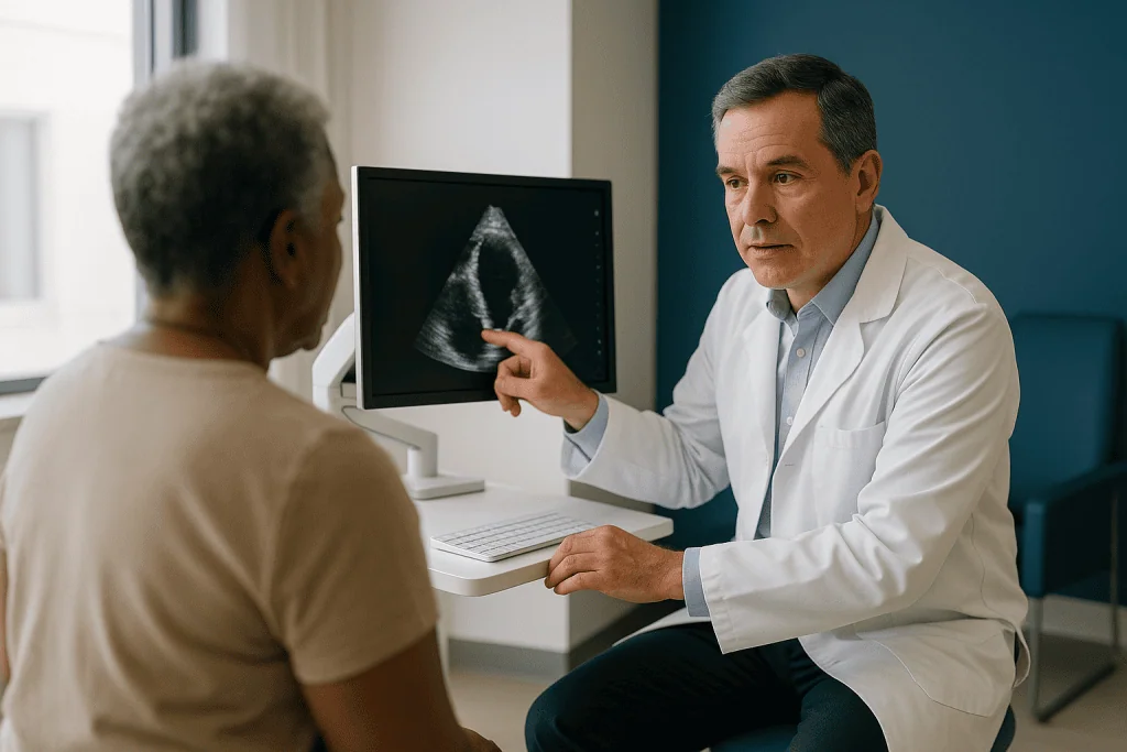
Sizing is a different problem. The aortic annulus, the ring of tissue at the base of the valve where the new device must seat, is not a circle. In most people it is oval, sometimes markedly so. A standard echo image is a two-dimensional slice, and a slice through an oval gives you one diameter that depends entirely on the angle you happened to cut. Measure the short axis and you will undersize; measure the long axis and you will oversize.
CT sidesteps the problem by capturing the whole ring in three dimensions. The reader can trace its full perimeter and calculate its area rather than guessing from a single width. Because the scan is gated to the heartbeat, the ring can also be measured at the moment in the cycle when it is largest, which is when a snug fit matters most.
There is a second gap echo cannot fill. Ultrasound does not travel well through calcium, and heavily calcified valves scatter the signal. CT sees calcium as bright white and shows exactly where the deposits sit, how bulky they are and whether they extend below the valve into the outflow tract. That distribution changes how a valve will expand and where it might leak.
How is the aortic annulus measurement done on CT?
The measurement begins with a small act of geometry. Using the three-dimensional dataset, the reader finds the three lowest points where the valve leaflets attach to the aortic wall and defines a flat plane passing through them. That plane is the annulus. Everything else in valve sizing is measured relative to it.
Within that plane, the software or the reader traces the inner edge of the ring. From the trace come three numbers that appear in nearly every planning report: the perimeter, the area, and the longest and shortest diameters. Different valve designs are sized by different numbers, which is one reason the same annulus may be matched to differently labelled devices, and why the choice of device and size belongs to the implanting team rather than to any single measurement.
Timing matters as much as tracing. The ring stretches slightly when the heart contracts and relaxes slightly when it fills. Most planning protocols measure in systole, the contraction phase, when the annulus is at its largest, because a valve chosen against the smallest dimension could be left with gaps around it.
The reader then moves upward from the ring, measuring the sinuses of Valsalva, the three rounded pouches just above the valve, and the height of the two coronary artery openings. They move downward, looking at the left ventricular outflow tract, the funnel of muscle leading into the valve, for calcium that might prevent the device from expanding evenly. A good report reads like a survey of a small, complicated room, and it is usually reviewed by more than one pair of eyes before the heart team meeting.
What can go wrong if the TAVI valve size is off?
This is the part patients rarely hear explained, and it is the honest reason the scan is non-negotiable. A transcatheter valve does not get sewn in. It holds its place by pressing outward against the old valve and the annulus, like a spring-loaded curtain rod braced in a window frame. The fit is the fixation.

Choose a valve too small for the ring and the rod does not brace. The most common consequence is paravalvular leak, blood slipping backwards around the outside of the new valve rather than through it. Small leaks are often tolerated; larger ones make the heart work harder and are associated with poorer outcomes in the clinical literature, which is why heart teams treat them as something to design out rather than manage afterwards. In the extreme, an undersized valve can shift from its position.
Choose one too large and the opposite risk appears. Excessive force against a calcified, brittle ring can tear the tissue, an injury called annular rupture. It is uncommon, but it is serious, and CT is the main tool for spotting the combination of a small ring and bulky calcium that makes it more likely. An oversized frame can also press on the electrical wiring that runs just beneath the valve, one reason some people need a permanent pacemaker after TAVI.
Notice the pattern. Both errors flow from the same missing information, and both are largely preventable at the planning stage. That is why a heart team will sometimes repeat a CT that is technically imperfect rather than proceed on a best guess. No one is being cautious for the sake of it; the measurement is the safety mechanism.
How does the CT choose the TAVI access route: femoral or another way?
The folded valve travels inside a delivery sheath, a tube that is thin by surgical standards yet still needs an artery wide enough to accept it and smooth enough not to tear. The femoral artery, the main vessel running through the groin at the top of each thigh, is the preferred entry point because it is close to the skin, easy to close, and allows the procedure to be done with local anesthetic and light sedation in many centers, as both the NHS and the Mayo Clinic describe.
The CT sweep of the pelvis and legs decides whether that route is open. Readers check the minimum diameter of each iliac and femoral artery along its entire length, look for tight kinks, and grade the calcium. A vessel that is narrow in only one short segment, or that is ringed by a hard cuff of calcium, can be enough to redirect the plan.
When the groin will not serve, the team looks at the alternatives the scan has already imaged.
| Access route | Where the catheter enters | What the CT is checking | Typically considered when |
|---|---|---|---|
| Transfemoral | Femoral artery at the groin | Vessel width, tortuosity, calcium | Default route in most planning pathways |
| Subclavian or axillary | Artery beneath the collarbone | Diameter, angle into the aorta | Leg vessels too small or diseased |
| Transcarotid | Neck artery | Vessel health, brain supply | Selected centers, when other routes fail |
| Transaortic | Small chest incision to the aorta | Aortic calcium, safe entry zone | Peripheral arteries unsuitable |
| Transapical | Tip of the heart via a small chest cut | Heart position, lung overlap | Now used less often |
Which route is chosen, and whether an alternative is offered at all, depends on the team’s experience and the individual anatomy. The CT does not make that decision; it makes it possible.
Why do coronary artery height and calcium matter so much?
Just above the aortic valve, two small openings lead into the coronary arteries, the vessels that feed the heart muscle itself. When a transcatheter valve expands, it pushes the old, calcified leaflets outward and upward. If a leaflet is long and the coronary opening sits low, the displaced leaflet can be pressed across that opening like a hand over a hose. Coronary obstruction during TAVI is rare, but it is one of the most dangerous things that can happen in the procedure, and it is almost entirely predictable from the CT.
Readers therefore measure the distance from the annular plane to each coronary opening and compare it with the length of the leaflet that will be pushed toward it. They also look at how wide the sinuses are, because a roomy sinus gives the leaflet somewhere to go. When the numbers look tight, the team can choose a different valve design, plan protective techniques, or in some cases decide that surgery is the safer option.
The same images serve a second purpose. Many people with aortic stenosis also have narrowing in the coronary arteries. In some centers the planning CT is read for coronary disease as well, and a clear scan may spare a patient the separate invasive angiogram that has traditionally been part of the workup. Where the coronary images are limited by calcium or heart rate, the invasive test is still done; the CT complements it rather than replacing it by default.
Finally, the team thinks ahead. A person receiving a valve today may need a coronary stent years from now. The scan helps predict whether the new valve frame will make those arteries harder to reach, which can influence device choice in younger patients.
Who is usually offered TAVI, and who is usually asked to wait?
TAVI is a treatment for severe aortic stenosis that is causing symptoms, most commonly breathlessness, chest tightness on exertion, dizziness or fainting. The Mayo Clinic and the NHS both describe it as an option for people in whom open-heart surgery carries elevated risk because of age or other health problems, and in many health systems it is now discussed alongside surgery for a broader group. The choice between the two is made by a heart team, a group that typically includes cardiologists, cardiac surgeons, imaging specialists and anesthetists reviewing the case together.
The planning CT is part of that conversation, not something that follows it. A person may be referred for the scan while the team is still weighing surgery against TAVI, precisely because the images inform both options.
Some people are asked to wait, and the reasons are usually practical rather than a sign of bad news.
- Kidney function is reduced, and the team wants to protect the kidneys before giving contrast, or to time the scan with extra fluid.
- There has been a previous reaction to contrast dye, so premedication or a modified protocol needs arranging.
- An active infection, including dental infection, needs treating first because it raises the risk of infection on any new valve.
- The heart rate is irregular or fast enough that the gated images would blur, and the team prefers to settle it first.
- The stenosis is severe on echo but has not yet caused symptoms, in which case guidelines generally support close monitoring rather than immediate intervention.
Waiting is uncomfortable when you have been told your valve is failing. It helps to know that in most of these situations the pause is about doing the scan well, so that the plan built on it can be trusted.
CT scan before TAVI and your kidneys: what the contrast dye asks of you
Contrast dye is the one part of the planning CT that carries a real, if small, medical trade-off. The iodine-based agent is filtered out by the kidneys, and in people whose kidneys are already under strain it can occasionally cause a temporary dip in function known as contrast-associated acute kidney injury. MedlinePlus and the Mayo Clinic both list this among the recognized risks of CT angiography.
Aortic stenosis and chronic kidney disease keep company more often than chance would predict, partly because both become common with age and partly because they share risk factors such as high blood pressure and diabetes. That overlap is why a blood test measuring kidney function, usually reported as an estimated glomerular filtration rate, is checked before the scan is booked.
What the team does with that number varies. In people with healthy kidneys the scan proceeds normally. In people with reduced function, options include giving fluid through a vein before and after the scan, using a lower contrast volume with newer scanner protocols, spacing the CT apart from other contrast studies such as the invasive coronary angiogram, and rechecking kidney function afterwards. In a small number of people the team may decide the information is best gathered another way.
Certain medicines interact with this picture. Some diabetes tablets and some blood pressure medicines can be paused around contrast exposure on a clinician’s instruction; others should never be stopped without advice. The right course depends on your kidney results and your full medication list, which is why the instruction should come from your prescribing clinician or the radiology team rather than from a general article. If you are unsure about any tablet on the morning of the scan, phone and ask before you skip it.
TAVI CT scan preparation: how the day usually goes
Most of the preparation is administrative. Expect to be asked about previous contrast reactions, asthma, kidney problems, diabetes medicines and whether you might be pregnant. Bring a current list of your medicines. The department will tell you whether to eat or drink beforehand; instructions differ between hospitals, and following yours matters more than any general rule.
On arrival, a nurse or radiographer places a cannula in a vein, usually in the arm, and attaches ECG electrodes to the chest. Some centers give a medicine from the beta-blocker class to slow the heart if it is running fast, because a steadier, slower rhythm produces sharper gated images of the coronary arteries. Whether that is used, and whether it is right for you, is a decision for the team on the day, informed by your other conditions.
You lie on your back with arms above your head. The scanner is a large ring, not a tunnel, so people who dislike enclosed spaces generally manage well. A short scout image is taken first. Then the contrast is injected by a pump while the table moves through the ring, and you are asked to hold your breath for a handful of seconds, sometimes more than once.
The warm flush from contrast spreads from the chest to the pelvis and fades within a minute. A metallic taste and a passing sensation of needing to pass urine are common and harmless. Afterwards you sit for a short observation period, drink fluids if allowed, and can usually go home the same day, as the Mayo Clinic describes for CT angiography generally.
The images are not read while you wait. Reconstruction and measurement take time, and the report is written to be discussed at the heart team meeting rather than handed to you in the corridor.
Is the radiation from CT angiography before TAVI a concern?
Any CT scan uses X-rays, and a gated cardiac study combined with a full chest-to-thigh sweep is among the larger diagnostic CT examinations. It is fair to ask about it, and it is fair to expect a straight answer.
The straight answer has two parts. First, the radiation from a single diagnostic CT is generally described by the Mayo Clinic and MedlinePlus as carrying a small theoretical increase in lifetime cancer risk, an effect that becomes less relevant with advancing age because any such cancer would take many years to develop. Most people undergoing TAVI planning are in their seventies or older. Second, the alternative is not a lower-radiation scan; it is proceeding with less information about a procedure whose main preventable complications are anatomical. When the two are set side by side, imaging specialists and cardiologists across guideline bodies have concluded that the benefit is clear.
Modern scanners help. Faster detectors can capture the heart in a single beat, ECG gating can be set to expose only during the phase that matters, and dose-reduction software rebuilds sharp images from lower exposures than were needed a decade ago. Departments also tailor the protocol to body size.
What you can reasonably ask is whether your scan will be done on a low-dose protocol and whether it will combine the heart and vascular imaging in one session rather than two. What is not reasonable, in this context, is declining the scan on radiation grounds without a discussion, because the decision it informs is a one-way door. Once a valve is chosen and implanted, there is no going back to check the measurements.
What happens in the weeks after the CT scan?
The scan is the pivot of the pathway, and the weeks that follow tend to have a rhythm. First comes the heart team meeting. Your images, echo, blood tests and general health are reviewed together, and a recommendation is formed: TAVI, surgery, or in some cases continued monitoring. You are then seen in clinic to hear that recommendation, ask questions and give consent. Timing between scan and clinic varies with local scheduling; if it stretches, ask what the expected wait is rather than assuming silence means something is wrong.
Other tests may be slotted in. An invasive coronary angiogram is still common where the CT could not clear the coronary arteries. A dental check is often requested because untreated dental infection is a recognized source of infection on artificial valves. Lung function tests or a frailty assessment may be added for people with other health conditions.
On the day of the procedure itself, the NHS describes TAVI as typically taking a couple of hours, with most people staying in hospital for a few days afterwards. Many transfemoral procedures are done under local anesthetic with sedation. Afterwards you lie flat for a period while the artery entry site seals, and you are monitored for heart rhythm changes, which is why a temporary pacing wire is sometimes left in place for the first day or two.
Recovery after TAVI is generally described by the NHS and the Mayo Clinic as quicker than after open surgery, with most people resuming light activity within days and building up over the following weeks, guided by their team. Blood-thinning medicines are usually prescribed for a defined period to reduce clot risk on the new valve; the type and duration are decided by the cardiologist based on your other conditions. A follow-up echo checks the valve’s position and function, closing the loop the CT opened.
What people often get wrong about the TAVI planning CT
It is a repeat of the heart scan I already had. Almost never. The diagnostic echo and the planning CT answer different questions with different physics. One measures blood flow through a narrowed valve; the other measures the ring, the arteries and the calcium to fit a device.
Being sent for the scan means TAVI has been decided. Not necessarily. The heart team often orders the CT while still comparing TAVI with surgery, because the anatomy affects both decisions. A scan is a step in deciding, not the decision itself.
Valve size is chosen like a shoe size from a standard chart. Manufacturers do publish sizing ranges, but the ring is oval, the calcium is uneven and the coronary heights differ from person to person. The team weighs perimeter, area, calcium pattern and coronary position together, and sometimes two experienced readers will legitimately favor different sizes. That debate is part of good planning.
If the groin arteries are too small, TAVI is off the table. Alternative routes exist, as the table above shows. Whether one is suitable is a judgment for the team, but a narrow femoral artery redirects the conversation rather than ending it.
The dye will damage my kidneys. For most people the risk is small and temporary, and teams have several ways of reducing it. Where kidney function is genuinely fragile, the plan is adapted rather than abandoned.
Results should be available before I leave the department. The images need reconstruction and specialist measurement, and their meaning depends on the rest of your case. Expect the findings in clinic, presented as part of a plan, not as a printout at the desk.
Questions to ask your care team about CT planning for TAVI
A good consultation leaves you understanding not just what will happen but why. These questions are phrased to open that conversation; you do not need to ask them all, and your team may answer several before you reach them.
- Will the CT cover both my heart and the arteries in my chest and legs in a single visit, or will I need two scans?
- What did my kidney blood test show, and does it change how the contrast will be given?
- Should I take my usual medicines on the morning of the scan, and which ones, if any, need pausing on your instruction?
- Will you be able to assess my coronary arteries from this CT, or will I need a separate angiogram as well?
- How large and how oval is my annulus, and does that make one type of valve more suitable than another?
- How high are my coronary openings, and is there any concern about obstruction?
- Are my femoral arteries suitable for the standard groin approach? If not, what route are you considering and why?
- Did the scan show anything unexpected elsewhere, such as in the lungs or aorta, that needs following up separately?
- How long is the usual interval between this scan and the heart team decision in your service?
- If my scan is technically limited, would you repeat it, and what would that involve?
Write the answers down or bring someone who will. The measurements themselves are not the point; what matters is that you leave understanding how they shaped the plan, and that the plan was made for your anatomy rather than for the average patient.
When to call your doctor
Three situations deserve prompt attention around the time of a TAVI planning CT.
After the contrast injection. Mild warmth, a metallic taste and slight nausea usually settle within minutes. Contact the department or your doctor the same day if you develop a spreading rash, itching or hives, or if you notice reduced urine output, unusual swelling or a marked drop in how much you are passing over the following two days, which can signal kidney strain. Seek emergency care immediately for swelling of the face, lips or throat, wheezing, difficulty breathing or feeling faint, as these can indicate a severe allergic reaction.
While waiting for the heart team decision. Severe aortic stenosis can change quickly. Call your cardiology team the same day if breathlessness gets noticeably worse, if you wake at night short of breath, or if your ankles swell more than usual. Treat chest pain that does not settle with rest, fainting or near-fainting, or sudden severe breathlessness as emergencies and call emergency services rather than waiting for a scheduled appointment.
After TAVI, if the procedure goes ahead. Bleeding or a rapidly enlarging lump at the groin entry site, a leg that becomes cold, pale or numb, fever, or a new fast or very slow heartbeat with dizziness all need urgent contact with your team. Sudden weakness on one side, facial drooping, slurred speech or confusion are signs of stroke and require an immediate emergency call.
None of these lists is exhaustive. If something feels wrong and you cannot explain it, the right response is to phone the team that knows your case. They would far rather hear from you about a false alarm than miss a real one.
Frequently asked questions
How long does a CT scan before TAVI take?
The scanning itself takes only minutes, and the whole visit, including cannula placement, preparation and a short observation afterwards, is typically well under an hour according to the Mayo Clinic’s description of CT angiography. Reading and measuring the images takes longer and happens after you leave, which is why results are usually given in clinic as part of the heart team’s plan rather than on the day.
What is the aortic annulus measurement on CT used for?
It determines the size of the replacement valve. The annulus is the ring of tissue at the base of the aortic valve where the new device seats. CT traces its full perimeter and area at the point in the heartbeat when it is largest, and those figures are matched against the sizing ranges of different valve designs. The final choice rests with the implanting team, who also weigh calcium and coronary height.
Why do I need a CT of my legs for a heart valve?
Because the valve usually travels to the heart through the femoral artery at the groin. The CT checks that the arteries in the pelvis and thighs are wide enough, not too twisted and not too calcified to accept the delivery sheath safely. If they are unsuitable, the same scan shows whether an alternative route, such as an artery beneath the collarbone, could be used instead.
Can a CT scan before TAVI harm my kidneys?
It carries a small risk. The iodine contrast dye is cleared by the kidneys and can occasionally cause a temporary drop in kidney function, most often in people whose kidneys are already impaired. Teams check kidney blood tests beforehand and may give extra fluid, reduce contrast volume or space the scan apart from other dye studies. For most people the effect is small and reversible, and the plan is adapted rather than abandoned.
What is TAVI CT scan preparation like, and can I eat beforehand?
Preparation is light. You will be asked about kidney problems, diabetes medicines, previous contrast reactions and asthma, and should bring a medicine list. Whether you eat or drink beforehand depends on your department’s instructions, which vary, so follow the letter you received. A cannula is placed in an arm vein, ECG electrodes are attached and you will hold your breath briefly during the scan. Most people go home the same day.
Does needing the CT scan mean TAVI has been decided for me?
Not necessarily. The heart team often requests the planning CT while still deciding between TAVI and open surgery, because the anatomy it reveals informs both options. In some people the scan shows features that favor surgery; in others it confirms a catheter route is feasible. The decision is made at the team meeting after the images, echo, blood tests and your overall health have been reviewed together.
What is ECG gating and why does the scan need it?
ECG gating links the scanner to your heartbeat so that images are captured at the same phase of each cycle, freezing the moving valve into a sharp picture. Without it, the annulus would blur and could not be measured to the millimeter. Gating also allows the team to measure the ring during systole, when it is largest, which is the dimension most planning protocols use for choosing valve size.
Why is the TAVI access route usually femoral?
The femoral artery at the groin is close to the skin, large enough in most adults to carry the delivery sheath, and straightforward to close afterwards. The NHS and Mayo Clinic both describe the groin approach as allowing many procedures under local anesthetic with sedation rather than general anesthesia. When the CT shows the leg arteries are too narrow or heavily calcified, the team considers other routes shown in the same scan.
Will the CT also check my coronary arteries?
Often, yes. In many centers the gated heart images are read for narrowing in the coronary arteries, and a clear result may spare you a separate invasive angiogram. Where heavy calcium or a fast heart rate limits the images, the invasive test is still needed. The CT also measures how high the coronary openings sit above the valve, which helps the team avoid coronary obstruction during the implant.
What happens if my heart rate is irregular during the scan?
Irregular or fast rhythms can blur gated images, particularly of the coronary arteries. Modern scanners cope better than older ones, and some centers give a beta-blocker on the day to steady the rate if it is safe for you. In other cases the team may accept slightly limited coronary images and rely on the annulus measurements, which are more forgiving, or arrange an invasive angiogram to fill the gap.
References
- NHS: Aortic valve replacement
- Cleveland Clinic: Transcatheter Aortic Valve Replacement (TAVR)
- MedlinePlus: Aortic stenosis
This article is for general information only and is not a substitute for professional medical advice. Please consult a qualified doctor about your individual situation.

More from the Blog

What Is Inflammation? Friend, Foe and the Chronic Middle
Inflammation is the immune system's built-in response to injury and infection, marked by redness, heat, swelling, pain, and loss of function. Short-term (acute) inflammation…

When Will You Hear Your Echocardiogram Results, and Who Explains Them?
Echocardiogram results are usually not final on the day of the scan. The sonographer records the images, a cardiologist or trained physician reviews and…

Groin Bruising After Valvuloplasty: What Is Normal and When to Call Your Cardiologist
Some groin bruising after balloon valvuloplasty is normal. The catheter sheath used is wider than for a routine angiogram, and blood-thinning medicine given during…

Angina Treatment in Older Adults and People With Diabetes: What Changes in the Plan
Angina treatment in older adults and people with diabetes uses the same building blocks as for anyone else: quick-relief medicines, daily preventive medicines, risk-factor…

Cardiac Amyloidosis Follow-Up: What Is Monitored and When to Contact Your Team Early
Cardiac amyloidosis follow up combines regular blood tests (NT-proBNP, troponin, kidney function and, for the AL type, free light chains) with repeat echocardiograms, ECGs…

Max Heart Rate and Heart Rate Recovery: What the Numbers Say About Fitness and Heart Health
Max heart rate is the fastest your heart can beat during all-out effort; most people estimate it as 220 minus age, though real values…