

Why Photos of Your Flares Help: Preparing for an Atopic Dermatitis Visit When Skin Looks Calm
Key Takeaways
- The NHS notes that some people with atopic eczema flare two or three times a month, which is why calm skin on appointment day is common rather than unusual.
- There is no laboratory test for atopic dermatitis; Mayo Clinic explains the diagnosis and severity assessment rest on examining the skin and taking a history, so dated photos fill the gap when the flare has faded.
- On brown and Black skin, active eczema often appears brown, purple or gray rather than red, so side lighting and a baseline shot of unaffected skin matter more than color.
- Two photos per site, one at arm's length for spread and one close for texture, in window light with a scale object, give a clinician nearly everything an image can offer.
- Weeping fluid, yellow crusts, spreading pain or clusters of painful blisters with fever are reasons to call the same day, not reasons to add another photo to the album.
- Images sent through a secure patient portal usually join your medical record under the same confidentiality rules as written notes, and you can ask how to have one removed.
Atopic dermatitis tends to settle between flares, so the skin a clinician examines on appointment day often does not reflect your worst weeks. Dated, well-lit photographs of active flares give the care team objective evidence of pattern, severity and distribution, help them judge how the current plan is working, and inform any adjustments. Photos add to an in-person examination; they never replace it.
The rash that kept you awake for three nights last week has, of course, faded to a faint pink smudge by the time you sit on the paper-covered couch. You point at your elbow. “It was much worse than this,” you say, and you can hear how unconvincing it sounds even to yourself. The clinician nods kindly, because they have heard it many times and know it is true.
This mismatch between the skin you live in and the skin that turns up to appointments is one of the most ordinary frustrations of atopic dermatitis. It also has an ordinary fix. Atopic dermatitis consultation photos, taken during a flare and brought along on your phone, let the team see what you saw at 2 a.m., not the tidied-up version at 2 p.m.
This article explains what a good set of images shows, how to take them, what else to bring, and where a photo stops being enough and an in-person or urgent review takes over.
Why does eczema always look calm on the day of the appointment?
Atopic dermatitis, the long-term form of eczema that usually begins in childhood, is a relapsing condition. The NHS describes it as a pattern of quieter periods interrupted by flare-ups, and notes that some people have flares as often as two or three times a month. A flare is a stretch of days when the skin becomes more inflamed, itchier and sometimes weepy or cracked. Between flares the skin may look nearly normal, or simply dry.
Appointments are booked weeks ahead. Flares are not. The result is a statistical accident that feels personal: the odds favor your skin being in a quieter phase on the one morning someone is paid to look at it. Add the fact that you probably used your treatments more diligently in the days before the visit, and the calm-skin appointment becomes almost predictable.
This matters because the diagnosis and, more importantly, the assessment of severity rest heavily on what the clinician sees. Mayo Clinic notes there is no laboratory test for atopic dermatitis; the diagnosis comes from examining the skin and taking a careful history. Severity scoring tools used in dermatology likewise depend on visible signs such as redness, thickening, scratch marks and oozing. When those signs are absent, the team is left estimating from memory and description, both of which shrink a flare in the retelling.
Calm skin is good news for you on the day. It is poor evidence for the decision being made about the next three months. A handful of photographs closes that gap without anyone having to guess.
How atopic dermatitis consultation photos change what happens in the room
Here is what actually happens when you hand over a phone with a dozen dated images. The clinician scrolls through them the way they would scan the skin itself, but with the advantage of time travel. They can compare the elbow crease in week one with the same crease in week four. They can see that the rash on your neck was there before you changed detergent, not after. They can note that the flares cluster on the hands, which raises different questions than flares on the trunk.

Three things typically shift. First, the conversation about severity becomes anchored to evidence rather than adjectives. “Bad” means something different to everyone; a photo of raw, fissured skin across both wrists does not. Second, the discussion about treatment response becomes more precise. If images show a flare settling within a few days of a prescribed topical treatment, a medicine applied to the skin surface, that is reassuring. If they show flares recurring in the same spot within days of stopping, the pattern itself becomes a data point for the team to weigh.
Third, the photos often surface details you would not think to mention. Clinicians commonly spot scratch marks, a crusted patch that could suggest infection, or a distribution that hints at contact with an irritant. Cleveland Clinic lists common triggers including harsh soaps, certain fabrics, sweat and stress; a photo timeline can help connect a flare to a plausible cause without anyone relying on recall.
None of this replaces the examination. The clinician will still look at your skin, feel its texture and ask about sleep and itch. The photos simply widen the window they are looking through, from one morning to several weeks.
What a clinician is actually looking for in your flare pictures
Knowing what the trained eye seeks helps you take images that answer the right questions. Dermatology assessment tends to focus on four visible features, each of which a decent photo can capture.
Redness and its extent. Erythema, the medical word for redness caused by increased blood flow, signals active inflammation. How far it spreads across a body region tells the team how much skin is involved. Bear in mind that redness reads differently on darker skin, which the next-but-one section covers.
Thickening and texture. Skin that has been scratched for months becomes leathery and lined, a change called lichenification. Mayo Clinic describes this thickened, cracked appearance as a hallmark of longstanding disease. Side lighting shows texture far better than a flash aimed straight on.
Oozing, crusting and blisters. Weeping fluid or honey-colored crust can point to bacterial infection, while clusters of small painful blisters may indicate a viral infection called eczema herpeticum. NHS guidance treats both as reasons for prompt review, so a photo showing these features is one to flag rather than file.
Scratch marks and distribution. Excoriations, the linear marks left by fingernails, are a proxy for itch intensity and lost sleep. The map of where flares occur (flexures, hands, face, eyelids) helps distinguish atopic dermatitis from contact reactions, and Mayo notes that patch testing may be considered when a contact allergy is suspected.
A photo that shows one region clearly, in honest light, with a sense of scale, serves all four purposes. Twenty blurry close-ups serve none. The aim is a small, well-organized set your clinician can read in under a minute.
How to photograph eczema so the image is medically useful
You do not need a good camera; you need good habits. A few techniques separate a picture that helps from one that gets a polite glance.
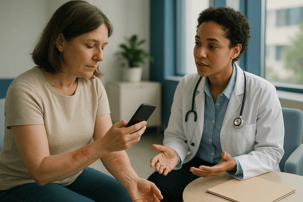
Use daylight where you can. Stand near a window with the light falling across the skin rather than behind you. Indoor bulbs cast yellow or blue tones that distort redness, and a direct flash flattens texture and bleaches color. If evening is the only time the flare is visible, turn on every light in the room and switch the flash off.
Take two shots per area: one from about arm’s length to show location and spread, one closer to show surface detail. Keep the phone parallel to the skin so the whole patch is in focus. Rest your elbow on a table if your hand shakes.
Include a sense of scale. A coin, a ruler or simply the whole hand in frame lets the team judge size. Keep a consistent background, ideally a plain sheet or wall, so color comparisons between weeks are fair.
Name the file or caption it with the date and body site. Most phones record the date automatically, but writing “left inner elbow, day 3 of flare, before cream” removes ambiguity. A before-and-after pair around a treatment application can be genuinely informative, provided you note the timing.
Resist filters, portrait mode and any automatic beautifying setting. These smooth skin and adjust tone, precisely the features the clinician needs to see unaltered.
Finally, photograph the calm periods too. An image of the same elbow when it is quiet gives the team a baseline, and it demonstrates the contrast that words struggle to convey.
Keeping an eczema flare photo diary between visits
A single flare photo is a snapshot. A diary is a story, and stories are what clinicians use to make sense of a relapsing condition. The format can be as simple as a dated album on your phone paired with a note on each image.
Aim to capture three moments per flare: the first day you notice it worsening, the peak, and the point at which it has clearly settled. Recording these three anchors over several flares lets the team estimate how long your flares typically last and how quickly they respond to treatment, without you having to reconstruct it from memory. Mayo Clinic advises tracking possible triggers alongside symptoms for exactly this reason.
Alongside each photo, jot down a handful of facts:
- An itch score from 0 to 10, and whether it disturbed sleep.
- What you applied that day, described as you understand it (moisturizer, prescribed cream), and roughly when.
- Anything new: a different soap, a woolen jumper, a stressful week, a cold, hot weather, heavy sweating.
- Whether the skin wept, crusted or cracked.
Do not try to log everything every day; that habit rarely survives a fortnight. A diary that captures flares reliably is worth far more than a comprehensive one that stops after ten days.
Before the appointment, spend five minutes curating. Pick the 8 to 12 images that tell the story best, put them in date order, and consider a short written summary: how many flares since the last visit, where, and what seemed to help or hurt. Handing this over at the start of the consultation, rather than scrolling for it midway, keeps the limited time focused on decisions.
Photographing eczema on brown and Black skin: why redness is not the whole story
Much of the classic teaching on eczema describes red, inflamed patches. On brown and Black skin, inflammation frequently does not look red at all. Mayo Clinic notes that areas appearing red on white skin may look brown, purple or gray on Black or brown skin, and the NHS makes the same point about darker skin tones. Photographs that fail to account for this can lead a viewer to underestimate how active a flare is.
Several practical adjustments help. Light matters even more: side lighting that picks out slightly raised, rougher or shinier patches reveals inflammation that color alone hides. Comparison shots are valuable, so include an unaffected area of the same limb in the frame or in a companion image, allowing the team to see the shift in tone relative to your baseline.
Look for, and photograph, features that show well across all skin tones. Small raised bumps around hair follicles (a follicular pattern) are common in darker skin. Thickened, lined skin from repeated scratching photographs well with oblique light. Dry, ashen scaling can be more visible than any color change.
Pigment changes after a flare also deserve documentation. Areas may become darker (post-inflammatory hyperpigmentation) or lighter (hypopigmentation) as the inflammation fades, and these marks can persist for months. Cleveland Clinic lists such discoloration among the recognized effects of eczema. Photographing these areas helps the team distinguish old activity from new, and reassures you that a lingering dark patch is a footprint of a flare rather than a flare itself.
Auto white balance on phones sometimes shifts skin tone toward orange or gray. Where possible, include a white or neutral object in the frame so the clinician can mentally correct for it.
Who benefits most from photos, and when a photo is not enough
Almost anyone with atopic dermatitis gains something from a flare album, but some situations make it close to essential.
People whose flares are short and unpredictable benefit most, since they are the ones most likely to arrive with calm skin. So do those whose eczema affects areas they would rather not undress for routinely, such as the buttocks or genital region, where a photo taken privately at home can spare repeated examinations while still informing care. Adults with hand eczema, whose skin may look acceptable in the morning and fissured by evening, find photos useful for showing the daily swing. Parents of young children, whose skin changes fast and who struggle to describe itch, have an obvious use for a dated record.
Photos also help when a treatment change is being considered. Decisions about stepping up or down a plan sit with the prescribing clinician, but they are easier to make when the record shows how many flares occurred and how long each lasted.
There are, equally, moments when a photo should not be the plan. If skin is weeping, crusting, rapidly spreading, or painful rather than itchy, NHS guidance is to seek review promptly, because these can signal infection that needs examination and sometimes swabbing. Clusters of painful blisters with fever fit the picture of eczema herpeticum, which is treated as urgent. A photo can accompany that call; it cannot substitute for it.
Photos are also a poor tool for judging skin that needs to be touched to assess, such as suspected thickening, or for a first-ever rash where the diagnosis is still open. In those cases the team will usually ask to see you in person, calm skin or not.
What to bring to a dermatologist for eczema: a one-page checklist
Photos are the headline item, but they work best alongside a small kit of facts. The table below sets out what most clinicians find useful and why it saves time in the room.
| Item | What it tells the team | Practical tip |
|---|---|---|
| Dated flare photos, 8 to 12 images | Pattern, severity, distribution and duration of flares | Order by date; label body site |
| Photos of calm skin | Your personal baseline for comparison | Same site, same light |
| Itch and sleep notes | Symptom burden that skin alone cannot show | A 0 to 10 score is enough |
| List of everything you apply | What has been tried and how it was used | Bring the tubes or photograph the labels |
| Trigger observations | Possible irritants or allergens worth exploring | Note timing relative to flares |
| Other diagnoses and medicines | Conditions such as asthma or hay fever, which commonly accompany eczema | A current list from your pharmacy or records |
| Your top three questions | Keeps the visit focused on what matters to you | Write them down beforehand |
Bringing the actual products you use, or clear photos of their labels, is more helpful than it sounds. People often describe a moisturizer as “the cream” and a prescribed treatment as “the other cream,” and the team cannot judge a plan they cannot identify. Mayo Clinic and the NHS both emphasize that emollients, the thick moisturizers that repair the skin barrier, are the foundation of management, so clarity about how and when you use them shapes the whole conversation.
Wear loose clothing that lets you show affected areas quickly, and skip heavy makeup on facial eczema that day so the clinician can see the skin. If the flare has faded, the photos carry the evidence; your job is simply to make them easy to find.
What the weeks after the visit usually look like
A consultation ends with a plan, and the plan is only as good as the follow-up. This is where the photo habit pays off a second time.
In the first days, most plans center on the skin barrier and on calming inflammation. The NHS describes emollients as the everyday mainstay and topical anti-inflammatory treatments as the tool for flares, used according to the clinician’s instructions. Continue the diary through this period, photographing the treated areas so that the response, or the lack of one, is visible rather than remembered.
Over the following weeks, the pattern of flares is what the team wants to learn. Are they fewer? Shorter? Shifting to new sites? Mayo Clinic notes that even when treatment goes well, flares can return, so the goal is control rather than the absence of every symptom. Photographs turn a vague sense of “a bit better” into a set of comparable images from the same elbow under the same window light.
Follow-up timing varies with severity and the treatment chosen; some teams ask to review within a few weeks of a change, others at longer intervals when things are stable. Whatever the schedule, ask at the end of the visit what the team wants to see documented before you return. Some will specifically request photos of a newly treated area; others may ask you to note how many flare days occur per month.
Many services now offer a route to send images between visits, sometimes through a secure patient portal. Ask whether yours does and what the expected response time is, so a photo that shows worsening skin does not sit unread. A photo sent through a portal is for routine review; anything urgent still needs a phone call.
Who sees my atopic dermatitis consultation photos, and how are they stored?
Handing over images of your skin, sometimes of intimate areas, is a reasonable thing to feel cautious about. Clinical photography, the practice of taking or receiving medical images for care, sits under the same confidentiality rules as the rest of your record, but it helps to understand the details.
Photos you show on your own phone during a visit generally stay on your phone unless the clinician asks to add them to your record. Images you upload through a patient portal or send at the team’s request usually become part of your medical file, viewable by the clinicians involved in your care and subject to the same access controls and retention rules as written notes. If a team member wants to photograph you in clinic, they should ask your consent, explain where the image will be stored, and tell you if it might be used for teaching or research, which requires separate agreement.
A few practical protections are within your control:
- Frame photos to show the affected skin without identifying features where possible; a rash on the abdomen does not need your face in it.
- Use the route your service recommends rather than a general messaging app, which may store copies on external servers.
- Ask how to have an image removed if you later change your mind.
- Keep your own copies organized in a folder rather than mixed into holiday pictures, and consider your phone’s lock settings.
For children and teenagers, consent involves the parent and, as appropriate for age, the young person. Many teens are comfortable with photos of an elbow and far less so with photos elsewhere. Their view should be sought and respected, and the team will usually offer alternatives such as examination in clinic.
Photos of a child's eczema: what parents should know
Atopic dermatitis is largely a childhood condition. The NHS notes that it affects around one in five children and one in ten adults in the UK, and that most cases begin before the age of five. Children also cannot always say where or how much they itch, which makes a parent’s dated photo record unusually valuable.
Where eczema appears changes with age, and the team will read your photos with that in mind. MedlinePlus describes the typical infant pattern of cheeks, scalp and outer limbs, shifting toward the elbow and knee creases, wrists and neck in older children. A photo of the same site over months can show this migration as clearly as any description.
Practical points for photographing a wriggling child: a second adult to hold a favorite toy at window height buys you two seconds of stillness; bath time, before creams go on, offers consistent light and bare skin; and a photo of the child asleep often reveals scratch marks and disturbed bedding that daytime hides. Note how many times they woke, because itch-related sleep loss is one of the main burdens the team will want to reduce.
Children are also more prone to infected eczema and to eczema herpeticum, the viral infection described earlier. If a photo shows weeping, crusting, clusters of small blisters, or the child is feverish and unwell, that image is a reason to call the same day, not to wait for the appointment.
Finally, involve the child at a level that suits their age. School-aged children can often point out which patches bother them most, and teenagers may prefer to take and manage their own photos. Preparation for a visit is an early lesson in describing their own health, one they will carry into adult care.
What people often get wrong about eczema photos and appointments
“If it looks fine today, there’s no point going.” Calm skin is exactly when the longer conversation about prevention, barrier care and flare planning can happen without the distraction of urgent itch. The photos supply the flare; you supply the questions.
“More photos are better.” Sixty near-identical close-ups bury the useful ones. A curated dozen, in date order and labeled, is read; a scroll of hundreds is skimmed.
“The clinician can tell how bad the itch was from the picture.” Itch is invisible. Scratch marks hint at it, but your notes on sleep and an itch score carry information no image can. Pair the two.
“Photos mean I don’t need to be examined.” Texture, warmth and tenderness are felt, not seen, and infection often needs a swab. Photos widen the window; they do not close the door to the room.
“I should stop my treatment so the flare shows properly.” Never adjust a prescribed plan to stage a flare. Beyond the discomfort, it distorts the record the team is trying to read. Photograph honestly, including what you applied, and let the prescriber interpret. Any change to treatment is theirs to decide.
“Redness is the measure.” On darker skin, active eczema may look brown, purple or gray, and lingering pigment change may look like an active flare when it is not. Baseline shots and side lighting matter more than color.
“Eczema is contagious, so I should hide it.” Cleveland Clinic and the NHS are clear that atopic dermatitis is not infectious. Documenting it openly with your team is part of managing it, not something to be embarrassed about.
Questions to ask your care team
A good set of photographs earns you a better conversation. These questions help you spend it well. Write down the answers, or ask whether they can be added to your visit summary.
- Looking at these photos, how would you describe the severity and pattern of my eczema, and has it changed since my last visit?
- Is there anything in these images that concerns you, such as signs of infection or a distribution that suggests a contact reaction?
- Which body sites or features would you like me to photograph before the next review, and how often?
- Is there a secure way to send you images between visits, and what should I expect in terms of response time?
- How should I use the moisturizers and prescribed treatments in relation to each other, and what does a good response look like over the coming weeks?
- What triggers, if any, do you think my photo diary points toward, and is testing for contact allergy worth considering?
- What signs in the skin should prompt me to call rather than wait for the next appointment?
- How will my photos be stored, who can see them, and can I ask for them to be removed?
- For a child: how might the pattern change with age, and what should I photograph as they grow?
- What would make you consider a different approach, and what evidence would you want to see in my record first?
Notice that several questions ask the team to interpret rather than you to guess. That is the point. Photos give you standing in the conversation, but the reading of them, and any decision that follows, belongs to the clinician who knows your full history. If an answer is unclear, ask for it in plain words; a good team will not mind.
When to call your doctor
Most flares of atopic dermatitis are uncomfortable but not dangerous, and a photo can wait for the next scheduled visit. Some changes should not wait. NHS guidance on atopic eczema and infected eczema, together with Mayo Clinic advice, points to the following red flags.
Contact your care team the same day if the skin is weeping clear or yellow fluid, developing yellow or honey-colored crusts, or showing small yellowish-white spots within the eczema. Do the same if a patch becomes hot, swollen, increasingly painful rather than itchy, or is spreading quickly. These features can indicate bacterial infection, which often requires examination and treatment decided in person.
Seek urgent care if you or your child develop clusters of small, painful blisters or punched-out sores over eczema, especially with fever, shivering, or feeling generally unwell. This pattern suggests eczema herpeticum, a viral infection that the NHS treats as an emergency because it can spread rapidly. Eye involvement, with a painful red eye or changes in vision, also needs prompt assessment.
Call promptly, though less urgently, if a flare is not settling with the plan you were given, if itch is preventing sleep for several nights in a row, if the eczema is affecting mood, work or school, or if you notice hair loss, nail changes or a rash appearing in a new pattern that does not look like your usual eczema.
Photographs help in every one of these situations; take them before you call so the team can see what you are describing. They do not, however, replace the call. When the skin looks infected or the person feels unwell, the image is a companion to urgent review, never a substitute for it, and the decision about what happens next rests with the treating clinician.
Frequently asked questions
Do atopic dermatitis consultation photos really change what the doctor decides?
They inform the decision rather than make it. Photos let the clinician see severity, distribution and duration of flares that are invisible on a calm day, so the assessment rests on evidence instead of recollection. Any change to treatment remains the prescriber’s judgment, weighing your history, examination and the images together. Think of photos as expanding the consultation from one morning to several weeks.
How do I photograph eczema on my own back or scalp?
Ask someone you trust to take the picture, or use a mirror and the phone’s timer with the front camera. For the scalp, part the hair with a comb and shoot in bright daylight, since flash flattens scale and redness. Two attempts at different angles usually give one usable image. If no one is available, tell the team; they can examine or photograph the area in clinic.
What should an eczema flare photo diary include besides pictures?
A date, the body site, an itch score from 0 to 10, whether sleep was disturbed, what you applied that day, and anything new such as a soap, fabric, illness or stressful event. Mayo Clinic recommends tracking possible triggers alongside symptoms. Three photos per flare, at onset, peak and resolution, are enough; a short diary that lasts beats a detailed one that stops after ten days.
What should I bring to a dermatologist for eczema apart from photos?
Bring the products you apply, or clear photos of their labels, a list of other medicines and conditions such as asthma or hay fever, your trigger notes, and your top three questions written down. Wear loose clothing so affected areas are easy to show, and skip heavy makeup on facial eczema. Clinicians can only assess a plan they can identify, so naming the actual creams matters more than most people expect.
Is there a simple eczema appointment preparation routine I can follow the night before?
Spend ten minutes curating your photos into a dated album of 8 to 12 images, add a two-line summary of how many flares you have had and where, gather your product tubes into a bag, and write three questions. Apply your usual treatments as prescribed; never stop them to stage a flare. In the morning, wear clothing that lets the clinician see the usual sites quickly.
Should I stop using my cream so the flare is visible at the visit?
No. Stopping a prescribed treatment to provoke a flare causes avoidable discomfort and distorts the record your team is trying to interpret. Photograph the flare when it happens naturally, note what you applied and when, and let the clinician read the response. Decisions about starting, stopping or changing any treatment belong to the prescriber, made with your full history in front of them.
Can I just send photos instead of attending in person?
Sometimes, for routine review, if your service offers a secure route and the clinician agrees. Photos cannot convey texture, warmth or tenderness, and infected skin often needs a swab, so the team may still ask to see you. Weeping, crusting, spreading pain, blisters or fever always warrant a call and, usually, in-person assessment. Ask your team which situations suit remote images and which do not.
Why does my eczema look brown or purple rather than red in photos?
On brown and Black skin, inflammation frequently shows as brown, purple or gray tones instead of the red described in older textbooks, a point Mayo Clinic and the NHS both make. The flare is no less active. Use side lighting to reveal raised or rough texture, include unaffected skin for comparison, and photograph any lingering dark or light patches so the team can distinguish old flares from new ones.
How are photos of a child's eczema handled, and does my child have a say?
Photos shared with the care team are held under the same confidentiality rules as the rest of the medical record, and consent comes from the parent with the child’s views sought as appropriate for age. Many teenagers are happy to have an elbow photographed and reluctant about other areas; that preference should be respected, and the team can offer examination in clinic instead.
How many photos should I bring, and how should I organize them?
Aim for 8 to 12 images in date order, each labeled with the body site and a note on treatment timing, plus one or two baseline shots of calm skin at the same sites. Keep them in a dedicated folder rather than scattered through your camera roll. A set the clinician can read in under a minute is far more useful than a long scroll of near-duplicates.
References
- NHS: Atopic eczema
- MedlinePlus: Atopic dermatitis
- Cleveland Clinic: Eczema
- NIH NIAMS: Atopic dermatitis
This article is for general information only and is not a substitute for professional medical advice. Please consult a qualified doctor about your individual situation.

More from the Blog

What a Congenital Nevus Check Involves: Dermoscopy, Photo Mapping and Tracking Change
A congenital nevus check is a skin examination of a birthmark mole. A clinician inspects the whole nevus and the surrounding skin, uses a…
How Does Phototherapy Help Atopic Dermatitis? What Sessions Involve and Who May Be Offered Them
Phototherapy for atopic dermatitis exposes affected skin to carefully measured ultraviolet light, usually narrowband UVB, in a clinic cabinet several times a week for…
A Daily Routine That Supports Atopic Dermatitis Treatment: Bathing, Moisturizing and Nail Care
A supportive atopic dermatitis daily routine rests on three anchors: a short, lukewarm bath or shower with a gentle, fragrance-free cleanser; a generous layer…
Is Coconut Oil Good for Your Hair? The One Oil That Penetrates
Coconut oil can genuinely help hair, and the reason is unusual: its main fatty acid, lauric acid, is small enough to penetrate the hair…
Peptides in Skincare: What Signal Peptides Do and What the Studies Show
Peptides in skincare are short chains of amino acids applied topically to signal skin cells, most often to encourage collagen production or soften expression…
How to Get Clear Skin: What Actually Works, According to Dermatology Trials
Dermatology trials show that clear skin comes mainly from treating acne consistently: a gentle cleanser, benzoyl peroxide, an adapalene retinoid, daily sunscreen, and patience…