Quick answer
Pneumothorax is a collapsed lung caused by air collecting between the lung and chest wall, which can lead to sudden chest pain and shortness of breath. Treatment depends on the size and cause and may range from observation and oxygen support to needle aspiration, chest tube drainage, or surgery, with diagnosis and follow-up guided by imaging and respiratory assessment.
What is pneumothorax?
Pneumothorax (pronounced noo-moh-THOR-aks) is the medical term for a collapsed lung. It happens when air leaks into the space between the lung and the chest wall, an area called the pleural space. Normally, this space contains only a thin film of fluid, and slight negative pressure keeps the lung expanded against the chest wall. When air enters the pleural space, that pressure balance is lost, and part or all of the lung collapses inward. If you have searched for “what is pneumothorax,” the short answer is this: it is air where air should not be, pressing on the lung and preventing it from filling properly with each breath.
A pneumothorax can be small, affecting only a portion of one lung, or large, causing the entire lung to collapse. It can develop suddenly in an otherwise healthy person, or it can occur as a complication of lung disease, a chest injury, or a medical procedure. Doctors often group pneumothorax into several types:
- Primary spontaneous pneumothoraxoccurs without any obvious lung disease, often in tall, thin young adults, and more commonly in men who smoke.
- Secondary spontaneous pneumothoraxoccurs in people with an existing lung condition, such as chronic obstructive pulmonary disease (COPD, a long-term disease that narrows the airways), asthma, cystic fibrosis, or lung infections.
- Traumatic pneumothoraxcaused by an injury to the chest, such as a rib fracture, a fall, a car accident, or a stab or gunshot wound.
- Iatrogenic pneumothoraxan unintended result of a medical procedure, such as a lung biopsy, insertion of a central intravenous line, or mechanical ventilation (breathing support with a machine).
- Tension pneumothoraxa life-threatening emergency in which air keeps entering the pleural space but cannot escape. Rising pressure pushes on the heart and major blood vessels and can quickly become fatal without immediate treatment.
Pneumothorax can affect people of any age. Spontaneous cases are most common in adolescents and young adults, while cases linked to underlying lung disease tend to occur in older adults. Understanding the type of pneumothorax matters, because it influences both treatment and the likelihood of the problem returning.
Symptoms of pneumothorax
Pneumothorax symptoms usually begin suddenly. Many people describe a sharp, stabbing pain on one side of the chest that came on out of nowhere, sometimes at rest or during light activity. The most common signs include:
- Sudden chest paintypically sharp and on one side, often worse when breathing in deeply or coughing.
- Shortness of breathranging from mild breathlessness to severe difficulty breathing, depending on how much of the lung has collapsed.
- Rapid breathingthe body tries to compensate for reduced lung function.
- Rapid heartbeatthe heart works harder to move oxygen around the body.
- Dry coughsome people develop a persistent, non-productive cough.
- A feeling of tightness in the chest or a sense that one side is not moving normally with each breath.
How severe these symptoms feel depends on the size of the pneumothorax and the person’s overall lung health. A young, otherwise healthy person with a small collapse may notice only mild chest discomfort and slight breathlessness, and in some cases the symptoms fade on their own even before the air leak has fully healed. In contrast, someone with existing lung disease may become severely short of breath even from a small pneumothorax, because their lungs have little reserve.
Warning signs of a tension pneumothorax are more dramatic and constitute a medical emergency. These may include severe and worsening breathlessness, bluish discoloration of the lips or skin (cyanosis, a sign of low oxygen), extreme rapid heartbeat, low blood pressure, confusion, dizziness, or loss of consciousness. Anyone with these symptoms needs emergency care immediately.
It is worth noting that pneumothorax symptoms can resemble other serious conditions, including a heart attack or a blood clot in the lung. Sudden chest pain with shortness of breath should always be evaluated by a medical professional rather than self-diagnosed.
Causes and risk factors
Pneumothorax causes vary by type. In a spontaneous pneumothorax, air usually escapes from a small, weakened area on the surface of the lung. Tiny air-filled blisters called blebs or larger ones called bullae can form on the lung surface; when one ruptures, air leaks into the pleural space. Why these blisters form in otherwise healthy people is not fully understood, though body build and smoking appear to play roles.
Common causes and contributing factors include:
- Ruptured blebs or bullaethe usual cause of primary spontaneous pneumothorax.
- Chronic lung diseasesCOPD is the most common condition behind secondary spontaneous pneumothorax; asthma, cystic fibrosis, interstitial lung disease (scarring of lung tissue), and severe pneumonia can also weaken lung tissue.
- Chest traumabroken ribs, penetrating wounds, or blunt injuries from accidents or sports.
- Medical procedureslung biopsies, needle procedures near the chest, central line placement, and mechanical ventilation can occasionally puncture the lung.
- Pressure changesscuba diving, flying in unpressurized aircraft, or high-altitude activities can trigger a pneumothorax in susceptible people.
Several risk factors make pneumothorax more likely:
- Smokingone of the strongest modifiable risk factors; risk rises with the amount and duration of smoking.
- Sex and body typeprimary spontaneous pneumothorax is more common in men, particularly those who are tall and thin.
- Agespontaneous cases without lung disease often occur between the late teens and early thirties; cases linked to lung disease are more common later in life.
- Family historysome people inherit a tendency toward pneumothorax, and certain rare genetic conditions affect connective tissue in the lungs.
- Previous pneumothoraxhaving had one collapsed lung significantly increases the chance of another, on the same or the opposite side.
- Underlying lung diseaseany condition that damages or weakens lung tissue raises risk.
Diagnosis
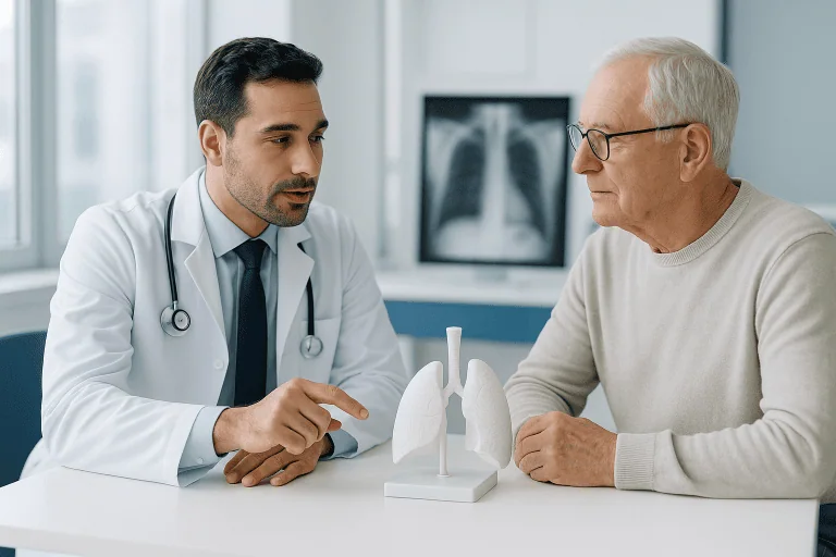
Pneumothorax diagnosis begins with a medical history and a physical examination. Your doctor will ask when the symptoms started, whether you have had a chest injury or recent medical procedure, whether you smoke, and whether you have any lung conditions or a previous collapsed lung. During the examination, the doctor listens to the chest with a stethoscope; over a collapsed lung, breath sounds are often reduced or absent on the affected side. The doctor may also tap gently on the chest, since a chest filled with extra air can produce a hollow sound.
Imaging is essential to confirm the diagnosis:
- Chest X-raythe standard first test. Air in the pleural space and the edge of the collapsed lung are usually visible on an upright X-ray. Doctors also use the X-ray to estimate the size of the pneumothorax, which helps guide treatment decisions.
- Chest CT scana computed tomography scan gives a much more detailed picture. It can detect small pneumothoraces that a plain X-ray may miss, identify blebs or bullae, and reveal underlying lung disease.
- Ultrasoundincreasingly used, especially in emergency and trauma settings, because it can be performed quickly at the bedside and is sensitive for detecting air in the pleural space.
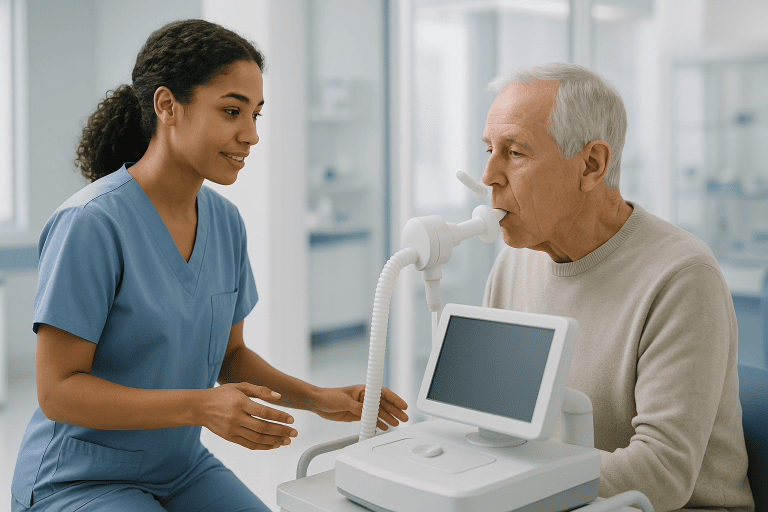
Additional tests may include a measurement of blood oxygen with a small finger sensor (pulse oximetry) or an arterial blood gas test, which analyzes oxygen and carbon dioxide levels in a blood sample. An electrocardiogram (ECG, a recording of the heart’s electrical activity) is sometimes done to rule out heart-related causes of chest pain. In a suspected tension pneumothorax, doctors may begin emergency treatment based on the clinical picture alone, without waiting for imaging, because delay can be dangerous.
Treatment options
Pneumothorax treatment depends on the size of the air leak, the severity of symptoms, whether it is a first episode or a recurrence, and whether the person has underlying lung disease. The goals are to remove the air from the pleural space, allow the lung to re-expand, and, where appropriate, reduce the chance of the problem coming back. A full overview of the condition and its management is available on the pneumothorax treatment page.
Observation (watchful waiting)
A small pneumothorax in a person with mild or no symptoms often heals on its own, because the body gradually reabsorbs the air. In such cases, your doctor may recommend monitoring with repeat chest X-rays, rest, and avoidance of strenuous activity and air travel until the lung has fully re-expanded. Supplemental oxygen is sometimes given, as it can help the trapped air be reabsorbed more quickly. Observation is generally reserved for stable patients, and any worsening of symptoms should prompt urgent reassessment.
Needle aspiration
For a larger pneumothorax or one causing noticeable symptoms, a doctor may perform needle aspiration. A thin needle or small catheter is inserted through the chest wall, usually under local anesthetic (numbing medication), and the excess air is withdrawn with a syringe. This can allow the lung to re-expand without a longer hospital stay, though in some cases the air re-accumulates and further treatment is needed.
Chest tube (chest drain)
When the pneumothorax is large, symptoms are significant, or aspiration has not worked, doctors typically place a chest tube. This is a flexible tube inserted between the ribs into the pleural space and connected to a one-way valve or a drainage system. It allows the trapped air to escape continuously while the lung re-expands, which may take several days. Chest tube placement usually requires hospital admission so the medical team can monitor progress with repeated imaging.
Surgery and pleurodesis
Surgery may be recommended when an air leak does not close, when a pneumothorax recurs, when both lungs are affected, or when a person’s occupation or lifestyle makes recurrence especially risky (for example, pilots or divers). The most common approach is video-assisted thoracoscopic surgery (VATS), a keyhole procedure in which the surgeon uses a small camera and instruments inserted through tiny incisions. The surgeon can remove or staple leaking blebs and often performs pleurodesis: a procedure that deliberately creates gentle scarring between the lung surface and chest wall so they stick together, making future collapse much less likely. Pleurodesis can also be done chemically, by introducing an irritant substance such as sterile talc through a chest tube. In some situations, an open surgical approach (thoracotomy) is needed. Your surgical team will discuss which option is appropriate for your circumstances.
Treating the underlying cause
There is no medication that reverses a pneumothorax itself, but medicines are often part of overall care: pain relief while the chest heals, treatment for any underlying lung disease, and antibiotics if infection is present. Long-term management of conditions such as COPD or asthma, together with stopping smoking, is important for lowering the risk of recurrence. Ongoing lung care is typically coordinated by a respiratory medicine team; at Acibadem, this condition is managed within the pulmonology department, often in cooperation with thoracic surgeons when procedures are needed.
Living with pneumothorax and outlook
Most people who experience a pneumothorax recover well, particularly when the collapse is small and treated promptly. Recovery time varies: a small pneumothorax managed with observation may resolve within one to two weeks, while recovery after a chest tube or surgery generally takes longer, and full return to normal activity depends on individual healing and any underlying lung disease.
Recurrence is a genuine concern. A meaningful proportion of people who have one spontaneous pneumothorax will experience another, most often within the first years after the initial episode, and the risk rises further after a second event. Procedures such as pleurodesis substantially reduce, though do not entirely eliminate, the chance of recurrence.
Practical steps that may support recovery and lower future risk include:
- Stop smokingthis is the single most important change; continued smoking markedly increases the chance of another collapse.
- Follow activity guidanceavoid heavy lifting and strenuous exercise until your doctor confirms the lung has fully healed.
- Delay air travelflying with an unresolved pneumothorax is dangerous because cabin pressure changes can enlarge the air pocket; your doctor will advise when flying is safe again.
- Reconsider scuba divingafter a spontaneous pneumothorax, diving is often discouraged permanently unless definitive surgical treatment has been performed, because the pressure changes underwater carry serious risk. Discuss this with a lung specialist.
- Attend follow-up appointmentsrepeat imaging confirms healing and helps detect any recurrence early.
People with underlying lung disease may have a more guarded outlook, and managing that disease well becomes a central part of preventing further episodes. Your medical team can give you a realistic picture based on your specific situation; no doctor can guarantee that a pneumothorax will never return.
Frequently asked questions
What is pneumothorax in simple terms?
Pneumothorax means a collapsed lung. Air leaks into the space between the lung and the chest wall, and the pressure of that trapped air prevents the lung from expanding normally when you breathe. It can affect part of a lung or the whole lung, and it may occur suddenly in healthy people, after a chest injury, or as a complication of lung disease or a medical procedure.
Can a pneumothorax heal on its own?
A small pneumothorax often heals without invasive treatment, because the body slowly reabsorbs the trapped air over days to weeks. However, this should always happen under medical supervision, with follow-up imaging to confirm the lung has re-expanded. Larger collapses, significant symptoms, or worsening over time usually require a procedure such as needle aspiration or a chest tube.
How serious is a pneumothorax?
Seriousness ranges widely. A small spontaneous pneumothorax in a healthy young person is often manageable and heals well. At the other extreme, a tension pneumothorax, where pressure builds continuously and compresses the heart and blood vessels, is a life-threatening emergency requiring immediate treatment. People with existing lung disease are generally at higher risk of complications, which is why every suspected pneumothorax deserves prompt medical evaluation.
What are the first pneumothorax symptoms people usually notice?
The most typical early symptoms are sudden, sharp chest pain on one side and shortness of breath. The pain is often worse with deep breathing or coughing. Some people also notice a fast heartbeat, rapid breathing, or a dry cough. Because these symptoms overlap with heart and other lung emergencies, sudden chest pain with breathlessness should be assessed by a doctor without delay.
What does pneumothorax treatment usually involve?
Treatment depends on the size of the collapse and your symptoms. Options range from careful observation with rest and follow-up X-rays, to removing the air with a needle, to placing a chest tube that drains the air over several days. If the leak does not close or the pneumothorax keeps returning, surgeons may perform a keyhole procedure to repair the lung and carry out pleurodesis, which helps prevent future collapses.
How long does recovery from a collapsed lung take?
Recovery varies with the size of the pneumothorax and the treatment used. A small collapse managed with observation may resolve within about one to two weeks, while recovery after a chest tube or surgery typically takes longer. Most doctors advise avoiding strenuous exercise, heavy lifting, and air travel until imaging confirms the lung has fully healed, and they will tell you when each activity is safe to resume.
Can a pneumothorax come back after treatment?
Yes, recurrence is possible, especially after a spontaneous pneumothorax and particularly in people who continue to smoke or who have underlying lung disease. The risk of another episode increases after each recurrence. Procedures such as pleurodesis or surgical repair of leaking areas on the lung considerably reduce this risk, which is why they are often recommended after a second episode or in higher-risk situations.
When to see a doctor
Any sudden chest pain accompanied by shortness of breath should be evaluated by a medical professional promptly, even if the symptoms seem to ease, because a partially collapsed lung can worsen. If you have already been diagnosed with a pneumothorax and are being observed, contact your medical team if your breathing becomes more difficult or your pain increases.
Seek emergency care immediately, do not wait, if you or someone near you experiences any of the following red-flag signs:
- Severe or rapidly worsening shortness of breath, or struggling to speak in full sentences.
- Sudden, intense chest pain that does not ease, especially after a chest injury.
- Bluish lips, face, or fingertips, which suggest dangerously low oxygen levels.
- Very fast heartbeat, dizziness, or fainting, which may signal falling blood pressure.
- Confusion or extreme drowsiness alongside breathing difficulty.
- Symptoms after chest trauma, such as a fall, accident, or blow to the chest, even if they seem mild at first.
These signs can indicate a tension pneumothorax or another life-threatening chest emergency, both of which require immediate treatment. When in doubt, it is always safer to be examined; a doctor can quickly determine with an examination and imaging whether a pneumothorax is present and how urgently it needs to be treated.
Medically reviewed by the Acıbadem International Medical Board September 2, 2026
See our medical review board →
Update history
- PublishedJune 14, 2026
- Medical review approvedSeptember 2, 2026
- Last content updateSeptember 2, 2026