CT Scan Ivp: Preparation, Procedure and Results
CT IVP combines X-ray-based CT imaging with contrast material to assess the urinary tract. It may be used for blood in the urine, suspected kidney stones, recurrent urinary infections, blockage or urinary tract masses.
Key Takeaways
- CT IVP combines X-ray-based CT imaging with contrast material to assess the urinary tract.
- It may be used for blood in the urine, suspected kidney stones, recurrent urinary infections, blockage or urinary tract masses.
- Preparation often includes discussing kidney function, allergies, pregnancy possibility and current medicines with the imaging team.
- The scan itself is usually painless and commonly takes about 30 to 60 minutes, although appointment time may be longer.
- Most people resume usual activities after the exam, unless their care team gives different instructions.
A CT scan IVP, also called CT intravenous pyelogram or CT urography, is an imaging examination of the urinary tract performed with CT scans and usually intravenous contrast dye. It provides detailed views of the kidneys, ureters and bladder and can help clinicians investigate urinary symptoms or abnormalities seen on other tests.
CT Scan IVP: What It Is and How It Works
A CT scan IVP is a computed tomography intravenous pyelogram. It is also commonly called CT urography or a CT IVP exam. The examination uses a CT scanner to take detailed cross-sectional images of the urinary tract: the kidneys, ureters (the tubes that carry urine to the bladder) and bladder. In many cases, contrast material containing iodine is injected into a vein so that urine-producing and urine-draining structures can be seen more clearly.
Unlike a traditional intravenous pyelogram, which relies on a series of standard X-rays, CT IVP produces highly detailed images from several angles. Images may be obtained before contrast, shortly after injection, and later as contrast passes through the kidneys and into the urine. These phases can help the radiologist assess stones, narrowing, blockage, inflammation, injury and certain growths.
The examination is a diagnostic test, not a treatment. Its results are interpreted alongside symptoms, medical history, urine tests and, when needed, other imaging. A CT IVP can be especially useful when a clinician needs a broad and detailed view of the urinary system.
Why a CT IVP May Be Recommended
A clinician may recommend a CT scan IVP for visible or microscopic blood in the urine, flank or abdominal pain, suspected urinary stones, repeated urinary tract infections, or a possible urinary obstruction. It may also be used to investigate an abnormality found on ultrasound, to assess the urinary tract after injury, or to monitor some known urological conditions.
The test can identify many possible explanations for urinary symptoms, but it does not by itself establish every diagnosis. For example, a stone, an enlarged structure or an area that needs closer assessment may lead to further urine testing, cystoscopy, laboratory work or follow-up imaging. Relevant symptoms can overlap with conditions such as kidney stones, which require individualized clinical assessment.
CT IVP is not appropriate for every person. Ultrasound, MRI, a non-contrast CT scan or another study may be preferable for some patients, particularly where radiation exposure or iodinated contrast requires special consideration. The referring clinician and radiology team select the examination based on the clinical question and the person’s health history.
How Should I Prepare for a CT IVP?

CT IVP prep instructions can vary by hospital and by the type of scan planned. The imaging department may ask the patient not to eat for a few hours before an examination involving contrast, while clear fluids may often be allowed. Patients should follow the instructions provided by their own radiology team rather than relying on general guidance alone.
Before the appointment, patients should tell the team about previous reactions to contrast material, asthma or significant allergies, kidney disease, diabetes, thyroid conditions, pregnancy or a possible pregnancy, and breastfeeding. They should also provide a complete medication list, including over-the-counter medicines and supplements. Some people may need a blood test to check kidney function before iodinated contrast is given.
Patients are usually asked to remove metal items around the abdomen and pelvis and may change into a gown. It is helpful to bring prior scan reports or images when available, along with identification and referral information. The team may advise a short period of hydration before or after the test, depending on the individual’s health needs.
- Ask whether fasting is needed and for how long.
- Report any past contrast reaction before the appointment.
- Tell the team about kidney problems and all regular medicines.
- Notify staff promptly if pregnancy is possible.
CT IVP Procedure: Step by Step
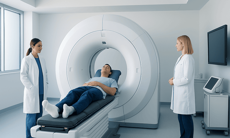
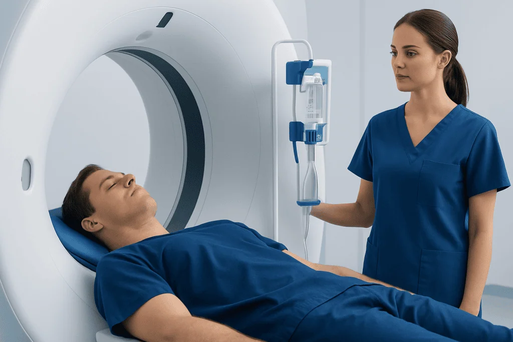
At the start of the CT IVP procedure, a radiology professional reviews safety questions and confirms the reason for the examination. If contrast is planned, a small intravenous line is placed in the hand or arm. The patient lies on a padded table that moves slowly through the wide, ring-shaped CT scanner. The scanner is open at both ends and is not usually enclosed like a traditional MRI machine.
Initial images may be taken without contrast. During injection, some people notice a brief warm sensation, a metallic taste in the mouth or a feeling similar to passing urine; these sensations usually fade quickly. The radiographer can see and communicate with the patient throughout the scan. Remaining still and following brief breathing instructions helps produce clear images.
Several image sets may be acquired at timed intervals. This allows the radiologist to observe how contrast is filtered by the kidneys and travels through the ureters to the bladder. The term “CT IVP protocol CPT code” or “CT IVP procedure code” may appear in scheduling, billing or referral documentation, but coding terminology does not describe a patient’s diagnosis or change the care experience. The exact imaging protocol is selected by the radiologist according to the medical question.
How Long Does a CT IVP Take?
The scan portion of a CT IVP is often completed within 30 to 60 minutes. However, the full appointment may take longer because of registration, safety checks, intravenous line placement, possible blood tests, contrast preparation and waiting for delayed images after contrast has moved through the urinary tract.
Timing differs between patients. A delayed phase may be particularly useful when the clinician is investigating possible blockage or needs a clearer view of how urine drains. The imaging department can provide the most accurate estimate when the appointment is scheduled.
Patients should allow enough time to avoid feeling rushed, especially if they need assistance with mobility or have other appointments on the same day. Most individuals can leave shortly after the scan unless they are asked to stay for observation because of a specific medical concern.
Is CT IVP Painful?
A CT IVP is generally not painful. Lying still on the examination table can be mildly uncomfortable for people with back pain or limited mobility, but staff can help with positioning and supports. Inserting the IV line may cause a brief pinch, similar to a blood draw.
When iodinated contrast is injected, a temporary warm flush, metallic taste, mild nausea or a sensation of needing to urinate can occur. These effects are usually short-lived and are not typically a sign of an allergic reaction. Patients should immediately tell the radiographer if they develop itching, hives, swelling, difficulty breathing, dizziness or any unexpected discomfort.
Serious contrast reactions are uncommon, and imaging teams are trained to recognize and manage them. For people with a known prior contrast reaction, the care team may recommend an alternative test, special preparation or a different contrast approach where clinically appropriate.
What Should I Expect After a CT IVP?
After a CT IVP, the IV line is removed and most patients can return home and resume normal activities the same day. Unless fluid intake is restricted for a medical reason, drinking water after contrast may support normal hydration. The radiology team will give specific aftercare instructions if they differ from this general advice.
There is no recovery period from the scan itself. Temporary bruising or tenderness at the IV site can occur and usually settles. People should seek urgent medical advice if they develop symptoms of a delayed allergic-type reaction, such as widespread rash, facial swelling, wheezing or breathing difficulty, after leaving the imaging department.
A radiologist reviews the images and sends a report to the referring clinician. Results may be available the same day in some settings, but timing varies. The referring clinician explains what the findings mean, whether more testing is needed and whether a urology consultation or a treatment such as kidney stone treatment may be appropriate.
Benefits, Risks and When to Seek Medical Care
The main benefit of CT IVP is its detailed view of the urinary tract. It can detect many causes of urinary symptoms and help guide timely next steps. CT uses ionizing radiation, so the examination is requested when its expected diagnostic value outweighs the radiation exposure. Modern CT techniques aim to use the lowest practical radiation dose while obtaining useful images.
Iodinated contrast can occasionally cause allergic-like reactions and may require added caution in people with reduced kidney function. Patients should share their health history openly so the team can reduce risks and select the most suitable test. A CT IVP does not replace medical evaluation for new or worsening symptoms.
Medical care should be sought promptly for severe flank or abdominal pain, fever with urinary symptoms, inability to pass urine, visible blood in urine, repeated vomiting, or symptoms that are rapidly worsening. Emergency care may be needed for severe pain with fever, confusion, fainting or signs of a serious allergic reaction. Acibadem International’s multidisciplinary specialists and JCI-accredited hospitals assess urinary conditions and coordinate diagnostic and treatment planning for international patients.
Frequently asked questions
What is the difference between a CT IVP and a regular CT scan?
A CT IVP is a CT examination tailored to evaluate the kidneys, ureters and bladder. It commonly includes contrast-enhanced and delayed images to show how contrast moves through the urinary tract. A regular CT scan may examine other body areas or use a different imaging sequence depending on the clinical question.
Can a CT IVP show kidney stones?
Yes. CT IVP can identify stones in the kidneys or ureters and can show whether a stone is associated with swelling or impaired urine drainage. In some situations, a non-contrast CT is preferred specifically for suspected stones, so the clinician will choose the most appropriate examination.
Do I need contrast for a CT IVP?
Many CT IVP examinations use iodinated contrast because it helps outline the urinary collecting system and bladder. Whether contrast is used depends on the reason for the scan, kidney function, allergy history and other individual factors. The radiology team will explain the plan before the test.
Can I eat and drink before a CT IVP?
Instructions differ between imaging centers and protocols. Some patients are asked to avoid food for several hours before contrast-enhanced CT, while water may be permitted. Patients should follow the specific directions from their imaging department and ask if anything is unclear.
When will I receive my CT IVP results?
A radiologist reviews the images and prepares a report for the referring clinician. The timing can range from the same day to several days, depending on the facility and urgency of the request. The clinician who ordered the scan should explain the results and any recommended next steps.
Is a CT IVP safe during pregnancy?
CT uses radiation, so it is generally avoided during pregnancy unless the expected medical benefit is important and other tests are not suitable. Anyone who is pregnant or could be pregnant should tell the clinician and radiology team before the examination. They can discuss safer alternatives or necessary precautions.
References
- Radiological Society of North America
- American College of Radiology
- National Institute of Diabetes and Digestive and Kidney Diseases
- National Health Service
- U.S. Food and Drug Administration
This article is for general information only and is not a substitute for professional medical advice. Please consult a qualified doctor about your individual situation.
Explore treatments in Turkey — costs, top hospitals & a free quote
JCI-accredited · board-certified surgeons · reply within 24h
Add us as a Preferred Source to see more of our trusted health content across Google Search, AI Overviews and Discover.