

Diverticula Bladder Ultrasound: Preparation, Procedure and Results

A bladder diverticulum is a pouch that extends outward from the bladder wall and may be found during evaluation of urinary symptoms. Ultrasound can show diverticula, bladder stones, urine left after voiding and signs that further urology assessment may be needed.
Key Takeaways
- A bladder diverticulum is a pouch that extends outward from the bladder wall and may be found during evaluation of urinary symptoms.
- Ultrasound can show diverticula, bladder stones, urine left after voiding and signs that further urology assessment may be needed.
- Patients are commonly asked to arrive with a comfortably full bladder, unless their imaging center gives different instructions.
- The scan is painless, uses sound waves rather than radiation, and people can return to normal activities immediately afterward.
- Treatment depends on symptoms, recurrent infections, stones, obstruction and the cause of incomplete bladder emptying.
A diverticula bladder ultrasound is a noninvasive imaging test used to examine pouch-like areas in the bladder wall, called diverticula, and to assess how well the bladder empties. It usually requires a comfortably full bladder, takes only a short time, and does not involve radiation or recovery time.
Overview: what a diverticula bladder ultrasound shows
A diverticula bladder ultrasound is a painless scan that uses high-frequency sound waves to create images of the bladder. It can identify a bladder diverticulum, which is a small pouch that pushes outward through a weaker area of the bladder wall. The examination can also help assess the bladder wall, detect stones or sediment, and measure urine remaining after urination.
Diverticula may be discovered when a person is being evaluated for urinary tract infections, trouble emptying the bladder, blood in the urine, frequent urination or lower abdominal discomfort. Some cause no symptoms and are found incidentally. Ultrasound is often an early imaging test because it is quick, does not use ionizing radiation and can be performed before and after the bladder is emptied.
An ultrasound result does not always establish why a diverticulum developed or whether treatment is necessary. A urologist considers the scan together with symptoms, medical history, urine testing and, when appropriate, further tests such as cystoscopy or CT imaging.
Is diverticulum in the bladder serious?
A bladder diverticulum is not automatically serious. Small diverticula that empty well and do not cause symptoms may only need observation and periodic review. However, urine can sometimes collect in a pouch after the rest of the bladder empties, creating conditions that may contribute to recurrent urinary infections, bladder stones or persistent urinary symptoms.
The clinical importance depends on the diverticulum’s size, number, contents and effect on bladder emptying, as well as the underlying cause. In adults, acquired diverticula are often related to long-term high pressure inside the bladder, sometimes from an obstruction to urine flow or a bladder-emptying problem. Finding and treating that cause is an important part of care.
Less commonly, a diverticulum may need closer investigation if there is blood in the urine, a suspicious wall change, repeated infection, stones or concern for a growth within the pouch. These possibilities do not mean they are present, but they explain why a urology assessment and follow-up plan are important.
How the ultrasound works and who may need it

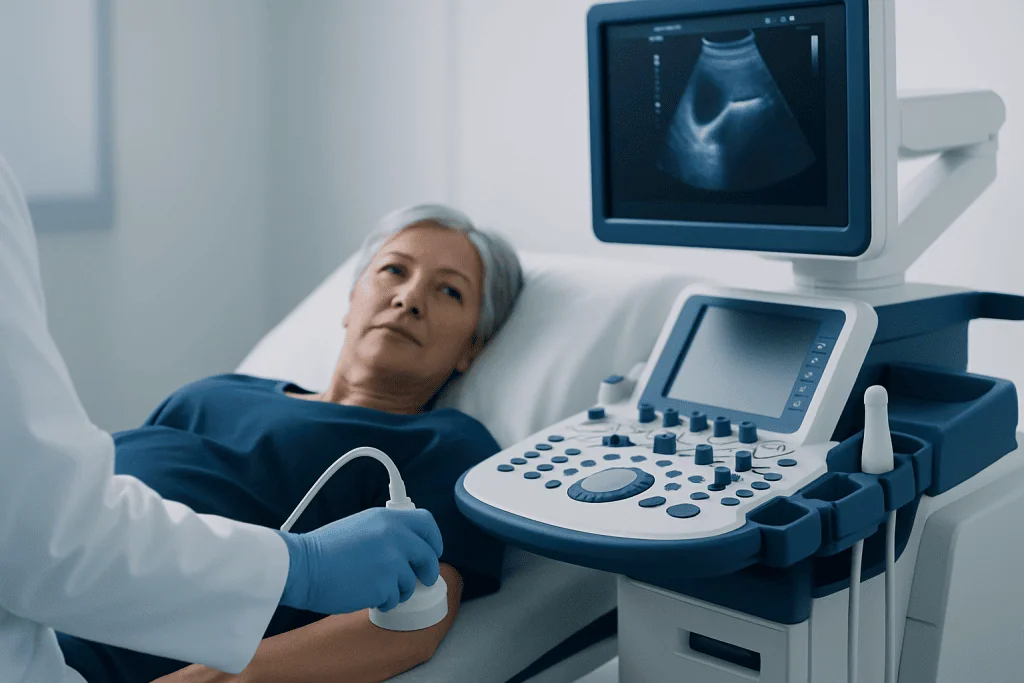
During ultrasound, a trained sonographer places a small handheld device called a transducer on gel-covered skin over the lower abdomen. The transducer sends sound waves into the body and receives returning echoes. A computer converts these echoes into live images of the bladder and nearby structures.
The test may be appropriate for adults or children with suspected bladder diverticula, incomplete bladder emptying, recurring urinary tract infections, urinary frequency, difficulty starting urination, weak urine flow or unexplained lower urinary tract symptoms. It may also be used after a diverticulum has been identified to monitor changes or help guide additional evaluation.
In many cases, images are obtained with the bladder full and then repeated after the person urinates. Comparing these images helps estimate the post-void residual, meaning the amount of urine left in the bladder after voiding. This information can help a clinician understand whether poor emptying may be contributing to symptoms or diverticula.
What do I need to do before a bladder ultrasound?
Preparation is usually straightforward. The imaging center may ask the patient to drink water before the appointment and avoid urinating until after the first set of images is taken. A full bladder provides a clearer ultrasound window and helps the clinician assess its shape, wall and contents.
Patients should follow the specific instructions provided by their clinic, as preparation can differ when the bladder scan is combined with a kidney, pelvic or abdominal ultrasound. Regular medicines are usually continued unless the healthcare team advises otherwise. It is helpful to tell the clinician about pregnancy, recent urinary procedures, inability to hold urine, urinary catheter use or severe urinary symptoms.
Comfortable two-piece clothing can make access to the lower abdomen easier. People who cannot comfortably hold urine should not force themselves to do so; they can contact the imaging department in advance. The team can often adapt the timing or approach to make the examination more comfortable.
How much water do I need to drink before an ultrasound of my bladder?
Many imaging centers advise drinking about 500 to 750 milliliters of water, roughly two to three glasses, around one hour before a bladder ultrasound. The patient is commonly asked not to urinate after drinking it. However, the exact amount and timing vary by age, body size, medical condition and the type of ultrasound being performed.
The goal is a comfortably full bladder, not pain. If the bladder becomes uncomfortably full before the appointment, the patient should notify the sonographer rather than leaving without guidance. In some cases, the team may take the initial pictures promptly or allow a small amount of urine to be passed before scanning.
People with heart failure, kidney disease, fluid restrictions or other conditions that limit fluid intake should ask their doctor or imaging center for individualized advice. They should not exceed a prescribed fluid limit simply to prepare for an examination.
Step by step: procedure, benefits, risks and recovery
At the appointment, the patient usually checks in and may be asked about symptoms and prior urinary tests. They lie on their back on an examination table while the sonographer applies clear gel to the lower abdomen. The transducer is moved gently over the skin to capture images of the full bladder and any visible diverticula.
The patient may then be asked to empty the bladder in a nearby restroom and return for repeat images. This second part can show how much urine remains in the bladder and whether urine collects within a diverticulum. The scan often takes about 15 to 30 minutes, although timing can vary if additional urinary tract images are needed.
The main benefits are that ultrasound is noninvasive, radiation-free and generally painless. Risks are minimal; the gel may feel cool and mild pressure from the transducer may be uncomfortable when the bladder is very full. There is no sedation, incision or special recovery period, and normal eating, driving, work and other usual activities can resume immediately.
A radiologist interprets the images and sends a report to the requesting clinician. The report may describe the number and size of diverticula, bladder wall appearance, stones or debris, and the post-void residual. Results should be discussed in context, as an ultrasound alone may not determine the best treatment plan.
Treatment options and how long recovery takes after surgery
Not every bladder diverticulum needs treatment. When there are no troublesome symptoms or complications, a urologist may recommend observation while addressing any contributing issue and monitoring symptoms. If there is infection, stones, persistent retention, obstruction or another complication, treatment is directed at both the diverticulum and its underlying cause.
Depending on the findings, care may include treatment of urinary infection, management of bladder outlet obstruction, medications for selected urinary conditions, endoscopic procedures or surgery to remove or repair the diverticulum. The choice depends on individual anatomy, symptoms, bladder function and overall health. Further evaluation may include urine tests, cystoscopy or specialized bladder-function testing.
How long does it take to recover from bladder diverticulum surgery? Recovery varies by the surgical approach and the person’s health. Minimally invasive procedures may allow an earlier return to light activities than open surgery, but a urinary catheter is often needed temporarily and full recovery may take several weeks. The surgeon provides individualized guidance about catheter care, activity, lifting, work, follow-up and warning symptoms.
Acibadem International’s multidisciplinary specialists and JCI-accredited hospitals can evaluate bladder diverticula and related urinary conditions for international patients, with care coordinated through urology, radiology and other relevant teams.
When to seek medical care
Patients should arrange a medical review for repeated urinary tract infections, new difficulty starting or maintaining urine flow, a weak stream, a feeling that the bladder does not empty, frequent urination, worsening urgency or persistent pelvic discomfort. These symptoms can have several causes, and assessment can help identify the most appropriate next steps.
Prompt medical care is important for visible blood in the urine, fever with urinary symptoms, inability to pass urine, severe lower abdominal pain, flank pain or vomiting. These symptoms may indicate infection, urinary retention, a stone or another problem requiring timely evaluation.
After any urologic procedure or surgery, the patient should follow the discharge instructions and contact the surgical team for worsening pain, fever, heavy bleeding, catheter problems, inability to drain urine or concerns about the surgical site. Seeking advice early can help the team address problems safely.
Frequently asked questions
Can an ultrasound diagnose a bladder diverticulum?
Ultrasound can often show a pouch extending from the bladder wall and can assess urine left after voiding. It may also identify stones or debris within the bladder or diverticulum. Depending on the findings, a urologist may recommend cystoscopy or other imaging to clarify the diagnosis and plan care.
Do bladder diverticula disappear on their own?
A formed bladder diverticulum generally does not disappear on its own. Some people have no symptoms and do not need surgery, especially if the pouch is small and empties well. Management focuses on symptoms, complications and any underlying cause of high bladder pressure or obstruction.
Does a bladder ultrasound hurt?
A bladder ultrasound is usually painless and does not require needles, anesthesia or radiation. The patient may feel mild pressure from the transducer and temporary discomfort from having a full bladder. They can tell the sonographer if they are uncomfortable.
Why is an ultrasound done before and after urinating?
The first images examine the bladder while it is full. Images after urination allow the clinician to estimate post-void residual urine and see whether urine remains in a diverticulum. This comparison can help evaluate bladder emptying.
Can a bladder diverticulum cause urinary tract infections?
It can contribute to infections if urine stays trapped in the pouch after urination. Stagnant urine may allow bacteria to grow more easily in some people. Recurrent infections should be assessed by a clinician to identify contributing factors and determine appropriate treatment.
Will I need surgery for a bladder diverticulum?
Surgery is not necessary for every diverticulum. It may be considered when there are significant symptoms, repeated infections, stones, retention, obstruction or other complications. A urologist can explain whether monitoring, treatment of the underlying cause or surgery is most appropriate.
References
- National Institute of Diabetes and Digestive and Kidney Diseases
- American Urological Association
- Radiological Society of North America
- European Association of Urology
This article is for general information only and is not a substitute for professional medical advice. Please consult a qualified doctor about your individual situation.
Ultrasound in Turkey — costs, top hospitals & a free quote
JCI-accredited · board-certified surgeons · reply within 24h
Add us as a Preferred Source to see more of our trusted health content across Google Search, AI Overviews and Discover.

Check-up & Preventive Medicine
Comprehensive health screenings designed for early detection and prevention.
3 specialists in this unitMore from the Health Library



Related Specialists

Dr. Şenol Durukan
Aesthetic Plastic and Reconstructive Surgery
Dr. Morena Taşdemir
Emergency Service
Dt. Memduha Merve Kinay
Oral & Dental Health
Dr. Nigar Aliyeva
Pediatric & Adolescent Psychiatry