Epididymis on Ultrasound: Preparation, Procedure and Results

A scrotal ultrasound does not use radiation and usually needs little or no preparation. The epididymis normally appears as a small structure behind and above each testicle, with no suspicious mass or increased blood flow.
Key Takeaways
- A scrotal ultrasound does not use radiation and usually needs little or no preparation.
- The epididymis normally appears as a small structure behind and above each testicle, with no suspicious mass or increased blood flow.
- Ultrasound can support evaluation for epididymitis, cysts, varicocele, fluid collections, trauma and testicular torsion.
- Sudden severe testicular pain needs urgent medical assessment, even if an ultrasound is planned.
- Results must be interpreted alongside symptoms, examination findings and, when needed, urine or blood tests.
An epididymis on ultrasound is assessed during a scrotal ultrasound, a painless imaging test that uses sound waves to examine the testicles, epididymis and surrounding tissues. It can help clinicians investigate scrotal pain, swelling, a lump, possible infection or concerns about blood flow.
Overview: What Does the Epididymis on Ultrasound Show?
An epididymis on ultrasound is viewed as part of a scrotal or testicular ultrasound. The epididymis is a soft, coiled tube that lies along the back and upper part of each testicle. It stores and carries sperm as they mature. Ultrasound creates real-time images using high-frequency sound waves, allowing a clinician to assess the epididymis, testicles, blood vessels and nearby scrotal tissues.
This examination is commonly requested for pain, tenderness, swelling, a palpable lump, injury, infertility assessment or changes noticed during self-examination. It can identify features that may fit with inflammation, a cyst, fluid around the testicle, enlarged veins or a testicular abnormality. Ultrasound findings are useful, but they do not replace a clinical examination or explain every cause of discomfort on their own.
Because urgent conditions can sometimes cause similar symptoms to less serious ones, timing matters. In particular, sudden intense testicular pain, especially with nausea or a high-riding testicle, should be assessed immediately for possible testicular torsion rather than managed at home.
How the Examination Works and Who May Need It
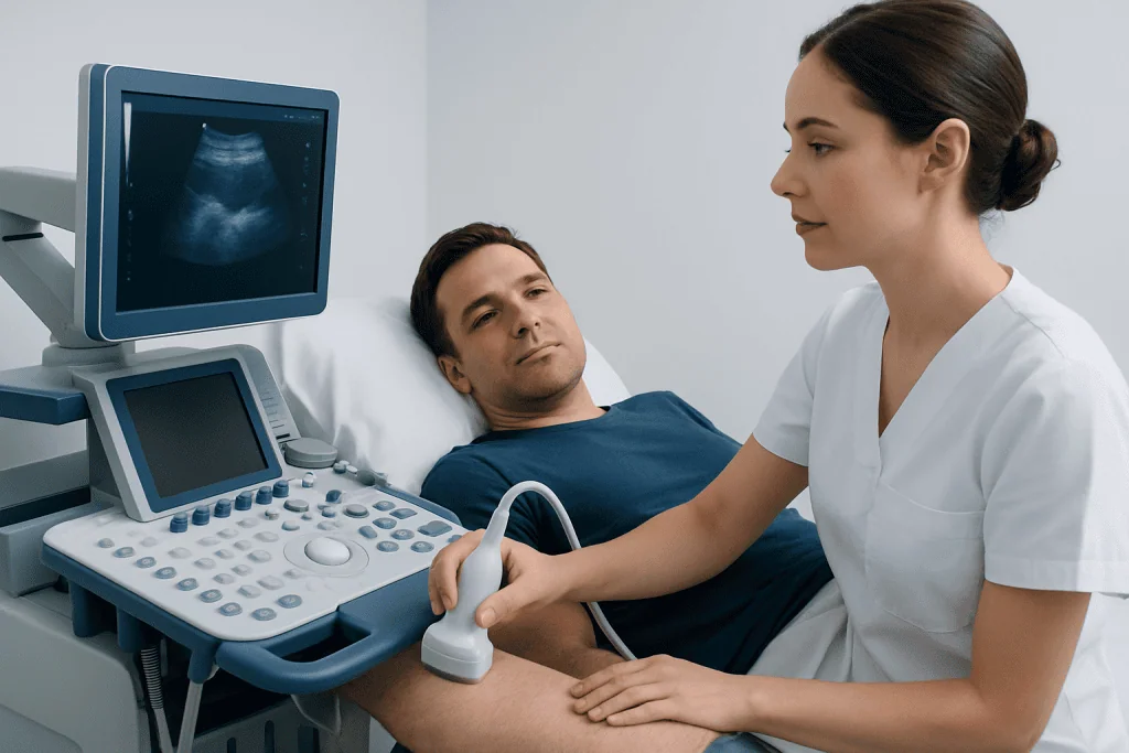
Ultrasound works by placing a small handheld device called a transducer against the skin. The transducer sends sound waves into the scrotal tissues and receives echoes back, which are converted into images. Doppler ultrasound may also be used to display and measure blood flow in the testicles and epididymis. This is particularly important when clinicians need to distinguish inflammation, which may increase blood flow, from torsion, which may reduce it.
A clinician may recommend a testicular ultrasound for persistent or unexplained scrotal symptoms, a new lump, swelling after an injury, suspected infection, fertility concerns or follow-up of a known benign finding. It may also be performed when the physical examination is limited by pain or swelling. The scan is generally suitable for adults and children because it does not expose the body to ionising radiation.
Common conditions considered during this assessment include inflammation of the epididymis, known as epididymitis, and other causes of testicular or scrotal discomfort. If symptoms suggest infection, a clinician may additionally request urine testing, tests for sexually transmitted infections when appropriate, or blood tests.
Preparation and Step-by-Step Procedure
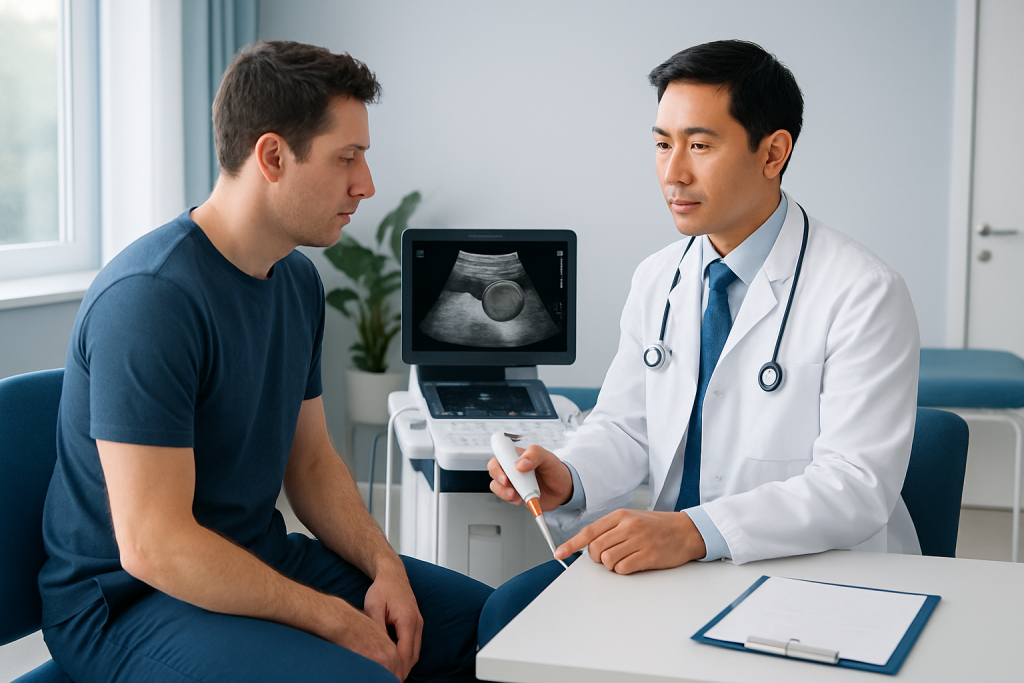
Most people do not need special preparation for a scrotal ultrasound. They can usually eat, drink and take regular medicines as normal unless their healthcare team gives different instructions. Wearing comfortable, easy-to-remove clothing may make the appointment simpler. It is helpful to tell the imaging team about recent injury, surgery, current symptoms, medicines and any previous imaging results.
During the test, the person lies on their back on an examination couch. The penis may be gently positioned upward and covered with a towel, while the scrotum is supported with another towel. A clear gel is placed on the skin to help the transducer make good contact. The sonographer moves the transducer carefully over both sides of the scrotum and may take still images and Doppler measurements for comparison.
The scan usually takes around 15 to 30 minutes, although timing can vary if more images are needed. It should not be painful, but pressure over an already tender area may feel uncomfortable. The sonographer may not discuss a final diagnosis during the appointment because the images are typically reviewed and formally reported by a radiologist or another qualified clinician.
Do I Need to Drink Water Before a Testicular Ultrasound?
No, a full bladder is usually not needed for a testicular or scrotal ultrasound. Unlike some pelvic or abdominal ultrasound examinations, this scan focuses on structures outside the abdomen, so drinking extra water does not normally improve the images.
People should follow the instructions provided by their own imaging centre, particularly if the appointment includes another examination on the same day. If no special instructions have been given, normal fluid intake is generally appropriate. Regular medicines can usually be taken with water unless a clinician advises otherwise.
If the person has urinary symptoms, such as burning, frequency or difficulty passing urine, they should mention these to the clinician. These symptoms may guide additional testing, but they do not usually change the basic preparation for the scrotal ultrasound itself.
What Is a Normal Epididymis on an Ultrasound?
A normal epididymis on ultrasound is generally seen as a small structure beside the testicle, most clearly at the upper pole and along its back edge. It is usually similar on both sides, without a solid mass, major enlargement or marked surrounding fluid. The imaging appearance may vary slightly between individuals and according to the equipment and scanning technique used.
On Doppler imaging, a normal epididymis has an expected level of blood flow. Increased blood flow together with enlargement and tenderness can support a diagnosis of inflammation, while a small fluid-filled epididymal cyst may appear as a well-defined, benign-looking area. A report may also note findings unrelated to the epididymis, such as a hydrocele, varicocele or testicular lesion.
A normal ultrasound result is reassuring, but it does not always identify the exact source of symptoms. A doctor considers the report together with the person’s history, physical examination and any laboratory results before recommending monitoring, treatment or specialist referral.
What if I Get Erect During a Testicular Ultrasound?
An erection during a testicular ultrasound can happen as an involuntary physical response to touch, anxiety or the cool or warm sensations of gel and room temperature. It is not unusual, and there is no need to feel embarrassed. Sonographers are healthcare professionals who perform intimate-area examinations respectfully and routinely.
The person can simply inform the sonographer if they feel uncomfortable or would like a brief pause. The examination can usually continue or resume once the person feels ready. The scan remains focused on obtaining medically useful images of the scrotal structures.
Privacy is an important part of the procedure. Only the area being examined should be exposed, and a towel or drape is used for support and coverage. A chaperone may be available according to local policy or upon request.
Is There Anything You Shouldn't Do Before an Ultrasound?
For a scrotal ultrasound, there are usually few restrictions. People generally do not need to fast, avoid exercise or stop routine medicines unless instructed by their healthcare professional. They should avoid applying thick creams, oils or powders to the scrotal skin shortly before the examination, as these may interfere with contact between the skin, gel and ultrasound probe.
It is also sensible not to delay urgent care in order to prepare for a scan. Sudden severe pain, rapid swelling, fever with significant scrotal tenderness, or pain after a substantial injury needs prompt assessment. In these situations, emergency clinicians decide which tests are required and how quickly they should be performed.
Anyone who has questions about privacy, cultural preferences, mobility needs or anxiety about the examination can contact the imaging department beforehand. Clear communication can help the team adapt the visit and make the process more comfortable.
Results, Recovery, Benefits and When to Seek Medical Care
There is no recovery period after a scrotal ultrasound. The gel is wiped away, and the person can usually return to normal daily activities immediately. There are no known radiation-related risks because ultrasound uses sound waves. The main limitation is that the scan may not provide a complete explanation for every symptom, and additional tests or follow-up may be needed.
The benefits include a non-invasive view of the testicles and epididymis, comparison between both sides and assessment of blood flow. Depending on the results, treatment may range from observation and supportive measures to medicines for an underlying infection or referral to a urologist. When a procedure is needed for a related urological concern, care should be based on the individual diagnosis rather than the ultrasound report alone.
Medical care should be sought urgently for sudden or severe testicular pain, pain with nausea or vomiting, major swelling, a scrotal injury, fever with worsening pain, or a new hard testicular lump. Non-urgent medical review is still advisable for discomfort lasting more than a few days, recurrent symptoms, a persistent lump or fertility concerns. Acibadem International’s multidisciplinary specialists and JCI-accredited hospitals can assess scrotal and testicular concerns for international patients when appropriate.
Frequently asked questions
Can an ultrasound diagnose epididymitis?
Ultrasound can show changes that support epididymitis, such as epididymal enlargement and increased blood flow on Doppler imaging. However, diagnosis also depends on symptoms, physical examination and, when indicated, urine or infection testing. Ultrasound is especially valuable when clinicians need to rule out other causes of acute scrotal pain.
Can an epididymal cyst be seen on ultrasound?
Yes. Ultrasound can usually identify an epididymal cyst or spermatocele as a fluid-filled structure near the epididymis. Many of these findings are benign and do not require treatment unless they are painful, large or causing other concerns. A clinician can explain what the size and appearance mean in the individual situation.
Does a testicular ultrasound hurt?
The examination is usually painless. The probe may apply mild pressure, which can be uncomfortable if the area is already sore or swollen. The person should tell the sonographer about pain so the examination can be performed as gently as possible.
How long does it take to get testicular ultrasound results?
Timing varies by healthcare setting. Images may be reviewed after the appointment by a radiologist, and the referring clinician then discusses the report and next steps. In urgent situations, results are often assessed more quickly to guide immediate care.
Can ultrasound rule out testicular torsion?
Doppler ultrasound can assess blood flow and is an important test when torsion is suspected. However, if symptoms strongly suggest torsion, urgent urological assessment is needed because treatment should not be delayed. A clinician interprets the ultrasound alongside the timing and severity of symptoms.
Should a person shave before a scrotal ultrasound?
Shaving is not normally necessary before a scrotal ultrasound. The transducer can obtain images through normal body hair with the help of ultrasound gel. If there are any special preparation needs, the imaging centre will provide instructions in advance.
References
- American College of Radiology
- Radiological Society of North America
- National Institute of Diabetes and Digestive and Kidney Diseases
- Urology Care Foundation
- National Health Service
This article is for general information only and is not a substitute for professional medical advice. Please consult a qualified doctor about your individual situation.
Ultrasound in Turkey — costs, top hospitals & a free quote
JCI-accredited · board-certified surgeons · reply within 24h

Check-up & Preventive Medicine
Comprehensive health screenings designed for early detection and prevention.
3 specialists in this unit