Kidney Stones: Severe Flank Pain and Treatment Choices

Kidney stones often cause sudden, intense pain in the side or back, sometimes with nausea, blood in the urine, or urinary urgency. Diagnosis usually involves urine tests, blood tests, and imaging such as ultrasound or CT, depending on the situation.
Key Takeaways
- Kidney stones often cause sudden, intense pain in the side or back, sometimes with nausea, blood in the urine, or urinary urgency.
- Diagnosis usually involves urine tests, blood tests, and imaging such as ultrasound or CT, depending on the situation.
- Treatment depends on stone size, location, symptoms, infection risk, and kidney function.
- Drinking enough fluids and making targeted dietary changes can reduce the chance of future stones.
- Fever, chills, uncontrolled pain, vomiting, or difficulty passing urine should be assessed urgently.
Kidney stones are hard mineral deposits that form in the urinary tract and may cause severe flank pain when they move or block urine flow. Many stones pass with supportive care, while others require medication or minimally invasive procedures.
Overview
Kidney stones are solid crystals that develop when minerals and salts in the urine become concentrated and join together. They may form inside the kidney and stay there, or they may travel into the ureter, the narrow tube that carries urine from the kidney to the bladder. When a stone blocks urine flow or irritates the urinary tract, it can cause a distinctive pattern of severe pain known as renal colic.
Stones vary widely in size. Some are as small as a grain of sand and pass without a person noticing, while others are large enough to cause blockage, infection, or kidney strain. The most common stones contain calcium, but other types include uric acid stones, struvite stones related to infection, and cystine stones linked to a rare inherited condition.
Although kidney stone pain can be intense, effective diagnostic and treatment options are available. The main goals are to relieve pain, confirm the stone’s size and position, protect kidney function, treat any infection, and reduce the likelihood of another stone in the future.
Symptoms of Kidney Stones
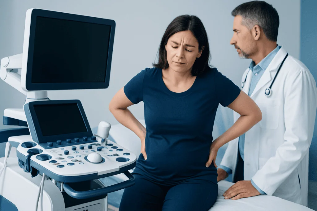
The classic symptom is sudden, severe pain in the flank, which is the side of the body between the ribs and the hip. The pain may move from the back toward the lower abdomen, groin, or genital area as the stone travels through the ureter. It often comes in waves, with periods of worsening and easing, because the ureter contracts as it tries to push the stone along.
Other symptoms may include nausea, vomiting, sweating, restlessness, and difficulty finding a comfortable position. Urinary symptoms are also common, especially when the stone is near the bladder. These may include burning with urination, frequent urination, urgency, or passing only small amounts of urine.
Blood in the urine can occur because the stone irritates the lining of the urinary tract. Sometimes the urine looks pink, red, or brown, while in other cases blood is detected only on a urine test. Fever, chills, or feeling very unwell may suggest infection, which requires prompt medical attention when a stone is present.
Causes and Risk Factors
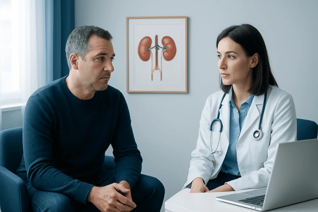
Kidney stones form when urine contains more stone-forming substances than it can dilute. Low fluid intake is one of the most important contributors because concentrated urine allows crystals to develop more easily. Hot climates, heavy sweating, and not replacing fluids can also increase risk.
Diet and medical conditions may play a role. A high intake of salt can increase calcium in the urine, while very high animal protein intake may raise uric acid levels and reduce protective citrate. Some people are more prone to stones because of recurrent urinary tract infections, gout, obesity, digestive disorders, certain bowel surgeries, or medications that affect urine chemistry.
Family history also matters. A person who has had one kidney stone has a higher chance of developing another, especially if the underlying cause is not addressed. Stone analysis and metabolic testing can help identify the specific pattern, allowing prevention advice to be tailored rather than based on guesswork.
- Calcium stones: often calcium oxalate or calcium phosphate; the most common type.
- Uric acid stones: linked to acidic urine and sometimes gout or high animal protein intake.
- Struvite stones: associated with certain urinary infections and may grow quickly.
- Cystine stones: caused by a rare inherited condition called cystinuria.
Diagnosis
A doctor usually begins with a description of the pain, urinary symptoms, medical history, and physical examination. Because several conditions can mimic kidney stone pain, including appendicitis, gallbladder disease, ovarian conditions, and abdominal problems, imaging is often needed to confirm the diagnosis and guide treatment.
Urine tests can check for blood, infection, crystals, and urine acidity. Blood tests may assess kidney function, signs of infection, calcium levels, uric acid, and other factors depending on the case. If a person catches or passes a stone, laboratory analysis can identify its type, which is very useful for prevention planning.
Imaging options include ultrasound, X-ray, and computed tomography. Ultrasound is commonly used because it avoids radiation and can show kidney swelling, larger stones, and some ureteral stones. Low-dose CT is highly accurate for many stones and is often used when the diagnosis is uncertain, symptoms are severe, or treatment planning requires precise stone location and size.
Treatment Options
Treatment depends on the stone’s size, location, degree of blockage, symptoms, infection risk, and the person’s overall health. Small stones may pass on their own with pain control, anti-nausea medication if needed, and adequate fluids. Doctors may also prescribe medication to help relax the ureter in selected cases, making passage easier.
Urgent treatment is needed if a stone is associated with infection, reduced kidney function, a single functioning kidney, uncontrolled pain, persistent vomiting, or complete blockage. In these situations, doctors may need to drain the kidney temporarily with a ureteral stent or a nephrostomy tube before definitive stone treatment. Antibiotics are used when infection is present, but drainage may also be necessary if urine is blocked.
When a stone is unlikely to pass or continues to cause problems, minimally invasive procedures can remove or break it. Shock wave lithotripsy uses focused sound waves to fragment certain stones so the pieces can pass in urine. Ureteroscopy involves passing a thin scope through the urinary tract to reach the stone, often using a laser to break it. Larger or complex kidney stones may be treated with percutaneous nephrolithotomy, a procedure performed through a small opening in the back.
The best choice is individualized. A urologist considers stone size, stone hardness, location, anatomy, kidney function, pregnancy status when relevant, bleeding risk, infection, and patient preference. Follow-up imaging may be recommended after treatment to confirm that the stone has passed or that fragments are not causing further blockage.
Prevention and Self-care
Prevention starts with producing enough urine throughout the day. Many people are advised to drink fluids regularly so urine stays pale rather than dark, but the right amount varies with climate, activity level, heart or kidney conditions, and medical advice. People with fluid restrictions should follow their doctor’s guidance rather than increasing fluids on their own.
Dietary prevention depends on stone type, but several general steps help many patients. Reducing excess salt is important because sodium can increase calcium loss into the urine. Moderate rather than excessive animal protein intake may be recommended for some stone formers. People with calcium oxalate stones may be advised to limit very high-oxalate foods while maintaining normal dietary calcium, because avoiding calcium completely can sometimes increase oxalate absorption.
Self-care during a suspected stone episode should be guided by medical advice, especially if pain is severe or symptoms are new. Some patients are asked to strain their urine to catch the stone for analysis. Over-the-counter pain medicines may not be appropriate for everyone, particularly people with kidney disease, stomach ulcers, blood thinner use, or certain heart conditions, so a clinician’s recommendation is safest.
- Drink fluids consistently unless a doctor has advised restriction.
- Limit excess salt and avoid highly processed salty foods.
- Ask whether stone analysis or 24-hour urine testing is appropriate after a stone episode.
- Do not start supplements or major diet changes without medical advice if stones are recurrent.
When to See a Doctor
Anyone with severe flank pain, especially pain that comes in waves or travels toward the groin, should seek medical evaluation. Prompt assessment is also important if there is blood in the urine, repeated vomiting, inability to keep fluids down, pregnancy, a known single kidney, or a history of kidney disease.
Fever, chills, confusion, weakness, or feeling seriously unwell with suspected kidney stone symptoms can indicate infection behind a blockage. This is treated as an urgent situation because infected, blocked urine may require rapid drainage as well as antibiotics. Difficulty passing urine or a marked decrease in urine output also needs urgent care.
People who have had kidney stones before should arrange follow-up to discuss prevention, especially if stones are recurrent, large, bilateral, or occur at a young age. Acibadem International’s multidisciplinary specialists and JCI-accredited hospitals diagnose and treat urinary stone disease for international patients, including imaging, urology care, and individualized follow-up planning.
Frequently asked questions
What does kidney stone pain feel like?
Kidney stone pain often starts suddenly in the side or back below the ribs. It may be very intense, come in waves, and move toward the lower abdomen or groin as the stone travels. Nausea, vomiting, urinary urgency, or blood in the urine may occur at the same time.
Can a kidney stone pass on its own?
Many small stones can pass without a procedure, especially if they are located lower in the ureter. Larger stones, stones causing infection, or stones that block urine flow may need medical or surgical treatment. A doctor can estimate the chance of passage based on imaging and symptoms.
How are kidney stones diagnosed?
Diagnosis usually includes a medical history, physical examination, urine testing, and sometimes blood tests. Imaging such as ultrasound or CT helps confirm the stone’s size and location. If the stone is passed, laboratory analysis can identify the stone type and guide prevention.
Is drinking more water always enough to prevent stones?
Good hydration is a key prevention step, but it may not be enough for everyone. Stone type, urine chemistry, diet, medical conditions, and medications can all influence recurrence risk. People with repeated stones often benefit from stone analysis and targeted prevention advice.
What treatments are available if a stone does not pass?
Treatment options include shock wave lithotripsy, ureteroscopy with laser fragmentation, and percutaneous nephrolithotomy for larger or complex stones. In urgent cases with infection or severe blockage, temporary drainage with a stent or nephrostomy tube may be needed first. The urologist chooses the approach based on the stone and the patient’s condition.
When is a kidney stone an emergency?
A suspected kidney stone should be assessed urgently if there is fever, chills, persistent vomiting, uncontrolled pain, difficulty passing urine, or known kidney disease. Infection with a blocked kidney requires prompt treatment. Pregnant patients or people with one functioning kidney should also seek urgent medical care.
References
- European Association of Urology
- American Urological Association
- National Institute of Diabetes and Digestive and Kidney Diseases
- Mayo Clinic
- BMJ Best Practice
This article is for general information only and is not a substitute for professional medical advice. Please consult a qualified doctor about your individual situation.
Explore treatments in Turkey — costs, top hospitals & a free quote
JCI-accredited · board-certified surgeons · reply within 24h
Check your numbers in seconds
BMI, calories, due date, blood pressure and 30+ more clinical calculators — free, instant, doctor-reviewed ranges.