MPFL Reconstruction Protocol: Procedure, Recovery and Results

MPFL reconstruction replaces or reinforces the medial patellofemoral ligament to help prevent recurrent kneecap dislocation. The operation is usually paired with a structured rehabilitation plan that gradually restores motion, strength, balance and confidence in the knee.
Key Takeaways
- MPFL reconstruction replaces or reinforces the medial patellofemoral ligament to help prevent recurrent kneecap dislocation.
- The operation is usually paired with a structured rehabilitation plan that gradually restores motion, strength, balance and confidence in the knee.
- Many patients begin protected walking soon after surgery, but brace use, crutches and weight-bearing instructions vary with the surgical plan.
- Recovery commonly takes several months; return to pivoting sports is usually considered only after strength and movement testing.
- Although outcomes are generally favorable for suitable patients, surgery carries risks including stiffness, infection, blood clots and ongoing instability.
An MPFL reconstruction protocol is an individualized plan for rebuilding the ligament that helps keep the kneecap stable, followed by staged protection, walking and rehabilitation. It is commonly considered for recurrent patellar dislocation when structured non-surgical care has not restored stability.
Overview: what is an MPFL reconstruction protocol?
An MPFL reconstruction protocol is the combined surgical and rehabilitation plan used to treat recurrent patellar instability, meaning repeated partial or complete dislocation of the kneecap. The medial patellofemoral ligament (MPFL) is a soft-tissue band on the inner side of the knee that helps guide the kneecap in its groove, especially during early knee bending. When it is torn or stretched after dislocations, the kneecap may continue to shift outward.
MPFL reconstruction does not usually repair the damaged ligament directly. Instead, a surgeon uses a tendon graft, often from the patient or a donor, to create a new stabilizing ligament. The protocol then protects the graft while movement, walking and muscle control are restored in stages.
Not every first kneecap dislocation requires surgery. Many people improve with bracing, activity modification and physiotherapy. Reconstruction may be recommended when dislocations recur, the knee remains unstable despite rehabilitation, or imaging shows an injury pattern that makes further instability more likely. Patellar instability can be reviewed in more detail in patellar dislocation information.
Who may be a candidate and how the knee is assessed
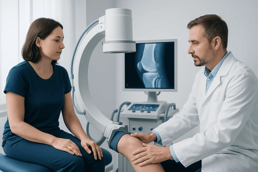
Candidacy is based on the person’s symptoms, activity needs, examination findings and knee anatomy. Typical concerns include repeated kneecap dislocations, a sensation that the kneecap slips or gives way, fear during stairs or turning, and inability to return safely to desired activities. A clinician will also ask about previous injuries, general health and goals for recovery.
Assessment usually includes a physical examination and X-rays. Magnetic resonance imaging (MRI) can identify MPFL injury, cartilage damage, loose fragments and other soft-tissue problems. In selected cases, CT or specialized X-ray measurements help assess the shape and alignment of the kneecap groove, the position of the tibial tubercle and rotational alignment of the leg.
These details matter because MPFL reconstruction is not identical for every knee. Some people have significant bony alignment differences, a very shallow trochlear groove, high-riding patella or cartilage injury. When present, the surgeon may discuss an additional procedure rather than an isolated reconstruction. The final plan should be individualized by an orthopedic knee specialist.
How MPFL reconstruction works: step by step
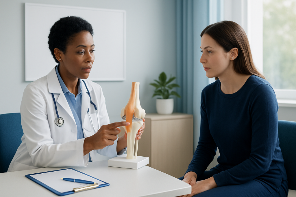
The procedure is generally performed under anesthesia. Before starting, the surgical team confirms the planned knee and procedure. Arthroscopy may be used to inspect the joint, assess cartilage and address loose bodies or associated injuries when needed.
A tendon graft is then prepared. It may be an autograft, using the patient’s own tendon tissue, or an allograft, using screened donor tissue. The surgeon makes small incisions at the inner side of the kneecap and around the inner part of the femur, places the graft in the appropriate anatomical position and secures it with surgical fixation devices.
Correct graft position and tension are important. The new ligament needs to support the kneecap without pulling it too tightly, which could limit motion or increase pressure on cartilage. The surgeon checks kneecap tracking through knee movement before closing the incisions. The operation may be performed alone or alongside alignment or cartilage procedures, which can change the recovery plan.
Patients undergoing MPFL reconstruction should receive specific instructions on wound care, bracing, pain management, weight bearing and physiotherapy before leaving the hospital or surgical center.
Recovery timeline: walking, rehabilitation and return to activity
Recovery is progressive rather than a fixed calendar. In the first days and weeks, the focus is swelling control, wound healing, safe use of crutches, gentle knee motion and activation of the quadriceps muscle. Many protocols use a knee brace initially, sometimes locked in extension for walking and adjusted as muscle control and range of motion improve.
Protected weight bearing often begins soon after isolated MPFL reconstruction, but the amount of weight allowed and the duration of crutch use vary. Patients should follow their own surgeon’s instructions, particularly if they have had an additional bone, cartilage or ligament procedure. Early physiotherapy commonly emphasizes full knee straightening, gradual bending, gait retraining and avoiding loss of quadriceps strength.
During the next several months, rehabilitation advances to closed-chain strengthening, hip and core control, balance work and movement retraining. Running, jumping, cutting and pivoting are introduced only when pain and swelling are controlled and testing shows adequate strength, stability and coordination. A broader knee treatment assessment may also be appropriate for people whose symptoms are driven by advanced joint degeneration rather than instability alone.
Return to sport is based on functional readiness, not time alone. It commonly takes several months, and higher-demand pivoting sports may require longer. Follow-up visits help the team monitor motion, patellar tracking, graft function and any symptoms that could slow progression.
How long does it take to recover from MPFL reconstruction surgery?
Most people need several months to recover from MPFL reconstruction surgery, while return to demanding sport often takes around six months or longer. The exact timetable depends on whether the surgery was isolated or combined with other procedures, the person’s preoperative strength, the condition of cartilage and bone, and consistent participation in rehabilitation.
Daily activities tend to improve before athletic activity. In the early phase, the knee may feel stiff, swollen and weak. Over time, walking becomes more natural, knee motion increases and muscle control improves. Returning too quickly to running, pivoting or jumping may increase symptoms or place unnecessary stress on the healing tissues.
A surgeon and physiotherapist use more than elapsed time to guide recovery. They may assess swelling, range of motion, walking pattern, quadriceps strength, single-leg control and confidence with sport-specific movements. This criteria-based approach supports a safer return to activity.
How long after MPFL surgery can you walk?
Many patients can begin walking with crutches and a brace shortly after isolated MPFL surgery, often on the same day or within the first few days. However, this is protected walking rather than an immediate return to normal walking. The knee may need to remain in a specific brace position, and the patient may be instructed to bear weight gradually.
Crutches are commonly used until the person can walk without a marked limp and has enough quadriceps control to keep the knee stable. For some patients, this occurs within a few weeks; for others, it takes longer. Swelling, pain, limited motion and additional surgical procedures can all affect progress.
It is important not to change weight-bearing or brace instructions based on another patient’s schedule. The treating surgeon’s plan takes priority because fixation method, graft choice and any simultaneous procedure can alter the restrictions needed for healing.
How painful is MPFL surgery recovery?
MPFL surgery recovery usually involves pain, swelling and tightness around the knee, particularly during the first days after surgery and when beginning rehabilitation exercises. The experience differs between individuals. Pain often becomes more manageable as early healing progresses, but rehabilitation can still cause temporary soreness as muscles are reactivated and motion increases.
The care team may recommend a combination of prescribed medicines, ice or cold therapy if suitable, elevation, compression and carefully paced activity. Medication choices should be individualized, especially for people with kidney disease, stomach ulcers, bleeding risk, allergies or other medical conditions. Patients should take medicines only as directed by their clinician.
Persistent or worsening pain should not simply be pushed through. New severe pain, rapidly increasing swelling, calf pain, fever, wound drainage, numbness or a change in foot color requires prompt medical advice. These symptoms do not always indicate a serious problem, but they should be assessed.
What is the success rate of MPFL reconstruction surgery?
MPFL reconstruction generally has favorable results in appropriately selected patients, with many reporting improved kneecap stability and a return to everyday activities. Published outcomes vary because studies include different patient ages, surgical methods, follow-up periods and associated anatomical factors. For this reason, a single success rate cannot reliably predict an individual outcome.
Results are influenced by accurate diagnosis, graft placement, rehabilitation, strength recovery and whether underlying bony alignment factors also need treatment. Some people may continue to have pain, apprehension, weakness or limits with high-demand sport despite a stable kneecap. Cartilage damage from earlier dislocations can also contribute to ongoing symptoms.
Possible complications include infection, bleeding, blood clots, stiffness, reduced motion, nerve irritation, persistent pain, fracture at a fixation site, recurrent instability or an overly tight graft. These risks are uncommon but important to discuss during informed consent. The surgeon can explain expected benefits and limitations in the context of the individual knee.
When to seek medical care
Medical assessment is advisable after a first kneecap dislocation, particularly if the knee remains swollen, cannot bear weight, locks, appears deformed or feels unstable. Urgent care is needed if the kneecap remains out of place, there is severe pain after an injury, the foot becomes cold or numb, or the person cannot move the knee normally.
After surgery, patients should contact their surgical team promptly for fever, increasing redness or drainage from the incision, uncontrolled pain, worsening swelling, calf tenderness, shortness of breath or chest pain. Emergency care is appropriate for chest pain, difficulty breathing or signs of a serious circulation problem.
Acibadem International’s multidisciplinary specialists and JCI-accredited hospitals assess and treat patellar instability for international patients, with surgical planning and rehabilitation coordinated around the individual’s needs.
Frequently asked questions
What does MPFL stand for?
MPFL stands for medial patellofemoral ligament. It is an important ligament on the inner side of the knee that helps prevent the kneecap from moving too far outward, especially as the knee starts to bend.
Is MPFL reconstruction the same as kneecap surgery?
MPFL reconstruction is one type of surgery for kneecap instability. It focuses on rebuilding a stabilizing ligament, but some patients also need procedures that address bone alignment, the trochlear groove, cartilage damage or loose fragments.
Will I need physiotherapy after MPFL reconstruction?
Physiotherapy is a central part of recovery for most patients. It helps restore knee motion, quadriceps strength, walking mechanics, balance and safe movement patterns before return to higher-level activity.
Can the kneecap dislocate again after MPFL reconstruction?
Recurrent instability can still occur, although reconstruction is intended to reduce that risk. The likelihood depends on surgical factors, rehabilitation, injury exposure and underlying knee anatomy, which is why careful assessment is important.
When can I drive after MPFL surgery?
Driving should resume only when the person can safely control the vehicle, is no longer impaired by sedating pain medicines and has been cleared by the surgeon. Timing varies according to the operated leg, vehicle type, knee control and recovery progress.
What should I avoid during early MPFL reconstruction recovery?
Patients should avoid activities that exceed their surgeon’s restrictions, including unsupported pivoting, jumping, running or deep knee bending when not yet permitted. They should also avoid stopping brace use, crutches or prescribed exercises without discussing it with the care team.
References
- American Academy of Orthopaedic Surgeons
- American Orthopaedic Society for Sports Medicine
- OrthoInfo by the American Academy of Orthopaedic Surgeons
- National Institute of Arthritis and Musculoskeletal and Skin Diseases
This article is for general information only and is not a substitute for professional medical advice. Please consult a qualified doctor about your individual situation.
Joint and spine care in Turkey — expert assessment & treatment
JCI-accredited · board-certified surgeons · reply within 24h