Osteoid Osteoma Treatment: How It Works, Results and What to Expect

Osteoid osteoma is a benign bone-forming tumor that commonly causes persistent pain, often worse at night. Anti-inflammatory medicines may control symptoms, but definitive treatment can be considered when pain persists or medication is unsuitable.
Key Takeaways
- Osteoid osteoma is a benign bone-forming tumor that commonly causes persistent pain, often worse at night.
- Anti-inflammatory medicines may control symptoms, but definitive treatment can be considered when pain persists or medication is unsuitable.
- CT-guided radiofrequency ablation is a commonly used minimally invasive treatment that destroys the lesion with heat.
- Most people return to light daily activities relatively soon after ablation, while sports restrictions depend on the treated bone and clinician guidance.
- Follow-up assesses pain relief, wound healing and, when needed, imaging findings to confirm successful treatment.
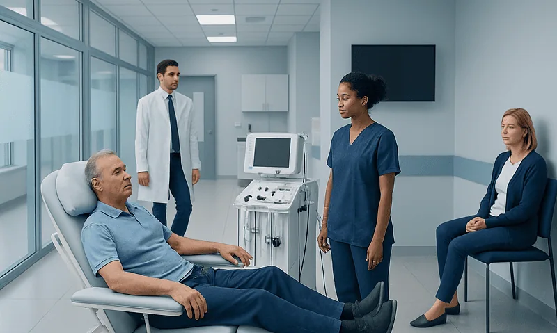
Osteoid osteoma treatment is tailored to the lesion’s location, symptoms and the person’s overall health. Image-guided thermal ablation is often preferred because it can target the small benign lesion precisely and usually involves a shorter recovery than open surgery.
Osteoid Osteoma Treatment: How It Works
Osteoid osteoma treatment aims to relieve pain by treating the small central area of the tumor, called the nidus. Osteoid osteoma is a benign, or non-cancerous, bone-forming tumor. Although it does not spread to other parts of the body, it can cause substantial and persistent pain and may interfere with sleep, school, work or activity.
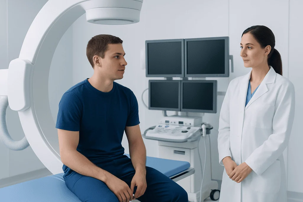
For many symptomatic lesions, image-guided ablation is the main definitive treatment. During this minimally invasive procedure, a specialist uses imaging—most often computed tomography (CT)—to guide a thin probe accurately into the nidus. Energy delivered through the probe heats and destroys the abnormal tissue while limiting effects on surrounding bone and soft tissues.
Image-guided osteoid osteoma treatment may use radiofrequency ablation, which produces controlled heat, or another thermal technique selected for the lesion’s site and proximity to important structures. Open surgical removal is now less commonly needed but remains useful in selected situations, including lesions that cannot be safely reached with a needle-based approach.
Some people manage symptoms with nonsteroidal anti-inflammatory drugs (NSAIDs), under medical guidance. These medicines often work particularly well because the nidus produces substances that contribute to pain and inflammation. However, long-term medication use may not be appropriate for everyone and does not remove the lesion.
Who May Be a Candidate for Treatment?
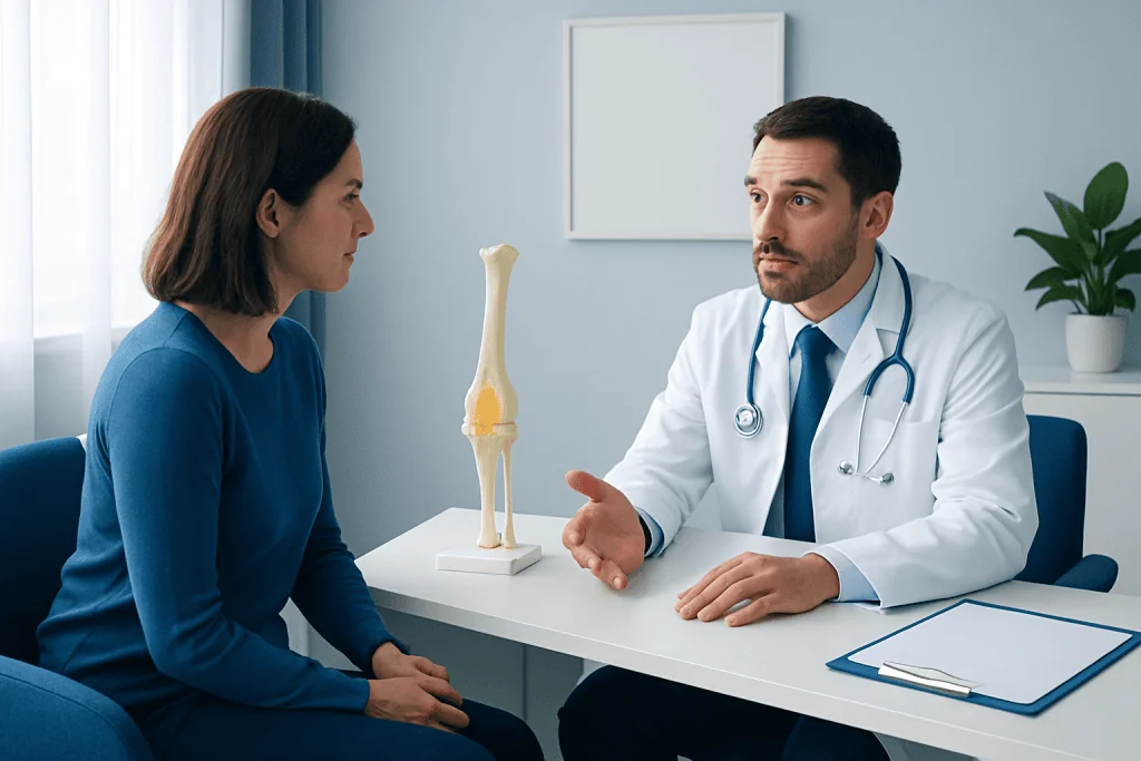
A treatment plan starts with confirming the diagnosis and locating the nidus precisely. A person may be considered for ablation when pain is ongoing, disrupts normal life, requires frequent pain medicine, or returns when medication is reduced. Treatment may also be reasonable when an individual prefers a definitive option after discussing the benefits and limitations with the care team.
Candidacy depends on the lesion’s size and location. Osteoid osteomas often occur in the long bones of the leg, particularly the femur or tibia, but can also develop in the spine, hands, feet or other bones. Lesions near nerves, joints, growth plates, blood vessels or skin may require especially careful planning or an alternative technique.
Orthopedic specialists, musculoskeletal radiologists, anesthesiologists and, when appropriate, pediatric specialists can work together to select the safest approach. Age alone does not determine eligibility. Children, adolescents and adults may all be assessed individually, with particular attention to protecting growing bone in younger patients.
Before treatment, clinicians review medical history, medication use, allergies, bleeding risk and any possibility of pregnancy. They also discuss anesthesia or sedation, expected activity restrictions and practical arrangements for going home after the procedure.
What Happens During Osteoid Osteoma Ablation?
Before the procedure, the patient may have blood tests or other pre-procedure assessments depending on the planned anesthesia and their health history. The team explains when to stop eating or drinking, which regular medicines can be taken, and whether medicines that affect bleeding need adjustment. These instructions should always come from the treating team.
During ablation, the person is positioned to provide safe access to the affected bone. Sedation, general anesthesia or another anesthesia plan may be used, depending on lesion location, age and anticipated comfort needs. The skin is cleaned and covered with sterile drapes, and the clinician makes a very small incision.
CT imaging guides a needle or probe into the nidus. Once position is confirmed, the abnormal tissue is treated with controlled heat for a planned period. In some cases, protective measures such as cooling or temperature monitoring are used to safeguard nearby nerves, skin or joint structures. The probe is removed and the small entry site is dressed.
The procedure is usually followed by a period of observation while the person wakes from anesthesia and pain control is reviewed. Many patients can go home the same day, but the appropriate observation period varies by procedure complexity, treated location and individual needs.
Benefits, Risks and Expected Results
The main expected benefit of definitive osteoid osteoma treatment is meaningful pain relief, often with less need for anti-inflammatory medicine. Because ablation targets the nidus through a small skin opening, it generally avoids the larger incision and bone removal that may be required with open surgery. This can support a more comfortable recovery for appropriately selected patients.
Pain improvement can begin quickly, although the timing differs among individuals. Temporary soreness at the treatment site is common after the procedure. If pain does not improve as expected, returns after initial relief, or changes in character, the care team may arrange a clinical review and imaging to check for incomplete treatment, recurrence or another cause.
All procedures carry risks. Potential complications include bleeding, infection, skin burn, injury to nearby nerves or blood vessels, a fracture in weakened bone, and pain that persists or recurs. The likelihood and type of risk depend strongly on the lesion’s location. A lesion near the spine or a joint requires tailored planning because nearby structures may be more vulnerable.
Open surgery may be advised if image-guided ablation is not technically safe or has not fully controlled the lesion. Surgical planning aims to remove the nidus while preserving bone strength and function; some operations may require a longer protected recovery than needle-based treatment.
Recovery Timeline and Follow-Up
Immediately after ablation, the treatment area may feel tender, and there may be mild bruising or swelling around the small incision. The care team provides individualized advice on wound care, showering, pain relief and warning signs to watch for. Someone else may need to accompany the patient home when sedation or anesthesia has been used.
Light daily activity is often possible within a short period, but the return to walking, work, school, driving and exercise should follow the clinician’s instructions. Weight-bearing limits are more likely when treatment involves a leg bone or a site where mechanical stress could increase discomfort or fracture risk. Running, jumping and contact sports may need to wait until the bone has recovered adequately.
Follow-up commonly focuses on symptoms, mobility and the healing of the entry site. In many cases, a clear improvement in the characteristic pain is an important sign of treatment response. Repeat imaging may be used when symptoms persist, when the diagnosis was uncertain, or when the lesion is in a location where detailed monitoring is helpful.
Recovery is individual. Patients should contact their clinical team promptly for fever, increasing redness or drainage at the incision, worsening pain, new numbness or weakness, or difficulty using the affected limb. These symptoms do not always indicate a serious problem, but they deserve timely assessment.
How Serious Is Osteoid Osteoma?
Osteoid osteoma is not cancer and does not metastasize. In that sense, it is generally not considered life-threatening. Its main impact is the pain it can cause, classically a deep ache that becomes worse at night and may improve with anti-inflammatory medication.
Even though it is benign, osteoid osteoma should be properly assessed because prolonged pain can affect sleep, mood, concentration and physical activity. When a lesion occurs close to a joint, it may lead to stiffness or altered movement. In children and adolescents, a lesion near a growth area may occasionally affect bone development if it remains untreated.
A confirmed diagnosis and an individualized treatment discussion can help prevent unnecessary disruption to everyday life. Persistent bone pain should not be self-diagnosed, since several conditions can produce similar symptoms.
How Quickly Does an Osteoma Grow?
Osteoid osteoma is typically slow-growing and usually small, often measuring less than 1.5 to 2 centimeters. The term “osteoma” may also describe other benign bone growths, which can behave differently depending on their type and location. Therefore, growth expectations should be based on the specific diagnosis rather than the name alone.
Although the lesion itself generally grows slowly, symptoms may become more noticeable over time because of inflammation around the nidus. This can make the condition feel as though it is progressing quickly, even when imaging shows only a small lesion. A clinician may compare imaging studies when needed to clarify changes.
Rapidly enlarging swelling, unexplained weight loss, fever, or pain that does not fit the expected pattern deserves medical evaluation. These symptoms are not typical features of osteoid osteoma and may point to another condition requiring assessment.
Is Osteoma Something to Worry About, and When to Seek Medical Care?
An osteoma or osteoid osteoma is often benign, but it should be evaluated when it causes symptoms or when the diagnosis is uncertain. Concern should focus less on assuming cancer and more on obtaining an accurate explanation for persistent pain, swelling, limited movement or a new bone-related finding on an imaging scan.
Medical assessment is advisable for pain that repeatedly wakes a person at night, lasts for several weeks, limits walking or sport, or improves only with regular pain medication. A clinician should also assess pain accompanied by warmth, redness, fever, numbness, weakness, or a change in bowel or bladder function, particularly if a spinal lesion is possible.
Orthopedic care may overlap with other bone and joint concerns. Depending on the diagnosis, related resources such as benign and malignant bone tumor evaluation and orthopedic surgery options may help patients understand the broader assessment process. At Acibadem International, multidisciplinary specialists in JCI-accredited hospitals evaluate and treat osteoid osteoma for international patients.
Frequently asked questions
At what age is osteoid osteoma most common?
Osteoid osteoma is most common in children, teenagers and young adults. It often develops during the first three decades of life, with many cases diagnosed in adolescence or early adulthood. However, it can occur at other ages, so persistent characteristic bone pain should still be assessed.
What is the first-line definitive osteoid osteoma treatment?
For many people with symptomatic osteoid osteoma, CT-guided thermal ablation, often radiofrequency ablation, is a commonly used definitive treatment. It targets and destroys the nidus through a small skin opening. The best method depends on the lesion’s location and the person’s individual circumstances.
Can osteoid osteoma go away without surgery or ablation?
Symptoms can sometimes be controlled with anti-inflammatory medicines, and osteoid osteoma may eventually resolve over time. However, this can take years, and regular medication may not be safe or acceptable for every person. A specialist can help weigh observation against definitive treatment.
How long does pain last after radiofrequency ablation?
Mild soreness at the insertion site can occur for several days after ablation. The characteristic osteoid osteoma pain often improves substantially soon after successful treatment, although the exact timing varies. Persistent or returning pain should be reported to the treating team.
Does osteoid osteoma treatment require an overnight hospital stay?
Many image-guided ablation procedures are performed with same-day discharge after a period of observation. Some patients may need longer monitoring based on the treated location, anesthesia used, age or other medical factors. The treating team will explain the expected plan before the procedure.
Can osteoid osteoma come back after treatment?
Recurrence or incomplete treatment is possible, though many patients obtain lasting pain relief after a successful procedure. If typical pain returns, clinicians may arrange repeat assessment and imaging. In some cases, repeat ablation or another treatment approach may be considered.
References
- American Academy of Orthopaedic Surgeons
- RadiologyInfo.org
- National Cancer Institute
- Orphanet
- Merck Manual Consumer Version
This article is for general information only and is not a substitute for professional medical advice. Please consult a qualified doctor about your individual situation.
Oncology care in Turkey — second opinion and treatment plan
JCI-accredited · board-certified surgeons · reply within 24h
Add us as a Preferred Source to see more of our trusted health content across Google Search, AI Overviews and Discover.