Pituitary Microadenoma MRI: Preparation, Procedure and Results
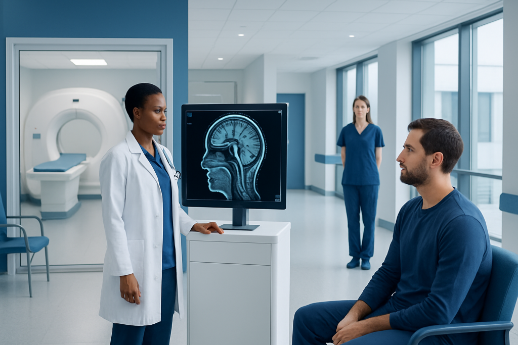
A pituitary microadenoma is a pituitary tumour measuring less than 10 millimetres and is usually non-cancerous. MRI does not use ionising radiation and is the preferred imaging test for evaluating the pituitary gland.
Key Takeaways
- A pituitary microadenoma is a pituitary tumour measuring less than 10 millimetres and is usually non-cancerous.
- MRI does not use ionising radiation and is the preferred imaging test for evaluating the pituitary gland.
- Contrast material may improve detection of very small adenomas, but it is not appropriate for every person.
- Most people do not need to fast for a pituitary MRI unless their imaging centre gives different instructions.
- MRI findings are interpreted alongside hormone blood tests, symptoms and, when needed, visual field testing.
A pituitary microadenoma MRI is a specialised magnetic resonance imaging scan that produces detailed pictures of the pituitary gland, often before and after contrast material is given. It can help clinicians identify a small pituitary adenoma, assess nearby structures and combine imaging findings with hormone tests and symptoms.
Overview: what a pituitary microadenoma MRI shows
A pituitary microadenoma MRI is a focused scan used to examine the pituitary gland, a small hormone-producing gland at the base of the brain. It helps doctors look for a microadenoma, meaning a pituitary adenoma smaller than 10 millimetres. These growths are commonly benign, and some are found incidentally while imaging is being done for another reason.
The scan uses a strong magnet and radio waves to create detailed images; it does not use X-rays or ionising radiation. Dedicated images are taken through the pituitary region, sometimes at several points after an intravenous contrast agent has been administered. This can make subtle differences between normal pituitary tissue and a small adenoma easier to see.
An MRI result is only one part of assessment. Endocrinologists consider the images together with symptoms and hormone testing, because a small lesion may or may not be responsible for a hormonal change. The MRI can also show the relationship of the gland to nearby structures, including the optic nerves and optic chiasm.
Why the scan may be recommended
A clinician may request pituitary MRI when blood or urine tests suggest excess or reduced pituitary hormone activity. It may also be used when a person has symptoms that could relate to pituitary hormone imbalance, such as unexplained menstrual changes, reduced libido, breast milk production when not pregnant or breastfeeding, changes in hand or foot size, easy bruising, persistent fatigue, or changes in weight.
Headaches are common and have many possible causes, so a headache alone does not necessarily indicate a pituitary adenoma. However, MRI may be appropriate when headaches occur alongside hormone abnormalities, visual symptoms or other clinical findings. Visual field testing may be recommended if a larger lesion is suspected to be close to visual pathways.
The same imaging technique may be used to monitor a previously identified lesion over time. The interval for repeat imaging varies according to the adenoma’s size, hormone activity, symptoms and prior treatment, so it should be decided with the treating specialist.
How should I prepare for a pituitary MRI?

Before the appointment, the patient should tell the MRI team about any implanted or retained metal, including pacemakers, neurostimulators, cochlear implants, aneurysm clips, medication pumps, previous surgery or metal fragments from an injury. Many modern implants can be scanned under specific conditions, but the radiology team must confirm the device details before the examination.
It is also important to report pregnancy, possible pregnancy, kidney disease, previous reactions to MRI contrast material and severe claustrophobia. The team can explain whether contrast is planned and whether any additional precautions are needed. People who are anxious in enclosed spaces may discuss coping strategies or, when appropriate, prescribed medication with the referring clinician in advance; they should arrange transport home if sedation is used.
Jewellery, watches, hairpins, hearing aids, removable dental items and clothing with metal components must be removed. The imaging centre may ask the patient to change into a gown. Bringing previous MRI images, reports and relevant hormone test results can help the radiologist and specialist compare findings accurately.
Do I need to fast for a pituitary MRI?
Most people do not need to fast for a pituitary MRI, including an MRI that uses intravenous contrast. They can usually eat, drink and take regular medicines as normal. The appointment instructions from the radiology department should always take priority, as individual circumstances may differ.
Fasting or altered medication instructions may be given if sedation is planned, if another procedure is scheduled on the same day, or if a clinician has specific concerns related to a person’s health. People with diabetes or other conditions requiring timed meals or medicines should ask for personalised instructions before the visit rather than changing their routine independently.
Hydration is generally helpful unless a clinician has advised fluid restriction. After contrast, usual drinking habits can typically be resumed, although patients with significant kidney disease should follow the plan provided by their medical team.
What happens during the procedure?
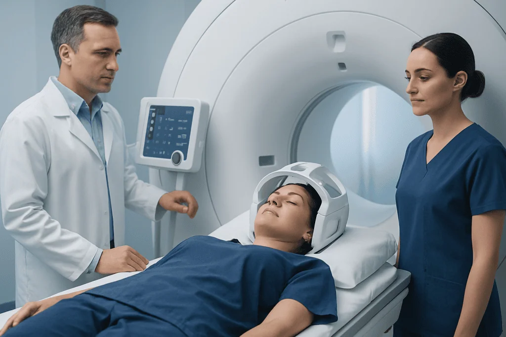
On arrival, a radiographer reviews safety questions and confirms the reason for the scan. If contrast is planned, a small intravenous cannula may be placed in an arm or hand. The patient then lies on a padded table with the head supported in a specialised head coil, which receives the MRI signals and helps produce sharp images of the pituitary area.
The table moves slowly into the scanner. During image acquisition, the machine makes rhythmic knocking or humming sounds, and ear protection is provided. The patient needs to lie as still as possible, but can communicate with the radiographer through an intercom. The staff can see and hear the patient throughout the examination.
A dedicated pituitary MRI commonly lasts about 30 to 60 minutes, although timing depends on the protocol and whether contrast sequences are needed. If contrast is used, it is injected during part of the scan. The injection may cause a brief cool sensation, but serious reactions are uncommon. MRI contrast agents are different from the iodine-based contrast commonly used for CT scans.
The main benefit of the examination is its high level of soft-tissue detail, which supports accurate diagnosis and treatment planning. Risks are low when MRI safety screening is followed. Potential concerns include discomfort from lying still, anxiety in the scanner, noise exposure without proper hearing protection, and uncommon reactions or kidney-related considerations associated with contrast.
Should I be worried about a pituitary microadenoma?
Finding a pituitary microadenoma does not automatically mean that urgent treatment is needed. Most pituitary adenomas are not cancerous, and microadenomas are small. Some do not produce excess hormones and may only require periodic clinical review, hormone testing and follow-up imaging when recommended.
The key question is whether the adenoma is affecting hormone production or causing symptoms. A hormone-producing adenoma may need treatment to control hormone excess and protect long-term health. Depending on the type of adenoma, options can include observation, medicines, surgery or, less commonly, radiation-based treatment.
A person should avoid drawing conclusions from an MRI report alone. A specialist can explain whether the finding is likely to be clinically significant and whether it is related to symptoms. Further evaluation may include tests of prolactin, cortisol, thyroid-related hormones, growth hormone pathways and sex hormones, selected according to the clinical situation.
When treatment is needed, care may involve endocrinology, neuroradiology, ophthalmology and neurosurgery. For selected cases, pituitary tumour surgery may be considered after a detailed review of the lesion, hormone findings and individual health needs.
How long does it take to get results from a pituitary MRI?
The images are available immediately for the radiology team, but the formal interpretation takes longer. A radiologist reviews the sequences carefully, compares them with prior scans where available and prepares a report for the referring clinician. Timing varies by hospital and the urgency of the request, but results are often discussed within a few days.
In urgent situations, such as sudden severe symptoms with visual change or concern for pituitary apoplexy, imaging may be reviewed much more quickly. For non-urgent examinations, the patient should ask the imaging centre or referring clinician when and how the result will be communicated.
The report may describe the size and location of a lesion, whether it appears to enhance differently from normal pituitary tissue, and whether there is any contact with nearby structures. It cannot by itself determine hormone activity. The next step may be reassurance and surveillance, targeted blood tests, referral to an endocrinologist or discussion of treatment options.
When to seek medical care
Medical advice should be sought promptly for new or worsening symptoms that may suggest a significant hormonal problem, including persistent unexplained changes in menstrual cycles, breast discharge outside pregnancy or breastfeeding, marked changes in sexual function, new enlargement of hands or feet, or features of excess cortisol such as easy bruising and muscle weakness. These symptoms have several possible causes, and assessment can identify the appropriate tests.
Urgent assessment is important for sudden severe headache unlike usual headaches, sudden changes in vision, double vision, drooping of an eyelid, confusion, fainting, severe vomiting or symptoms of very low blood pressure. Although these symptoms are not always related to the pituitary gland, they require timely medical evaluation.
People with known pituitary lesions should attend recommended follow-up appointments and contact their clinician if new symptoms develop. Acibadem International’s multidisciplinary specialists in JCI-accredited hospitals support international patients with pituitary assessment and treatment planning when appropriate.
Frequently asked questions
What is the best MRI for a pituitary microadenoma?
A dedicated pituitary MRI protocol provides thin, detailed images through the pituitary gland and surrounding structures. It often includes images before and after gadolinium-based contrast material, which can improve detection of small adenomas. The exact protocol is selected by the radiology team and referring clinician.
Can a pituitary microadenoma be missed on MRI?
Very small lesions can be difficult to identify, particularly when their appearance is similar to normal pituitary tissue. MRI findings are therefore interpreted with hormone tests, symptoms and any previous imaging. If clinical suspicion remains high, a specialist may recommend repeat imaging or further endocrine evaluation.
Is contrast always needed for a pituitary MRI?
Contrast is often useful for evaluating suspected pituitary microadenomas because it may help distinguish a lesion from normal gland tissue. However, it is not required in every situation. The decision depends on the reason for imaging, prior scans, kidney function and individual safety considerations.
Can I drive home after a pituitary MRI?
Most people can drive and return to normal activities immediately after a routine MRI, including one with contrast. Driving may not be appropriate if sedation or medication for claustrophobia has been used. In that case, the patient should arrange for a responsible adult to take them home.
What does a pituitary microadenoma look like on MRI?
A microadenoma may appear as a small area within the pituitary gland that differs from surrounding tissue on particular MRI sequences or after contrast administration. Its appearance can vary, and not every small difference on MRI represents a clinically important adenoma. A radiologist and endocrinologist interpret the finding in clinical context.
What happens after a pituitary microadenoma is found?
The next steps usually include reviewing symptoms and measuring relevant hormone levels. Some microadenomas are monitored without immediate treatment, while others require medicines, surgery or another targeted approach depending on hormone activity and clinical effects. The care plan should be individualised by a qualified specialist.
References
- Endocrine Society
- Pituitary Society
- Radiological Society of North America
- National Institute of Neurological Disorders and Stroke
This article is for general information only and is not a substitute for professional medical advice. Please consult a qualified doctor about your individual situation.
Explore treatments in Turkey — costs, top hospitals & a free quote
JCI-accredited · board-certified surgeons · reply within 24h