Retinal Detachment Treatment: How It Works, Results and What to Expect
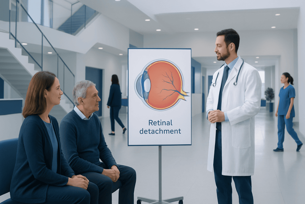
A detached retina requires prompt assessment by an ophthalmologist because delays can increase the risk of lasting vision loss. Treatment may include laser or freezing therapy, pneumatic retinopexy, scleral buckling, vitrectomy, or a combination of procedures.
Key Takeaways
- A detached retina requires prompt assessment by an ophthalmologist because delays can increase the risk of lasting vision loss.
- Treatment may include laser or freezing therapy, pneumatic retinopexy, scleral buckling, vitrectomy, or a combination of procedures.
- Surgery often restores the retina’s position, but visual recovery is variable and may be incomplete, especially when the macula was detached.
- Recovery commonly involves eye drops, activity limits, follow-up visits and, for some procedures, a required head position.
- New flashes, floaters, a shadow or curtain in vision, or sudden loss of sight should be assessed urgently.
Retinal detachment treatment is usually an urgent eye procedure that seals retinal tears and reattaches the retina to help preserve sight. The best approach depends on the type, size and location of the detachment, whether the macula is involved, and the person’s overall eye health.
Overview: how retinal detachment treatment works
Retinal detachment treatment aims to close the break that allowed fluid beneath the retina and to place the retina back against the back wall of the eye. The retina is the light-sensitive layer that sends visual information to the brain. When it separates from its supporting tissue, it cannot function normally and may be permanently damaged without timely care.
Most retinal detachments need a procedure rather than medicines alone. A retina specialist selects treatment according to the type of detachment, the number and location of retinal tears, the amount of retina involved, and whether the macula—the central area needed for detailed vision—has detached. Treatment is often performed urgently, although the exact timing is individualized.
Small retinal tears without detachment may sometimes be sealed in clinic using laser treatment or cryotherapy (controlled freezing). Once a detachment has developed, a surgical approach is usually needed to remove or redirect fluid, support the retina, and create a lasting seal around the tear.
Who may need treatment and how diagnosis guides the plan

An ophthalmologist evaluates suspected retinal detachment promptly. Assessment usually includes a detailed symptom history, vision testing, measurement of eye pressure, and a dilated examination of the retina. If the retina cannot be clearly seen because of bleeding or a dense cataract, an ultrasound scan may help confirm the diagnosis.
Retinal detachment is more likely after a retinal tear, eye injury, previous eye surgery such as cataract surgery, severe short-sightedness, certain inherited eye conditions, or a detachment in the other eye. Diabetes can also lead to a different form of retinal detachment when abnormal scar tissue pulls on the retina.
Not every person with flashes or floaters has a detachment, but these symptoms deserve assessment when they are new or worsening. A clinician also distinguishes retinal detachment from related conditions such as posterior vitreous detachment, which is common with age and may cause flashes and floaters but does not always require treatment.
- Rhegmatogenous detachment: fluid enters through a retinal tear or hole.
- Tractional detachment: scar tissue pulls the retina away, often in advanced diabetic eye disease.
- Exudative detachment: fluid collects beneath the retina without a tear, due to inflammation, vascular problems, or other causes.
Types of retinal detachment surgery: step by step

For a tear without detachment, laser photocoagulation creates small controlled burns around the break, forming a scar that helps secure the retina. Cryotherapy creates the same protective adhesion by applying freezing treatment from outside the eye. These procedures are generally performed with local anesthetic and may be used as part of a larger repair.
Pneumatic retinopexy involves injecting a small gas bubble into the eye. The bubble rises and presses against a suitable retinal tear while laser treatment or cryotherapy seals it. This option is mainly considered for selected tears in the upper part of the retina. The person must follow specific head-positioning instructions so the bubble supports the correct area.
Scleral buckling places a silicone band or small sponge on the outside of the eye. This gently indents the eye wall toward the detached retina, reducing traction and helping the retinal tear close. Vitrectomy removes the gel-like vitreous from inside the eye, addresses traction or scar tissue, and may use a gas bubble or silicone oil to support the retina while it heals. In complex cases, surgeons may combine techniques.
Procedures are usually performed in an operating room with local anesthesia and sedation or, in some cases, general anesthesia. The eye team gives individualized instructions about fasting, regular medicines, transport home and planned follow-up. The appropriate surgical technique is determined by the retina specialist rather than by patient preference alone.
Benefits, risks and what results can be expected
The central benefit of retinal reattachment surgery is the opportunity to preserve remaining vision and prevent further loss. In many cases, the retina can be successfully reattached anatomically. However, an attached retina does not always mean vision returns to its previous level, because vision also depends on how long the retina was detached, whether the macula was affected, and whether there is underlying retinal damage or scar tissue.
Possible risks include infection, bleeding, increased or decreased eye pressure, cataract development or progression after vitrectomy, corneal problems, double vision, recurrent retinal detachment, and the need for further procedures. Serious complications are uncommon but can affect vision. The surgeon discusses the individual balance of benefit and risk before treatment whenever circumstances allow.
Gas bubbles temporarily blur vision and prevent air travel or travel to high altitude until the gas has fully absorbed, because pressure changes can dangerously raise pressure inside the eye. People who have a gas bubble must tell all healthcare professionals, including anesthetists, because nitrous oxide anesthesia must be avoided while gas remains in the eye.
Recovery timeline and aftercare
Recovery after retinal detachment surgery varies by procedure and by the condition of the eye. Mild discomfort, redness, watering, swelling and blurred vision are common initially. The care team may prescribe antibiotic and anti-inflammatory eye drops and provide instructions for protecting the eye, bathing, sleeping, lifting, exercise and return to work.
After pneumatic retinopexy or vitrectomy with a gas bubble, head positioning can be an important part of treatment. The required position and duration are tailored to the tear location. Vision may remain substantially blurred while the gas is present; the bubble gradually becomes smaller and may appear as a moving line or circle in the field of view.
Many people resume light daily activities over days to a few weeks, but full healing and stabilization of vision may take weeks to several months. Follow-up appointments are essential because the retina may need monitoring for recurrent detachment, pressure changes, inflammation or cataract. Patients should not drive until their vision and clinician’s advice make it safe.
Does your eyesight get back to 100% after surgery for retina detachment?
Vision does not always return to 100% after retinal detachment surgery. The operation is designed first to reattach the retina and protect as much vision as possible. Visual improvement may continue gradually for months, but the degree of recovery differs greatly between individuals.
Results are generally more favorable when treatment occurs before the macula detaches. If the macula has been detached, central vision may remain less sharp or appear distorted even after successful repair. Other factors, including the duration and extent of the detachment, repeat detachments, scar tissue, age, cataract and other eye conditions, also influence the outcome.
It is common for vision to fluctuate during early recovery, particularly when a gas bubble is used. The retinal specialist can provide the most relevant outlook after examining the retina and monitoring healing over time.
Is detached retina surgery a big deal?
Detached retina surgery is a significant eye procedure because it treats a sight-threatening condition and requires careful postoperative monitoring. At the same time, these procedures are well-established and are performed by ophthalmic surgeons with specialized retinal training. The urgency reflects the importance of protecting the retina, not necessarily that every procedure is the same in complexity.
The level of surgery varies. Laser treatment for a small tear is often an outpatient procedure, while vitrectomy or scleral buckling is more involved and may require operating-room anesthesia, temporary changes to daily activities, and strict positioning. A person’s surgical plan is based on the anatomy of the detachment and their individual health needs.
At Acibadem International, multidisciplinary specialists in JCI-accredited hospitals assess and treat retinal conditions for international patients, with care plans coordinated around the required procedure and follow-up.
How long does it take to fully recover from a detached retina?
There is no single recovery period for a detached retina. Physical comfort often improves within one to two weeks, while healing inside the eye and visual recovery commonly take several weeks to several months. If silicone oil is used, a later procedure may be needed to remove it in selected cases, which can extend the treatment course.
Recovery may be longer after a large or complex detachment, when the macula was involved, or if scar tissue develops. Some people regain useful vision but continue to notice reduced detail, distortion, reduced peripheral vision or poorer night vision. Regular follow-up allows the surgeon to identify issues that may be treatable during recovery.
Following eye-drop, positioning and activity instructions can support healing. Patients should contact their eye team promptly if pain increases, vision worsens suddenly, new flashes or floaters appear, or a new shadow develops in the field of vision.
How painful is retinal reattachment surgery?
Retinal reattachment surgery is usually not painful during the procedure because anesthesia is used. Depending on the operation and the person’s circumstances, this may be local anesthesia with sedation or general anesthesia. Patients may feel pressure or movement, but they should tell the team if they feel pain or significant discomfort.
Afterward, mild to moderate soreness, grittiness, headache, redness and light sensitivity can occur for several days. These symptoms are often manageable with the aftercare plan and pain relief recommended by the clinical team. Severe or increasing pain is not something to ignore, especially if it occurs with nausea, marked redness, or worsening vision.
Because comfort and recovery differ by procedure, the retina specialist can explain what is typical for the planned operation and which symptoms require urgent contact.
When to seek medical care
New flashes of light, a sudden increase in floaters, a dark curtain or shadow, missing areas of vision, or sudden worsening of sight require urgent same-day eye assessment. These symptoms can occur with retinal tears or detachment, and prompt examination gives the best opportunity to protect vision.
After retinal treatment, urgent advice is needed for worsening vision, increasing pain, persistent nausea or vomiting, increasing redness or discharge, fever, or new flashing lights, floaters or a curtain-like shadow. People should use the emergency contact information provided by their eye team or seek emergency care if they cannot reach them.
Routine eye examinations are also important for people at increased risk, including those with high short-sightedness, previous retinal detachment, a family history of retinal detachment, significant eye trauma or certain diabetic eye complications.
Frequently asked questions
Can retinal detachment be treated without surgery?
A retinal tear that has not yet caused detachment may sometimes be treated with laser therapy or cryotherapy in an outpatient setting. A true retinal detachment usually needs a procedure to reattach the retina. The suitable approach depends on the type and extent of the detachment.
Is retinal detachment treatment an emergency?
Retinal detachment is considered an urgent eye condition because delayed treatment can increase the risk of permanent vision loss. New flashes, many new floaters, a curtain-like shadow, or sudden visual loss should be assessed the same day. The exact urgency and treatment timing are decided by an ophthalmologist after examination.
Can a retinal detachment come back after surgery?
Yes, a retinal detachment can recur, particularly when scar tissue develops or additional retinal breaks occur. Follow-up visits help the surgeon monitor healing and identify complications early. New visual symptoms after surgery should be reported promptly.
Why can’t people fly after some retinal detachment operations?
Air travel is unsafe while a gas bubble remains in the eye because reduced cabin pressure can cause the bubble to expand. This may lead to a dangerous rise in eye pressure and vision loss. The retina specialist will confirm when the gas has absorbed and flying is safe again.
Will a gas bubble affect vision after retinal surgery?
Yes, a gas bubble causes temporary blurred or blocked vision in the treated eye. As it gets smaller, people may notice a shifting line or circular edge in their vision. The bubble is gradually absorbed, but the timeframe varies according to the gas used and the eye’s healing.
What should someone avoid after retinal detachment surgery?
Restrictions vary by procedure, but may include avoiding air travel and high altitude with an intraocular gas bubble, strenuous lifting, vigorous exercise, eye rubbing and driving before vision is safe. Some patients must maintain a prescribed head position. Individual postoperative instructions from the surgical team should take priority.
References
- American Academy of Ophthalmology
- National Eye Institute
- Royal College of Ophthalmologists
- Mayo Clinic
- NHS
This article is for general information only and is not a substitute for professional medical advice. Please consult a qualified doctor about your individual situation.
Explore treatments in Turkey — costs, top hospitals & a free quote
JCI-accredited · board-certified surgeons · reply within 24h