Semilunar Valves vs Valve: Differences Explained

The heart has four valves: two semilunar valves and two atrioventricular valves. The aortic and pulmonary valves are the two semilunar valves.
Key Takeaways
- The heart has four valves: two semilunar valves and two atrioventricular valves.
- The aortic and pulmonary valves are the two semilunar valves.
- Semilunar valves have three pocket-like cusps and do not use chordae tendineae.
- Valve disease may cause narrowing, leakage, or both, sometimes without early symptoms.
- Evaluation may include a physical examination, echocardiogram, and other heart tests.
- Treatment ranges from monitoring and medicines to valve repair or replacement, depending on the valve problem.
Semilunar valves are two of the heart’s four valves: the aortic and pulmonary valves. Compared with atrioventricular valves, they sit at the exits of the heart’s pumping chambers and prevent blood from flowing backward after each heartbeat.
Overview: semilunar valves vs valve
In the comparison of semilunar valves vs valve, “valve” is the broad term for any structure that keeps blood moving in one direction. The heart has four valves. Semilunar valves are a specific pair: the aortic valve and pulmonary valve. They control blood leaving the heart’s two main pumping chambers.
The other two heart valves are called atrioventricular valves. The mitral and tricuspid valves sit between the upper chambers, called atria, and lower chambers, called ventricles. Although all four valves have the same basic purpose, their location, shape, and supporting structures differ.
Healthy valves open and close with each heartbeat in response to pressure changes. This coordinated movement helps the heart deliver oxygen-rich blood to the body and send oxygen-poor blood to the lungs. A valve can become narrowed, leaky, infected, or otherwise damaged, which may affect the heart’s workload over time.
How heart valves work
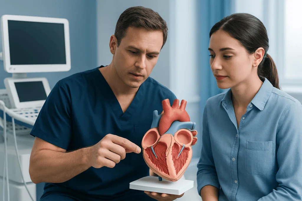
Blood returns from the body to the right atrium, passes through the tricuspid valve into the right ventricle, and then leaves through the pulmonary valve to travel to the lungs. After receiving oxygen in the lungs, blood returns to the left atrium, flows through the mitral valve into the left ventricle, and leaves through the aortic valve to supply the body.
Valves are passive structures: they do not actively contract. Instead, pressure differences on either side cause them to open or close. A valve opens when pressure behind it is greater than pressure ahead of it. It closes when the pressure reverses, helping stop backward flow.
The semilunar valves close shortly after blood is ejected from the ventricles. Their closure contributes to the second heart sound, often described as “dub.” The atrioventricular valves close at the start of ventricular contraction and contribute to the first heart sound, often described as “lub.”
What are the key differences between semilunar valves and atrioventricular valves?

The main difference is location. Semilunar valves are located where blood exits the ventricles: the pulmonary valve lies between the right ventricle and pulmonary artery, while the aortic valve lies between the left ventricle and aorta. Atrioventricular valves are positioned between the atria and ventricles.
They also differ in structure. Semilunar valves usually have three cup-shaped cusps that fill with a small amount of blood to seal the valve when it closes. The mitral valve has two leaflets, and the tricuspid valve generally has three. Atrioventricular valve leaflets are supported by chordae tendineae, thin fibrous cords attached to papillary muscles in the ventricles. Semilunar valves do not have these cords.
Timing is another difference. Atrioventricular valves open while the ventricles relax and fill with blood, then close as the ventricles contract. Semilunar valves open during ventricular contraction to allow blood out of the heart and close during ventricular relaxation to prevent blood returning to the ventricles.
- Semilunar valves: aortic and pulmonary; located at ventricular outflow tracts.
- Atrioventricular valves: mitral and tricuspid; located between atria and ventricles.
- Semilunar structure: pocket-like cusps without chordae tendineae.
- Atrioventricular structure: leaflets supported by chordae tendineae and papillary muscles.
What are the two types of semilunar valves?
The two semilunar valves are the pulmonary valve and the aortic valve. The pulmonary valve directs blood from the right ventricle into the pulmonary artery, which carries blood to the lungs. It prevents blood from returning to the right ventricle after each contraction.
The aortic valve directs oxygen-rich blood from the left ventricle into the aorta, the body’s largest artery. Because the left ventricle pumps at higher pressure than the right ventricle, the aortic valve works under substantial mechanical stress throughout life.
Both valves most often have three cusps. Some people are born with an aortic valve that has two cusps rather than three, known as a bicuspid aortic valve. This congenital difference may increase the likelihood of aortic valve narrowing or leakage over time and should be monitored by a cardiology team.
Is semilunar valve the same as aortic valve?
No. The aortic valve is one type of semilunar valve, but the term semilunar valve includes both the aortic valve and the pulmonary valve. Therefore, all aortic valves are semilunar valves, but not all semilunar valves are aortic valves.
The distinction matters because the two valves serve different circulation pathways. Aortic valve problems affect blood flow from the left side of the heart to the body. Pulmonary valve problems affect blood flow from the right side of the heart to the lungs.
Both types of valve disease can be assessed with an echocardiogram, an ultrasound examination that shows valve structure, movement, and blood flow. Further testing may be advised when a clinician needs more detail about the valve, heart muscle, coronary arteries, or major blood vessels.
Which heart valve is most likely to fail?
The aortic and mitral valves are most commonly affected by clinically significant valve disease in adults. The aortic valve is frequently affected by calcific aortic stenosis, a progressive stiffening and narrowing that becomes more common with age. The mitral valve may be affected by degeneration, prolapse, rheumatic disease, or changes in the heart muscle that prevent proper closure.
“Fail” can mean more than one thing. Stenosis means a valve does not open fully, restricting forward blood flow. Regurgitation, also called insufficiency, means a valve does not seal fully and allows blood to leak backward. Some people have both narrowing and leakage in the same valve.
Pulmonary and tricuspid valve disorders also occur. They may be present from birth, develop after certain heart or lung conditions, or result from infection or prior procedures. The likely cause, severity, symptoms, and effect on heart function are more important than simply identifying which valve is involved.
Diagnosis, treatment options, and recovery
A clinician may suspect valve disease after hearing a heart murmur or reviewing symptoms such as breathlessness, chest discomfort, fatigue, palpitations, dizziness, fainting, or ankle swelling. An echocardiogram is the main test for evaluating valve anatomy and function. Electrocardiography, chest imaging, exercise testing, cardiac CT or MRI, and cardiac catheterization may be used in selected situations.
Not every valve problem requires an immediate procedure. Mild disease without symptoms may be monitored with regular examinations and repeat imaging. Medicines can help manage blood pressure, fluid retention, irregular rhythms, or other related conditions, but they generally do not remove severe mechanical narrowing of a valve.
When a valve problem is severe, causing symptoms, or affecting heart function, repair or replacement may be considered. Options can include surgical valve repair, surgical valve replacement, or catheter-based approaches for suitable patients. The choice depends on the affected valve, anatomy, age, overall health, other heart disease, and personal preferences. A heart team discusses the expected benefits and limitations of each approach.
For a catheter-based procedure, assessment includes imaging and a review of vascular access, heart anatomy, kidney function, medicines, and surgical risk. During the procedure, a specialist guides equipment through a blood vessel or, in some cases, performs surgery through the chest. Recovery varies widely: catheter procedures may involve a short hospital stay and gradual return to usual activity, while open-heart surgery usually requires a longer recovery over several weeks. Potential risks include bleeding, infection, abnormal rhythm, stroke, kidney problems, clotting, leakage around a replacement valve, and the possible need for further treatment; the individual risk profile should be discussed carefully with the treating team.
Prevention, self-care, and when to seek medical care
Many congenital or age-related valve changes cannot be fully prevented. However, controlling blood pressure, avoiding tobacco, staying physically active within a clinician’s advice, following a balanced eating pattern, and managing diabetes and cholesterol support overall heart health. People with known valve disease should attend recommended follow-up visits, even if they feel well.
Good dental hygiene and routine dental care are also important. In certain higher-risk situations, clinicians may recommend antibiotics before selected dental procedures to reduce the risk of infective endocarditis. Antibiotics are not routinely needed for everyone with a heart murmur or valve condition, so individualized medical advice is essential.
Medical assessment is appropriate for new or worsening shortness of breath, reduced ability to exercise, chest pain, fainting, persistent palpitations, unexplained leg swelling, or unusual fatigue. Urgent medical care is needed for severe chest pain, fainting, sudden severe breathlessness, or symptoms suggesting a stroke, such as new facial weakness, arm weakness, speech difficulty, or confusion.
Acibadem International’s multidisciplinary cardiology and cardiac surgery specialists at JCI-accredited hospitals can assess and treat heart valve conditions for international patients. A qualified clinician can explain whether monitoring, medical management, or a valve intervention is appropriate for an individual situation.
Frequently asked questions
What does “semilunar” mean in heart valves?
The term refers to the half-moon or pocket-like shape of the valve cusps. When blood tries to move backward, these pockets fill and come together to close the valve.
How many semilunar valves does the heart have?
The heart has two semilunar valves. They are the aortic valve, which leads to the aorta, and the pulmonary valve, which leads to the pulmonary artery.
Can a person have a semilunar valve problem without symptoms?
Yes. Mild or gradually progressive valve disease may not cause noticeable symptoms at first. Regular examinations and echocardiograms can identify changes before symptoms become prominent.
What is the difference between aortic stenosis and aortic regurgitation?
Aortic stenosis occurs when the aortic valve becomes narrowed and restricts blood leaving the left ventricle. Aortic regurgitation occurs when the valve does not close completely, allowing blood to leak back into the left ventricle.
Can medicines fix a damaged heart valve?
Medicines may reduce symptoms or help manage related conditions such as high blood pressure, fluid buildup, or irregular heart rhythm. They do not usually correct severe structural narrowing or leakage, which may require valve repair or replacement.
When is valve replacement considered?
Valve replacement may be considered when valve disease is severe, produces symptoms, weakens the heart, or creates a significant risk of future complications. The timing and type of procedure are decided after individualized assessment by a cardiology and cardiac surgery team.
References
- American Heart Association
- American College of Cardiology
- European Society of Cardiology
- National Heart, Lung, and Blood Institute
- Mayo Clinic
This article is for general information only and is not a substitute for professional medical advice. Please consult a qualified doctor about your individual situation.
Explore treatments in Turkey — costs, top hospitals & a free quote
JCI-accredited · board-certified surgeons · reply within 24h
Add us as a Preferred Source to see more of our trusted health content across Google Search, AI Overviews and Discover.