Thermography and Breast Cancer Screening: How It Works, Results and What to Expect
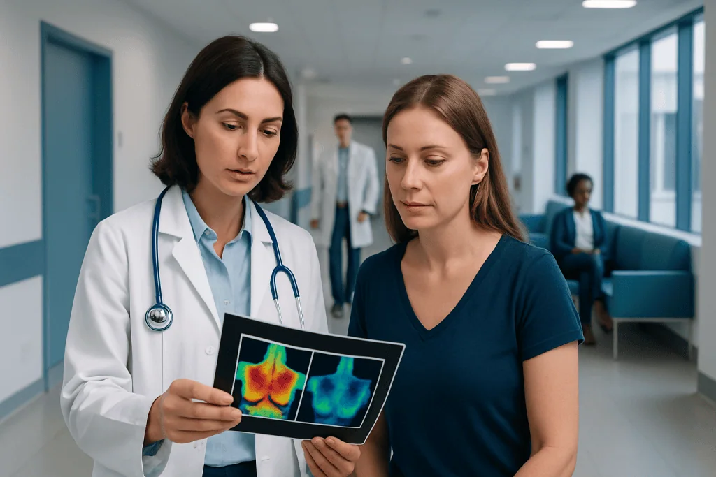
Breast thermography measures heat patterns on the skin; it does not create an image of breast tissue or identify a tumour directly. Thermography is not considered a reliable stand-alone screening or diagnostic test for breast cancer.
Key Takeaways
- Breast thermography measures heat patterns on the skin; it does not create an image of breast tissue or identify a tumour directly.
- Thermography is not considered a reliable stand-alone screening or diagnostic test for breast cancer.
- Mammography remains the main screening test for many people, with ultrasound or MRI used in selected situations.
- An abnormal thermography result requires appropriate medical assessment and standard breast imaging, not self-diagnosis.
- There is no recovery period after thermography, but preparation instructions can affect image quality.
- A new lump, nipple discharge, skin changes or persistent breast symptoms should be assessed promptly, regardless of thermography results.
Thermography and breast cancer screening involves taking infrared images of breast skin temperature patterns. It is noninvasive and radiation-free, but current medical guidance does not support it as a replacement for mammography or other recommended breast cancer screening tests.
Overview: What Thermography Can and Cannot Do
Thermography and breast cancer screening refers to the use of an infrared camera to map temperature differences on the surface of the breasts. The test is painless, does not use radiation and does not compress the breast. However, it is not an accepted substitute for mammography, breast ultrasound, breast MRI or clinical breast assessment.
The scan detects patterns of heat and blood flow near the skin. Some cancers may be associated with increased blood supply and altered temperature patterns, but these changes are not specific to cancer. Normal hormonal variation, inflammation, infection, recent exercise and benign breast conditions may also affect the images.
Health authorities and breast imaging specialists advise that thermography should not be used alone to screen for, diagnose or exclude breast cancer. A normal thermal image cannot reliably rule out cancer, and an abnormal image cannot confirm it. Standard breast cancer treatment planning begins only after appropriate imaging and, when needed, a biopsy has established a diagnosis.
How Breast Thermography Works
During breast thermography, a specialised infrared camera records heat emitted naturally from the skin. Software converts these measurements into colour images, often called thermograms. Warmer and cooler areas appear in different colours, allowing the reader to compare patterns between the two breasts and across different regions.
The technology is based on the idea that some abnormalities may change local circulation or metabolism. Although this principle is biologically plausible, temperature changes alone are not sufficiently specific or sensitive to identify breast cancer consistently. Small, deep or slow-growing cancers may not produce a detectable surface temperature pattern.
By comparison, mammography can detect some changes within breast tissue, including calcifications and masses, before they can be felt. Breast ultrasound can help assess a specific area or finding, while breast MRI may be recommended for selected people at higher risk. These tests answer different clinical questions and are interpreted alongside a person’s symptoms, risk factors and examination findings.
Who May Consider the Test and What Happens During It
Some people seek thermography because they prefer a test without radiation or breast compression. It may also be marketed to people with dense breasts or a family history of breast cancer. These concerns are understandable, but dense breast tissue and increased inherited risk require an individual screening plan from a qualified clinician; thermography does not replace recommended imaging in either situation.
At the appointment, the person usually undresses from the waist up and sits or stands in a temperature-controlled room for a short period to allow the skin temperature to stabilise. A technician takes several images from different positions without touching the breasts. The imaging itself is typically completed within minutes, although the whole visit may take longer.
There is no injection, anaesthesia or compression, and people can return to usual activities immediately. The report may describe symmetry or areas of increased or decreased heat. If a result is described as abnormal or concerning, it should be discussed with a breast specialist, who may recommend clinical examination and conventional breast imaging. Related breast conditions, including breast cancer, can only be properly assessed through evidence-based diagnostic pathways.
What Not to Do Before Breast Thermography?
Preparation instructions vary between providers, but the aim is to avoid temporary changes in skin temperature and circulation. A person should follow the imaging centre’s instructions carefully and tell the provider about pregnancy, breastfeeding, breast surgery, implants, skin conditions, recent injury and any current breast symptoms.
Common instructions may include avoiding strenuous exercise, hot baths, saunas, sun exposure, smoking and caffeine for a specified period before the appointment. People may also be asked not to apply deodorant, lotion, powder, perfume or topical medicines to the chest or underarm area on the day of testing unless their clinician advises otherwise.
It is also important not to postpone a medically recommended mammogram, ultrasound, MRI or specialist consultation in order to have thermography first. If there is a new breast lump or other concerning symptom, timely clinical evaluation is more important than preparation for any optional imaging test.
How Accurate Is Thermography for Detecting Breast Cancer?
Thermography is not accurate enough to be used as a stand-alone test for detecting breast cancer. It may miss cancers, including those that are small, deep within the breast or not associated with measurable surface heat changes. It can also show abnormal patterns in people who do not have cancer.
For these reasons, thermography cannot reliably confirm or rule out breast cancer. A false-negative result may create false reassurance and delay assessment, while a false-positive result may cause anxiety and lead to unnecessary follow-up testing. Major regulatory and professional organisations do not recommend thermography as a replacement for screening mammography.
People should ask their clinician which screening test is appropriate for their age, personal history, breast density and family history. If an imaging finding needs further clarification, a breast team may use targeted ultrasound, diagnostic mammography, MRI or image-guided biopsy. These approaches are designed to evaluate breast tissue directly rather than relying on surface temperature alone.
What Are the Benefits and Disadvantages of Thermography?
The practical benefits of thermography are that it is contact-free, painless and does not expose the person to ionising radiation. There is no recovery time, and the test may feel more comfortable for people who find breast compression difficult. It may be used by some providers as an adjunctive observation tool, but this role remains limited and should not change recommended screening.
The main disadvantages are its limited diagnostic reliability and the risk of misleading results. Thermography does not visualise internal breast structures, does not identify calcifications and cannot determine whether a temperature difference is caused by cancer, infection, hormonal changes or another benign reason.
It can also create additional expense and uncertainty if findings lead to repeat tests. Most importantly, relying on thermography instead of standard screening may delay the detection of breast cancer. A person considering the test should discuss its limitations with a doctor and continue evidence-based screening as recommended.
- Risks: false reassurance, false-positive results, delayed diagnosis and avoidable follow-up investigations.
- Benefit: a noninvasive temperature image, but not a validated replacement for breast cancer screening.
How Much Does Breast Thermography Cost?
The cost of breast thermography varies by country, city, clinic, the type of report provided and whether follow-up imaging is needed. It may not be covered by public or private health insurance because it is not considered a standard breast cancer screening test in many healthcare systems.
Before booking, it is reasonable to ask what is included in the quoted fee, who interprets the images, whether a doctor reviews the findings and what follow-up will be advised if the result is abnormal. It is equally important to consider whether the test would add useful information beyond the screening or diagnostic imaging already recommended by a clinician.
Cost should not be the deciding factor when there are breast symptoms or an elevated cancer risk. A healthcare professional can explain the most appropriate and efficient diagnostic route, which may include a clinical examination and established breast imaging tests.
When to Seek Medical Care
A person should arrange medical assessment promptly for a new breast or underarm lump, persistent focal breast pain, nipple discharge that is bloody or occurs without squeezing, nipple inversion that is new, or skin changes such as dimpling, thickening, redness or scaling. These symptoms are often caused by non-cancerous conditions, but they should be checked rather than monitored through thermography alone.
Medical advice is also appropriate after an abnormal thermography result, even if there are no symptoms. A breast clinician can determine whether diagnostic mammography, ultrasound, MRI or referral to a specialist is needed. A normal thermography result should not be used as reassurance if symptoms are present or routine screening is due.
Acibadem International’s multidisciplinary specialists and JCI-accredited hospitals support international patients with breast assessment, imaging and coordinated care. For people who require evaluation beyond screening, breast biopsy may be recommended when imaging findings need a definite tissue diagnosis.
Frequently asked questions
Can thermography replace a mammogram?
No. Thermography should not replace mammography or other recommended breast imaging tests. It does not reliably detect all breast cancers and cannot rule out cancer when the result is normal.
Is breast thermography painful?
Breast thermography is generally painless because it does not involve needles, radiation or breast compression. The person stands or sits while an infrared camera takes images from a short distance.
Can thermography detect breast cancer early?
Thermography may show surface temperature differences, but it cannot reliably detect early breast cancer. Some early cancers do not cause measurable heat changes, and many non-cancerous conditions can alter temperature patterns.
What happens if a thermography result is abnormal?
An abnormal result should be reviewed by a qualified doctor or breast specialist. Further assessment commonly involves a clinical examination and standard imaging, such as diagnostic mammography or targeted ultrasound, with biopsy only when indicated.
Do people with dense breasts need thermography?
Dense breasts can make mammogram interpretation more complex, but thermography is not the recommended solution. A doctor can advise whether supplemental imaging, such as ultrasound or MRI, is appropriate based on overall risk and local screening guidance.
Is there any recovery after breast thermography?
No recovery period is usually needed. People can resume normal activities immediately after the images are taken, although they should follow the provider’s preparation guidance before the appointment.
References
- U.S. Food and Drug Administration
- American College of Radiology
- American Cancer Society
- World Health Organization
- European Society of Breast Imaging
This article is for general information only and is not a substitute for professional medical advice. Please consult a qualified doctor about your individual situation.
Breast Cancer in Turkey — costs, top hospitals & a free quote
JCI-accredited · board-certified surgeons · reply within 24h
Add us as a Preferred Source to see more of our trusted health content across Google Search, AI Overviews and Discover.