

Visual Field Loss: When Brain or Optic Nerve Imaging Is Needed

Visual field loss can come from the eye, optic nerve, or brain pathways that carry visual information. A detailed eye examination and visual field test often help localize where the problem may be.
Key Takeaways
- Visual field loss can come from the eye, optic nerve, or brain pathways that carry visual information.
- A detailed eye examination and visual field test often help localize where the problem may be.
- MRI is commonly preferred when doctors need to evaluate the optic nerves, optic chiasm, brain, or pituitary region.
- Urgent assessment is important if field loss is sudden, painful, associated with weakness or speech changes, or affects both eyes in a matching pattern.
- Treatment depends on the cause and may involve eye care, neurological care, medicines, procedures, or monitoring.
Visual field loss means part of a person’s side, central, upper, or lower vision is reduced or missing. Imaging of the brain, optic nerves, or eye socket may be needed when the pattern or symptoms suggest a problem beyond the surface of the eye.
Overview
Visual field loss refers to a missing, blurred, dim, or distorted area within the full area a person can see while looking straight ahead. It may affect peripheral vision, central vision, one side of vision, the upper or lower field, or small patchy areas. Some people notice bumping into objects, difficulty reading, or trouble driving, while others discover the problem only during an eye examination.
Vision is created by a connected system. Light enters the eye and is focused on the retina, where visual information is converted into nerve signals. These signals travel through the optic nerve, cross partially at the optic chiasm near the pituitary gland, continue through the brain’s visual pathways, and are processed in the occipital lobes at the back of the brain. A problem at any point in this pathway can produce a recognizable pattern of field loss.
Because visual field loss can arise from common eye conditions such as glaucoma, but also from optic nerve inflammation, stroke, compression, or other neurological conditions, doctors decide on imaging based on the clinical picture. Brain or optic nerve imaging is not required for every person, but it becomes important when the findings suggest that the cause may be behind the eye or within the nervous system.
Symptoms and Patterns Patients May Notice
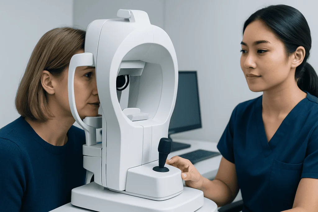
Symptoms can be subtle, especially when only one eye is affected or when the loss develops slowly. The brain may compensate for missing areas, so a person may not realize there is a blind spot until covering one eye, reading, driving, or navigating a busy environment. A formal visual field test can detect and map changes that are difficult to describe.
Common experiences include loss of side vision, a shadow or curtain-like area, missing words while reading, difficulty seeing steps or curbs, or needing to turn the head more often to scan the surroundings. Some people report dimming of vision, reduced color brightness, pain with eye movement, flashes, floaters, headaches, or double vision. These associated symptoms help the doctor decide whether the issue is likely to be retinal, optic nerve-related, or neurological.
The pattern of field loss is especially important. For example, gradual tunnel-like peripheral loss may suggest glaucoma or retinal disease. Loss of the same side of vision in both eyes can point to a problem in the brain’s visual pathways. Loss involving the outer halves of both visual fields may suggest pressure near the optic chiasm, an area close to the pituitary gland. These patterns are not diagnoses by themselves, but they guide the next step.
Common Causes and Risk Factors

Many causes of visual field loss begin in the eye. Glaucoma can damage the optic nerve and often affects peripheral vision before central vision. Retinal conditions, including retinal detachment, retinal vascular blockage, diabetic retinal disease, inherited retinal disorders, or macular disease, may create field defects or central blind spots. Eye examination with pupil dilation, retinal imaging, and optic nerve testing can often identify these causes.
Optic nerve conditions are another important group. Optic neuritis, ischemic optic neuropathy, compression of the optic nerve, trauma, and inflammatory or autoimmune diseases can impair the signal between the eye and brain. These conditions may cause reduced sharpness, decreased color perception, pain with eye movement, swelling or pallor of the optic disc, or a visual field pattern that does not match typical glaucoma.
Brain-related causes include stroke, transient ischemic attack, tumors, pituitary or parasellar masses, aneurysm, demyelinating disease, infection, migraine-related visual symptoms, and increased pressure around the brain. Risk factors vary by cause but may include older age, high blood pressure, diabetes, high cholesterol, smoking, irregular heart rhythm, autoimmune disease, cancer history, severe headaches, recent trauma, or a known neurological condition.
When Brain or Optic Nerve Imaging Is Needed
Imaging is considered when the history, examination, or visual field pattern suggests that the problem may involve the optic nerve behind the eye, the optic chiasm, or the brain. Magnetic resonance imaging, or MRI, is often the preferred test for the optic nerves and brain because it provides detailed soft-tissue images. MRI of the brain and orbits, sometimes with contrast material, may be requested to look for inflammation, compression, demyelination, tumors, or other structural causes.
Doctors may recommend imaging when visual field loss is unexplained after a complete eye examination, progresses despite appropriate eye treatment, affects both eyes in a pattern suggesting the brain pathways, or is associated with optic nerve swelling or unusual optic nerve appearance. Imaging is also commonly considered when there is reduced color vision, pain with eye movement, double vision, drooping eyelid, unequal pupils, new neurological symptoms, or headaches with concerning features.
Situations that may prompt urgent imaging or emergency assessment include:
- Sudden loss of part of the visual field, especially if it affects the same side in both eyes.
- Visual field loss with weakness, numbness, facial droop, speech difficulty, confusion, dizziness, or trouble walking.
- A new severe headache, headache with vision changes, or vision symptoms after head or eye trauma.
- Eye pain with significant vision loss or a swollen optic nerve noted by an eye specialist.
- Field loss with signs suggesting pressure near the optic chiasm, such as bitemporal visual field loss.
Computed tomography, or CT, may be used first in certain urgent situations, such as suspected bleeding, major trauma, or when MRI is not immediately available or not suitable. Additional vascular imaging, blood tests, or neurological evaluation may be needed depending on the suspected cause.
Diagnosis: What to Expect During Evaluation
The evaluation usually begins with a careful history. The doctor asks when the symptom started, whether it is stable or worsening, whether one or both eyes are affected, and whether there are headaches, eye pain, neurological symptoms, trauma, or previous eye disease. It is often useful for patients to describe whether the missing area is central, to one side, above, below, or patchy.
An eye examination may include visual acuity, pupil reactions, eye pressure measurement, eye movement testing, color vision, slit-lamp examination, and dilated examination of the retina and optic nerve. Optical coherence tomography, known as OCT, can measure the retinal nerve fiber layer and macula, helping doctors detect glaucoma, optic nerve damage, or retinal disease. Fundus photography or retinal imaging may document visible changes for comparison over time.
A formal visual field test, often automated perimetry, maps the areas a person sees and misses. The result helps distinguish patterns associated with glaucoma, retina disease, optic nerve disease, or brain pathway problems. Sometimes the test is repeated because fatigue, dry eye, poor fixation, or misunderstanding the instructions can affect reliability.
If imaging is needed, the request is tailored to the suspected location. MRI of the orbits focuses on the optic nerves and eye sockets, while MRI of the brain evaluates the visual pathways, pituitary region, and occipital lobes. Contrast material may be used when inflammation, tumor, infection, or active nerve involvement is suspected, but the decision depends on kidney function, allergies, pregnancy status, and the clinical question.
Treatment Options Depend on the Cause
Treatment is directed at the underlying reason for the visual field loss. In glaucoma, treatment may include prescription eye drops, laser procedures, or surgery to lower eye pressure and reduce the risk of further optic nerve damage. Lost field from glaucoma is often not reversible, so early detection and consistent follow-up are important.
Retinal causes require cause-specific care. Retinal detachment, retinal tears, diabetic eye disease, macular disease, or retinal vascular blockage are managed by ophthalmologists, often with retinal specialists. Treatment may involve laser therapy, injections, surgery, control of diabetes or blood pressure, or close monitoring, depending on the condition and timing.
Optic nerve and brain-related causes may require coordinated care between ophthalmology, neuro-ophthalmology, neurology, neurosurgery, endocrinology, or oncology. For example, optic neuritis may need evaluation for inflammatory or demyelinating disease. A pituitary or other compressive lesion may require monitoring, medication, or surgery. Stroke-related field loss requires urgent medical management and rehabilitation planning to reduce future risk and support adaptation.
Some visual field defects improve when inflammation, swelling, or compression is treated early, while others may remain stable or partially permanent. Low-vision rehabilitation, occupational therapy, driving advice, magnifiers, scanning strategies, and home safety adjustments can help people function better and maintain independence.
Prevention and Self-care
Not all causes of visual field loss can be prevented, but regular eye care can detect many problems before symptoms become obvious. People with glaucoma risk factors, diabetes, high myopia, a family history of eye disease, previous eye injury, or long-term steroid use may need more frequent eye examinations. Those who already have glaucoma or retinal disease should follow their monitoring schedule even when vision seems stable.
General vascular health also supports the eyes and brain. Managing blood pressure, diabetes, cholesterol, sleep apnea, and heart rhythm problems can reduce the risk of vascular events that may affect vision. Avoiding smoking, staying physically active as advised by a doctor, and taking prescribed medicines consistently are practical steps that benefit both eye and neurological health.
Patients can also protect themselves by taking visual symptoms seriously. If a person notices a new missing area in vision, it is helpful to check each eye separately, note the time symptoms began, and seek prompt medical advice. They should not drive if vision is suddenly reduced, if side vision is impaired, or if they feel unsafe judging traffic, pedestrians, or lane position.
When to See a Doctor
A person should arrange an eye examination if they notice persistent blind spots, loss of peripheral vision, difficulty reading because words disappear, or repeated bumping into objects on one side. Even if the symptom is mild, a structured examination can identify whether the cause is in the eye, optic nerve, or brain pathway. Early evaluation is especially important for people with glaucoma, diabetes, high blood pressure, or previous neurological disease.
Emergency care is appropriate for sudden visual field loss, a curtain-like shadow, severe eye pain, new neurological symptoms, a new severe headache, or vision changes after trauma. Sudden loss of the same side of vision in both eyes may indicate a brain pathway problem and should be assessed urgently. Prompt care can help doctors identify treatable causes and reduce the risk of further vision or health complications.
For international patients, Acibadem International provides access to multidisciplinary specialists and JCI-accredited hospitals where eye, neurology, radiology, and related teams can evaluate and treat visual field loss when advanced testing is needed. The right pathway depends on the individual’s symptoms, examination findings, and medical history.
Frequently asked questions
Does visual field loss always mean a brain problem?
No. Many cases are caused by eye conditions such as glaucoma, retinal disease, or optic nerve problems visible during an eye examination. Brain imaging is considered when the pattern, symptoms, or examination findings suggest the visual pathways behind the eye may be involved.
What type of imaging is most often used for optic nerve or brain causes?
MRI is commonly preferred because it shows the optic nerves, eye sockets, brain, and pituitary region in detail. Contrast may be used in some cases to help identify inflammation, tumors, infection, or active nerve changes. CT may be used first in urgent situations such as trauma or suspected bleeding.
Can a visual field test show where the problem is located?
A visual field test can provide strong clues by mapping the missing areas of vision. Certain patterns are more typical of glaucoma, retinal disease, optic nerve disease, or brain pathway involvement. The result is interpreted together with the eye examination and medical history.
Is visual field loss reversible?
It depends on the cause, severity, and how quickly treatment begins. Some causes, such as inflammation or certain compressive problems, may improve with timely care. Other causes, including advanced glaucoma or completed stroke, may leave lasting field loss, but rehabilitation strategies can help patients adapt.
When is visual field loss an emergency?
Sudden field loss should be treated as urgent, especially if it occurs with weakness, numbness, speech difficulty, confusion, severe headache, eye pain, or trauma. A curtain-like shadow or sudden loss of the same side of vision in both eyes also needs prompt medical assessment.
Can migraines cause temporary visual field symptoms?
Migraine aura can cause temporary visual changes such as shimmering lights, zigzag lines, or blind spots, usually resolving within a short period. However, new, unusual, prolonged, or one-sided visual symptoms should be assessed by a doctor to rule out other causes. People should not assume a first episode is migraine without medical advice.
References
- American Academy of Ophthalmology
- National Eye Institute
- American Association for Pediatric Ophthalmology and Strabismus
- National Institute for Health and Care Excellence
- European Glaucoma Society
This article is for general information only and is not a substitute for professional medical advice. Please consult a qualified doctor about your individual situation.
Explore treatments in Turkey — costs, top hospitals & a free quote
JCI-accredited · board-certified surgeons · reply within 24h
Check your numbers in seconds
BMI, calories, due date, blood pressure and 30+ more clinical calculators — free, instant, doctor-reviewed ranges.

More from the Health Library



Related Specialists

Assoc. Prof. Dr. Didem Öncel Yakar
General Surgery
Dr. Senem Almaç Deniz
Otorhinolaryngology
Dyt. Beyza Erdoğan Aksan
Nutrition & Diet
Dr. Alptekin Arifoğlu
Radiation Oncology