White Spot on Color of Eye: An Evidence-Based Guide for Patients

A white spot seen on the colored part of the eye is commonly located on the cornea rather than inside the iris. Possible causes include corneal abrasion, corneal ulcer, inflammation, scarring, calcium deposits, or a foreign body.
Key Takeaways
- A white spot seen on the colored part of the eye is commonly located on the cornea rather than inside the iris.
- Possible causes include corneal abrasion, corneal ulcer, inflammation, scarring, calcium deposits, or a foreign body.
- Pain, redness, discharge, light sensitivity, or reduced vision need prompt medical evaluation.
- Contact lens use, eye trauma, dry eye, and infection can raise the risk of serious corneal problems.
- Treatment depends on the cause and may include lubricating drops, medicines, removing a foreign body, or urgent specialist care.
A white spot on color of eye is often not actually on the iris itself but on the clear cornea in front of it, making the spot appear to sit on the colored part of the eye. Causes range from minor surface changes to infections, inflammation, injury, or scarring, so symptoms such as pain, redness, light sensitivity, or blurred vision should be assessed by an eye doctor.
Overview: what a white spot on the color of the eye usually means
A white spot on color of eye often worries patients because it seems to sit directly on the iris, the colored ring of the eye. In many cases, however, the spot is actually on the cornea, the clear dome-shaped surface at the front of the eye. Because the cornea lies over the iris, any cloudy, white, or gray mark on it can look like it is on the colored part.
Some causes are relatively mild, such as a tiny scratch, dryness-related surface damage, or an old scar. Others need faster attention, especially if the white area is new and comes with pain, redness, tearing, sensitivity to light, discharge, or blurred vision. A white corneal spot may sometimes reflect infection or inflammation, which can threaten sight if treatment is delayed.
Less commonly, the spot may relate to changes deeper in the eye or to a reflection or small deposit on the eye surface. Because different problems can look similar without magnification, the safest approach is a professional eye examination rather than self-diagnosis.
How the spot may look and the symptoms that can come with it
The appearance of a white spot can vary. It may look like a tiny dot, a cloudy patch, a sharply defined speck, or a larger opaque area. Some spots are bright white, while others appear grayish or slightly yellow. A patient may notice it in the mirror, or another person may point it out first.
Symptoms depend on the cause. A harmless old scar may cause little or no discomfort. By contrast, an active corneal problem often causes irritation or pain. Vision may remain normal if the spot is off to one side, but if it lies over the visual axis, even a small lesion can blur sight.
- Eye redness
- Pain, burning, or a feeling that something is in the eye
- Watering or discharge
- Sensitivity to light
- Blurred or hazy vision
- Difficulty keeping the eye open
- Swelling of the eyelids
If the spot appeared suddenly after contact lens wear, an eye injury, or exposure to dust or plant material, urgent assessment is especially important. These details help doctors narrow the cause quickly.
Common causes and risk factors

One of the most important causes is a corneal ulcer, an open sore on the cornea often linked to infection. Bacteria, viruses, fungi, and parasites can all affect the cornea, particularly in contact lens wearers or after an eye injury. A painful white spot with redness and reduced vision can suggest this type of problem and should be treated as urgent.
A corneal abrasion, or scratch on the eye surface, can also create a pale area while healing. In some cases, an ulcer or abrasion later leaves a scar. Dry eye disease may damage the corneal surface and contribute to small defects, while inflammation from underlying eye disease can produce cloudy spots. A trapped foreign body, such as metal, sand, or plant material, can cause a visible mark and trigger surrounding inflammation.
Other possibilities include calcium or lipid deposits in the cornea, congenital corneal changes, and less common inflammatory or autoimmune disorders. Some patients describing a “white spot on the iris” may actually have a lesion involving the front structures of the eye that needs specialist evaluation to distinguish it from surface disease.
- Contact lens wear, especially overnight use or poor lens hygiene
- Recent eye trauma or rubbing the eye after debris exposure
- Dry eyes or eyelid disorders
- Previous eye infection or surgery
- Immune system problems or inflammatory disease
- Exposure to chemicals, plant matter, or contaminated water
Because several of these causes overlap, clinicians may also evaluate related conditions such as corneal disease or urgent eye infections when symptoms suggest a more serious process.
How doctors diagnose the cause
Diagnosis begins with a careful history. The clinician asks when the spot appeared, whether there is pain or blurred vision, and whether the patient uses contact lenses, had recent trauma, or has a history of eye disease. Even details like swimming with lenses, using old eye makeup, or recent viral illness can be relevant.
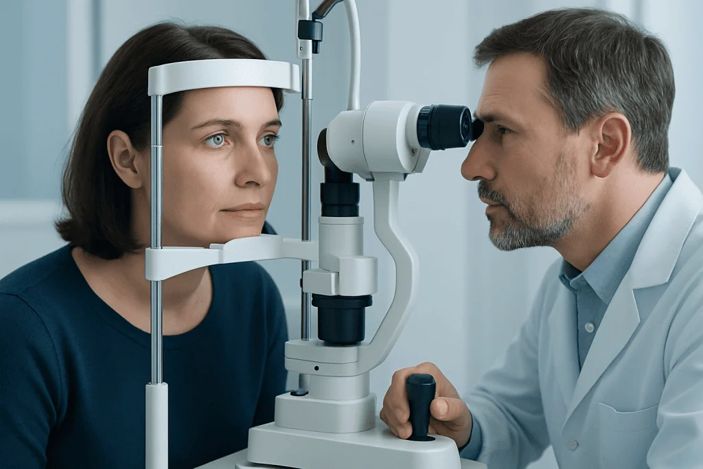
The eye is then examined under magnification, often with a slit lamp. This allows the doctor to determine whether the white area is on the cornea, within deeper structures, or due to surface debris. Fluorescein dye may be placed in the eye to highlight scratches, ulcers, and other defects on the corneal surface.
In selected cases, the doctor may measure vision, eye pressure, and tear film quality. If infection is suspected, a corneal sample may be taken for laboratory analysis, especially in severe or unusual ulcers. Imaging is not always needed, but specialist assessment is important when the diagnosis is uncertain or the lesion involves sight-threatening structures.
Patients should avoid wearing contact lenses and avoid using leftover prescription eye drops before assessment unless a doctor has advised them to do so. Some medicines, especially steroid drops, can worsen certain infections if used without proper diagnosis.
Treatment options depend on the underlying problem
Treatment is guided by the cause rather than by the white color alone. A simple superficial abrasion may improve with protective measures, lubricating drops, and close follow-up. A foreign body may need careful removal in clinic. If the eye is very dry, treatment may focus on restoring the tear surface and reducing irritation.
When infection is suspected, treatment may include prescription antibiotic, antiviral, or antifungal medication, sometimes quite urgently. Corneal ulcers should not be treated casually because delayed care can lead to scarring or lasting vision loss. If inflammation is involved, the doctor may use anti-inflammatory treatment, but only after infection has been excluded or addressed appropriately.
When a white spot represents a scar, treatment depends on whether it affects vision or comfort. Some scars can simply be monitored. More significant corneal damage may need specialist care, including procedures aimed at restoring the cornea. In selected cases, doctors may discuss options used in advanced corneal transplant care or more comprehensive eye treatment for persistent visual problems.
If the white area is related to a broader eye condition, management may involve ophthalmology evaluation by cornea or anterior segment specialists. Treatment plans are individualized based on the patient’s age, symptoms, examination findings, and visual needs.
Self-care, prevention, and what not to do
Good eye-surface habits can lower the risk of corneal irritation and infection. Contact lens users should wash hands before handling lenses, replace lenses and storage cases as advised, and never sleep in lenses unless specifically approved by an eye care professional. Lenses should not be exposed to tap water, hot tubs, or swimming pools unless proper precautions are used.
Protective eyewear is also important during gardening, construction, sports, and tasks involving metal, chemicals, or dust. Small injuries may seem minor at first, but they can introduce infection or create scratches that later become painful. Patients with dry eyes should follow their clinician’s recommendations to keep the eye surface healthy.
- Do not try to scrape or wipe off the spot
- Do not wear contact lenses until cleared by a doctor
- Do not use someone else’s eye drops
- Do not start steroid eye drops without medical advice
- Do not ignore a painful or rapidly appearing white spot
If symptoms are mild, preservative-free artificial tears may help comfort while waiting for medical advice, but they do not replace diagnosis. At the end of care planning, some international patients may choose centers such as Acibadem International, where multidisciplinary specialists in JCI-accredited hospitals evaluate and treat eye conditions.
When to seek medical care
A white spot on the colored part of the eye should be assessed soon if it is new, unexplained, or associated with symptoms. Same-day or urgent care is appropriate when there is pain, marked redness, light sensitivity, discharge, sudden vision change, or a history of contact lens wear. These features can point to corneal ulceration or another condition that should not wait.
Medical care is also important after any eye injury, chemical splash, or possible foreign body exposure. Even if the spot seems small, a lesion in the center of the cornea can affect vision significantly. Children, older adults, and people with reduced immunity may need earlier assessment because eye infections can progress more quickly or be harder to detect.
If the spot has been present for a long time without symptoms, it is still reasonable to mention it during a routine eye examination. A clinician can determine whether it is a stable scar or something that needs follow-up. Prompt evaluation generally leads to simpler treatment and better protection of vision.
Frequently asked questions
Is a white spot on the color of the eye always serious?
Not always. Some white spots are old corneal scars or minor surface changes, but others can signal infection, inflammation, or injury. Because it is difficult to tell the difference at home, a new or symptomatic spot should be evaluated.
Can a white spot be on the iris itself?
Sometimes patients describe it that way, but many white spots are actually on the cornea in front of the iris. The cornea is transparent, so a cloudy lesion there can appear to sit on the colored part of the eye. An eye exam helps locate it accurately.
What if the white spot does not hurt?
A painless spot can still deserve attention, especially if it is new or affecting vision. Some scars and deposits cause little discomfort, while some serious eye problems may not be very painful at first. If the cause is unclear, an eye doctor should assess it.
Should contact lenses be removed if a white spot appears?
Yes. Contact lenses should usually be removed right away and not worn again until an eye professional says it is safe. Continued lens wear can worsen irritation and increase the risk of infection.
Can I use over-the-counter eye drops first?
Lubricating artificial tears may help with comfort, but they do not treat infections, ulcers, or embedded foreign material. Patients should avoid using steroid eye drops unless specifically prescribed, because they can make some conditions worse. If there is pain, redness, or blurred vision, medical assessment is more important than self-treatment.
Will the white spot go away on its own?
It depends on the cause. A small abrasion-related change may heal, while a scar or deposit may remain. Infectious or inflammatory causes usually need directed treatment rather than waiting.
References
- American Academy of Ophthalmology
- National Eye Institute
- American Optometric Association
- Mayo Clinic
- Merck Manual
This article is for general information only and is not a substitute for professional medical advice. Please consult a qualified doctor about your individual situation.
Explore treatments in Turkey — costs, top hospitals & a free quote
JCI-accredited · board-certified surgeons · reply within 24h