Osteochondroma: Symptoms, Causes, and Treatment Options
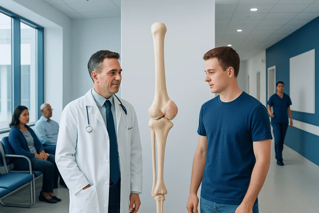
Osteochondroma is usually a noncancerous bone growth that develops near growth plates. Many osteochondromas cause no symptoms and are found incidentally on imaging.
Key Takeaways
- Osteochondroma is usually a noncancerous bone growth that develops near growth plates.
- Many osteochondromas cause no symptoms and are found incidentally on imaging.
- Symptoms can include a visible lump, pain with activity, limited joint motion, or pressure on nerves or blood vessels.
- Diagnosis typically relies on physical examination and imaging such as X-ray, with MRI used in selected cases.
- Treatment may be observation or surgery, depending on symptoms, location, and concern for complications.
- Medical review is important if the mass grows after puberty, becomes painful, or causes numbness, weakness, or circulation changes.
Osteochondroma is the most common benign bone tumor. It usually appears as a cartilage-capped bony growth near the ends of long bones and often causes no symptoms, but treatment may be needed if it causes pain, irritation, nerve or blood vessel compression, or continued growth after skeletal maturity.
What is osteochondroma?
Osteochondroma is a usually benign, or noncancerous, bone growth that forms on the outer surface of a bone. It is made of bone covered by a cap of cartilage and most often develops near the growth plates of long bones, especially around the knee, shoulder, or pelvis. In many people, it is discovered because of a firm lump or by chance on an X-ray done for another reason.
This condition often begins during childhood or adolescence while the skeleton is still growing. A person may have a single osteochondroma, which is more common, or multiple osteochondromas as part of an inherited condition called hereditary multiple exostoses. Most lesions stop growing when the bones finish maturing.
Although osteochondroma is not usually dangerous, its location matters. A growth that presses on a tendon, muscle, nerve, blood vessel, or joint can cause symptoms and may need treatment. Understanding whether a lesion is stable and harmless or causing mechanical problems is an important part of care.
How osteochondroma affects the body

An osteochondroma grows outward from the parent bone and remains connected to it. It can be broad-based, called sessile, or attached by a stalk, called pedunculated. Because it arises near the growth plate, it tends to appear near the ends of long bones, particularly the femur, tibia, and humerus, though it can also occur in the pelvis, shoulder blade, or other bones.
Some osteochondromas do not interfere with nearby structures at all. Others can create a rubbing effect on muscles or tendons, limit normal joint movement, or cause bursitis, which is irritation of a fluid-filled sac over the bony prominence. In less common situations, pressure on a nerve may lead to tingling or weakness, and pressure on a blood vessel may affect circulation.
Doctors also consider the small possibility of change over time. In adults, new growth, increasing pain without a clear reason, or changes in the cartilage cap may prompt closer assessment. This does not mean cancer is likely, but it is a reason for timely evaluation by an orthopedic specialist.
Symptoms and possible complications

Many people with osteochondroma have no symptoms. When symptoms are present, the most common sign is a hard, painless lump near a joint. Some people notice discomfort only when the area is bumped, during sports, or after repeated movement that causes friction over the growth.
Symptoms vary depending on size and location. A lesion near the knee may cause discomfort with bending, kneeling, or exercise. One near the shoulder blade may create snapping or restricted shoulder motion. Pelvic lesions may be harder to notice physically but may still cause local pain or mechanical symptoms.
Possible symptoms and complications include:
- A visible or palpable bony lump
- Pain with activity or pressure
- Reduced range of motion in a nearby joint
- Tendon or muscle irritation
- Bursitis over the lesion
- Numbness, tingling, or weakness if a nerve is compressed
- Swelling, circulation changes, or coldness if a blood vessel is affected
- Differences in limb growth or alignment, more often with multiple lesions
In children with multiple osteochondromas, bone growth can be affected more noticeably. This may lead to limb length differences, bowing, or joint alignment problems that need orthopedic monitoring. If symptoms overlap with other bone conditions, specialists may also consider possibilities such as bone cancer during evaluation, especially when imaging shows atypical features.
Causes and risk factors
Osteochondroma develops from abnormal growth near the growth plate, but it is not caused by anything a person did or did not do. In most cases, the exact reason a single osteochondroma forms is not fully understood. It is generally considered a developmental bone lesion rather than an overuse injury.
A major risk factor for multiple osteochondromas is hereditary multiple exostoses, an inherited condition linked to changes in genes involved in bone growth, especially the EXT genes. People with this condition may develop several lesions and are more likely to need long-term follow-up because of skeletal deformities or other complications.
Age is another important factor. Osteochondromas usually appear in childhood, adolescence, or the teenage years and tend to stop enlarging once growth plates close. A mass that begins growing for the first time in adulthood or one that clearly increases in size after skeletal maturity deserves medical assessment.
There are no well-established lifestyle causes, and routine exercise does not create osteochondroma. However, physical activity can make an existing lesion more noticeable if it rubs against nearby tissues. Family history, young age at presentation, and having multiple lesions are the main clinical clues that guide doctors.
How osteochondroma is diagnosed
Diagnosis begins with a medical history and physical examination. A doctor asks when the lump was first noticed, whether it has changed over time, and whether there is pain, limitation of motion, numbness, or weakness. The exam helps determine if the mass feels fixed to the bone and whether nearby joints, nerves, or blood vessels are affected.
Plain X-rays are usually the first and most informative imaging test. They often show a characteristic bony outgrowth that is continuous with the underlying bone. When the location is complex or symptoms suggest irritation of soft tissues, more detailed imaging may be recommended through MRI scanning or CT scanning. MRI is especially helpful for evaluating the cartilage cap and nearby nerves, muscles, or vessels.
Not every osteochondroma needs a biopsy. If the imaging appearance is typical and the lesion is stable, biopsy is often unnecessary. However, if the findings are unusual, the lesion is growing after skeletal maturity, or there is concern about change in the cartilage cap, a specialist may advise further evaluation and, in selected cases, tissue sampling.
Assessment is often led by orthopedic teams experienced in bone lesions, including orthopedic oncology when the diagnosis is uncertain or the lesion is in a sensitive location. The goal is to confirm that the growth is benign, identify complications, and plan observation or treatment appropriately.
Treatment options and follow-up
Treatment depends on symptoms, location, age, and imaging findings. If an osteochondroma is small, painless, and not causing functional problems, careful observation is often all that is needed. Follow-up may include periodic clinical review and imaging to confirm stability, especially in children who are still growing.
Surgery is considered when the lesion causes pain, repeated irritation, restricted movement, cosmetic concerns that significantly affect quality of life, or compression of a nerve or blood vessel. It may also be recommended if the lesion continues to grow after skeletal maturity or if imaging raises concern for malignant transformation, which is uncommon but important to assess.
The operation generally involves removing the bony growth along with its cartilage cap. The exact technique depends on the site and surrounding anatomy. For example, a lesion near a joint or major neurovascular structures may require more detailed planning, and recovery time varies with location and the extent of surgery.
After treatment, many patients do well, especially when the lesion is completely removed. Follow-up focuses on wound healing, return of movement, and monitoring for recurrence, which is uncommon when excision is complete. In patients with hereditary multiple exostoses, long-term orthopedic follow-up may be needed because new or changing lesions can develop while growth continues.
Prevention and self-care
There is no known way to prevent a single osteochondroma from forming. Because it usually arises from bone growth processes rather than lifestyle factors, self-care is aimed at symptom management rather than prevention of the lesion itself.
For mild symptoms, practical steps may help reduce irritation. These include modifying activities that repeatedly rub or press on the area, using protective padding when appropriate, and allowing time to rest after activities that trigger discomfort. If pain persists, a doctor can advise on suitable pain-relief measures and whether imaging is needed.
People with hereditary multiple exostoses may benefit from regular monitoring during growth years. Keeping track of changes in limb alignment, joint motion, or new lumps can make it easier to identify problems early. Structured follow-up also helps distinguish expected growth from signs that need closer evaluation.
If surgery is recommended, preoperative planning and rehabilitation are part of self-care as well. Understanding the procedure, expected recovery, and safe return to school, work, or sports can help patients feel prepared and supported throughout treatment.
When to seek medical care
Medical review is advisable for any persistent hard lump on or near a bone, especially in a child or teenager. Even though osteochondroma is usually benign, a proper examination and imaging are important to confirm the diagnosis and rule out other causes of a mass.
A person should seek prompt care if the lump becomes painful, seems to enlarge after puberty or adulthood, limits joint movement, or causes numbness, tingling, weakness, swelling, or circulation changes in the limb. These symptoms can mean the lesion is irritating nearby structures and may need treatment.
Specialist evaluation is also important for people with multiple bony lumps or a family history suggestive of hereditary multiple exostoses. Multidisciplinary assessment can help guide imaging, follow-up, and surgery if needed. Acibadem International’s multidisciplinary specialists and JCI-accredited hospitals diagnose and treat bone conditions for international patients.
Frequently asked questions
Is osteochondroma cancer?
Osteochondroma is usually a benign bone growth, not cancer. Most cases remain noncancerous throughout life, but doctors may investigate further if a lesion grows after skeletal maturity, becomes newly painful, or shows unusual imaging features.
Can osteochondroma go away on its own?
Osteochondroma does not usually disappear on its own. However, many lesions stop growing once the bones finish maturing and may never need treatment if they do not cause symptoms.
Does every osteochondroma need surgery?
No. Many osteochondromas can be safely observed if they are painless and not affecting movement, nerves, or blood vessels. Surgery is usually reserved for symptomatic lesions or those with concerning changes.
What is hereditary multiple exostoses?
Hereditary multiple exostoses is an inherited condition in which a person develops multiple osteochondromas. It can affect bone growth and alignment, so regular orthopedic follow-up is often recommended during childhood and adolescence.
How is osteochondroma different from other bone tumors?
Osteochondroma has a characteristic appearance as a cartilage-capped bony outgrowth connected to the parent bone. Imaging, especially X-ray and sometimes MRI, helps doctors distinguish it from other benign or malignant bone lesions.
Can a child with osteochondroma play sports?
Many children with osteochondroma can stay active, especially if the lesion is not painful or interfering with motion. A doctor may suggest activity modification if the growth is being repeatedly hit, rubbed, or causing symptoms during sports.
References
- American Academy of Orthopaedic Surgeons
- National Organization for Rare Disorders
- National Cancer Institute
- OrthoInfo
- MedlinePlus
This article is for general information only and is not a substitute for professional medical advice. Please consult a qualified doctor about your individual situation.
Joint and spine care in Turkey — expert assessment & treatment
JCI-accredited · board-certified surgeons · reply within 24h