Scans — Explained by Medical Evidence, Not Myths
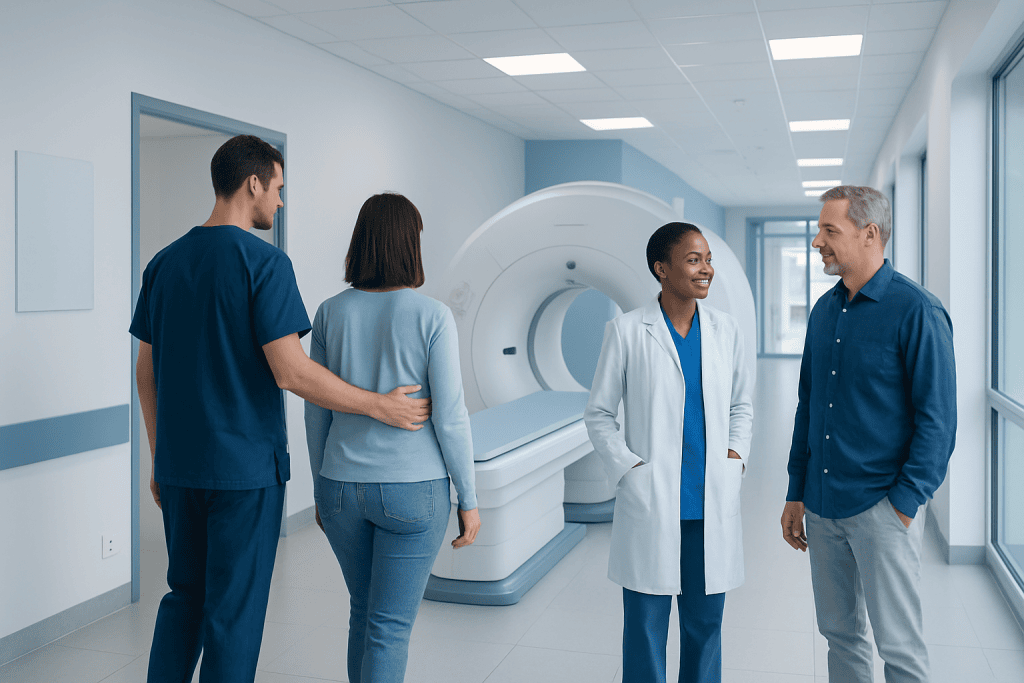
Scans include X-rays, ultrasound, CT, MRI, PET, and nuclear medicine, and each serves a different purpose. Not all scans use radiation; ultrasound and MRI do not, while X-rays and CT scans do.
Key Takeaways
- Scans include X-rays, ultrasound, CT, MRI, PET, and nuclear medicine, and each serves a different purpose.
- Not all scans use radiation; ultrasound and MRI do not, while X-rays and CT scans do.
- Doctors choose a scan based on symptoms, urgency, the body area involved, and the level of detail needed.
- Preparation may include fasting, removing metal items, or checking kidney function before contrast is used.
- Scan results often need to be interpreted alongside symptoms, examination findings, and lab tests.
Scans are medical imaging tests that help doctors look inside the body to diagnose symptoms, guide treatment, or monitor recovery. Different scans use different technologies, so the best choice depends on the health question being answered, the body part involved, and safety considerations such as radiation or contrast use.
What scans are and why doctors use them
Scans are tests that create pictures of structures inside the body. They are used to investigate symptoms, confirm or rule out a diagnosis, check for injury or disease, guide procedures, and monitor how a condition changes over time. In everyday use, the word “scan” often refers to many different imaging tests rather than one single exam.
Medical evidence does not support the common myth that one scan is always “best.” The most useful scan depends on the clinical question. For example, a chest X-ray may be enough to look for pneumonia, while an MRI may be more informative for soft tissues such as the brain, spinal cord, ligaments, or muscles. A CT scan is often valuable when fast, detailed imaging is needed, especially in emergencies.
Scans do not replace a doctor’s assessment. Imaging works best when combined with a person’s symptoms, medical history, physical examination, and sometimes blood tests or other investigations. A scan can reveal important findings, but it may also show incidental changes that are harmless and unrelated to the main symptom.
Main types of scans and how they differ
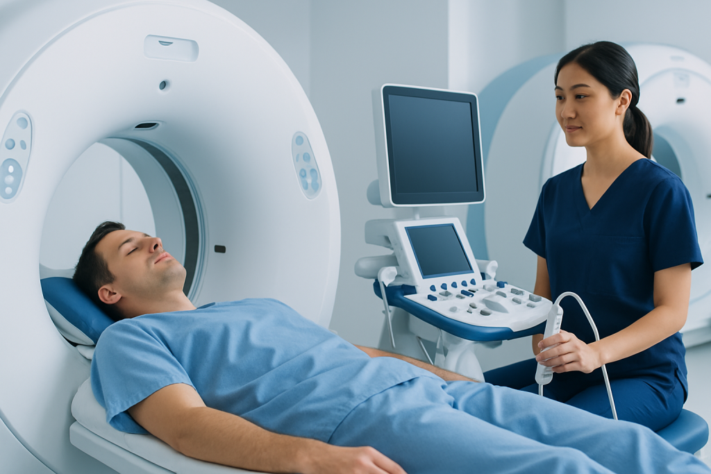
Different scans use different forms of technology to create images. Understanding the basics can make results and recommendations easier to follow. Some tests are quick and simple, while others give more detail but require more preparation.
Common types include:
- X-ray: Uses a small amount of radiation to image bones and some organs, especially the chest.
- Ultrasound: Uses sound waves, not radiation. It is often used in pregnancy, abdominal problems, blood vessels, and soft tissue assessment.
- CT scan: Uses X-rays taken from multiple angles to create detailed cross-sectional images. It is often used for trauma, chest and abdominal conditions, and some cancers.
- MRI scan: Uses a strong magnet and radio waves, not radiation. It is useful for the brain, spine, joints, and many soft tissues.
- PET scan: Uses a small amount of radioactive tracer to show metabolic activity and is often combined with CT in cancer assessment.
- Nuclear medicine scans: Use small amounts of radioactive material to assess how organs or tissues function.
There is no universal ranking among these tests. A simpler scan may be the right first step, while a more advanced scan may be reserved for cases where extra detail changes treatment decisions. In some situations, a doctor may recommend a staged approach, starting with an ultrasound or X-ray and moving to CT or MRI only if needed.
How doctors decide which scan is right

The choice of scan is guided by the person’s age, symptoms, medical history, urgency, and the part of the body being examined. Doctors also consider whether the goal is to detect a problem, define its extent, guide a procedure, or monitor treatment. For instance, severe headache with neurological symptoms may call for urgent brain imaging, while persistent joint pain may be better evaluated with MRI after an examination.
Safety matters too. If a person is pregnant, has kidney disease, a pacemaker, severe claustrophobia, or an allergy history related to contrast material, the imaging plan may need adjustment. Doctors try to use the lowest-risk test that still answers the clinical question accurately.
Imaging is also used differently in screening, diagnosis, and follow-up. Screening scans are offered to people without symptoms in selected situations where medical evidence shows benefit. Diagnostic scans are done to investigate a symptom or abnormal test. Follow-up scans may help track recovery after injury, assess response to treatment, or monitor known conditions such as cancer or stroke.
What to expect before, during, and after a scan
Preparation depends on the type of scan. Some tests require no preparation at all. Others may involve fasting for a few hours, drinking water to fill the bladder, avoiding metal objects, or having blood tests to check kidney function before contrast dye is given. The radiology team usually provides instructions in advance.
During the scan, the person may need to lie still on a table while the machine takes images. Some scans are completed in minutes, while MRI and certain nuclear medicine tests may take longer. Contrast material may be given by mouth, injection, or rarely through other routes to improve image detail. This is common in some CT scans and MRI examinations.
After the scan, most people can return to normal activities soon afterward. If sedation was used, someone may need to accompany the patient home. Results are usually reviewed by a radiologist, who sends a report to the referring doctor. It is common for the final interpretation to take longer than the scan itself, especially when images need detailed review or comparison with older tests.
Safety, radiation, contrast, and common myths
A frequent concern is whether scans are safe. In general, medically indicated imaging is considered safe when used appropriately. However, different scans have different considerations. X-rays, CT scans, PET scans, and many nuclear medicine tests involve radiation. Ultrasound and MRI do not use ionizing radiation.
Radiation exposure from a medically necessary test is usually kept as low as reasonably achievable. Doctors do not request radiation-based scans lightly, especially for children and younger adults, but they may be important when the benefit of diagnosis outweighs the small potential risk. Repeated imaging may require extra thought, so it helps to keep a record of recent scans and share it with the care team.
Contrast agents can improve image quality but may not be suitable for everyone. Some people need special caution because of kidney problems, prior contrast reactions, thyroid conditions, or implanted medical devices. MRI is not harmful because of “radiation,” but it may not be appropriate for some metal implants or devices unless they are confirmed as MRI-compatible. Ultrasound, including ultrasound imaging, is widely used because it is radiation-free and portable, but it may not visualize every structure clearly.
Another common myth is that scans always provide a definite answer. In reality, imaging can be normal despite symptoms, or it may show changes that need further interpretation. That is why scan findings should always be discussed with a qualified clinician who can place them in the full medical context.
What scan results can show—and what they cannot
Scan results may identify inflammation, infection, fractures, tumors, bleeding, blocked blood vessels, organ enlargement, fluid collections, structural abnormalities, or signs of wear and tear. They can also help guide biopsies, drainages, and treatment planning. In many cases, imaging is essential to reach a diagnosis quickly and accurately.
Still, a scan is a snapshot, not a complete health verdict. Some conditions are diagnosed mainly by symptoms, physical examination, or laboratory tests, with imaging used only to support the assessment. In other cases, findings may be “non-specific,” meaning they do not point to one single cause. Follow-up imaging or further tests may then be needed.
It is also common for scans to detect incidental findings. These are unexpected changes unrelated to the original reason for the test. Many incidental findings are harmless, but some need follow-up. This can understandably cause worry, so patients benefit from asking what a finding means, whether it is urgent, and whether any repeat imaging is recommended.
When to seek medical care
Medical care should be sought promptly if symptoms are severe, sudden, or worsening. This includes chest pain, shortness of breath, signs of stroke such as facial drooping or difficulty speaking, major injury, severe headache with neurological symptoms, heavy bleeding, or significant abdominal pain. In these situations, imaging may be needed urgently, but the priority is rapid clinical assessment.
Non-urgent review is appropriate for ongoing symptoms such as persistent pain, unexplained weight loss, repeated headaches, a new lump, prolonged cough, or changes in bowel or bladder habits. A doctor can decide whether a scan is needed and which one is most appropriate. Requesting a scan without clinical guidance may lead to unnecessary testing or results that are difficult to interpret.
Near the end of the care pathway, imaging may also help direct specialist treatment. For example, detailed scans can inform decisions around brain tumor surgery or other procedures. Acibadem International’s multidisciplinary specialists and JCI-accredited hospitals diagnose and treat conditions that may require imaging-based evaluation for international patients.
Frequently asked questions
Do all scans use radiation?
No. X-rays, CT scans, PET scans, and many nuclear medicine tests use ionizing radiation, while ultrasound and MRI do not. The type of scan is chosen based on the medical question and the safest effective option.
Which scan is the most accurate?
There is no single scan that is most accurate for every problem. Accuracy depends on what doctors are looking for, the body area involved, and the person’s health history. A simpler test may be best in one situation, while a more detailed scan is better in another.
Are scan results available immediately?
Sometimes a doctor may discuss an urgent finding quickly, but full results often take time. A radiologist usually reviews the images in detail and sends a report to the referring clinician. The final explanation should come from the doctor who requested the scan.
Is contrast dye always necessary?
No. Many scans are done without contrast. Contrast is used only when it is likely to improve the clarity of the images or help answer a specific diagnostic question.
Can a normal scan mean nothing is wrong?
Not always. Some health problems do not show clearly on imaging, especially in early stages or when symptoms are caused by functional rather than structural changes. Doctors interpret a normal scan alongside symptoms, examination findings, and other test results.
What if someone is claustrophobic or has metal implants?
This is especially relevant for MRI. The imaging team should be told in advance about claustrophobia, implanted devices, past surgeries, or any metal fragments. In many cases, alternative arrangements, safety checks, or a different scan can be considered.
References
- World Health Organization
- American College of Radiology
- Radiological Society of North America
- National Institute for Health and Care Excellence
- U.S. Food and Drug Administration
This article is for general information only and is not a substitute for professional medical advice. Please consult a qualified doctor about your individual situation.
Explore treatments in Turkey — costs, top hospitals & a free quote
JCI-accredited · board-certified surgeons · reply within 24h

More from the Health Library



Related Specialists

Dr. Mithat Ulay
Aesthetic Plastic & Reconstructive Surgery
Dr. Reşat Gökağaçlı
Anesthesiology
Melisa Yay
Physical Medicine & Rehabilitation
Demet Orhan
Physical Medicine & Rehabilitation