Knee Pain Location Chart Explained: Common Triggers and Red Flags
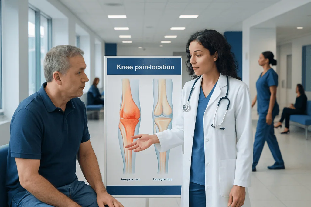
The location of knee pain can offer useful clues, but it does not confirm a diagnosis on its own. Front knee pain is often linked to the kneecap, tendon irritation, or cartilage wear.
Key Takeaways
- The location of knee pain can offer useful clues, but it does not confirm a diagnosis on its own.
- Front knee pain is often linked to the kneecap, tendon irritation, or cartilage wear.
- Inner, outer, or back knee pain may involve ligaments, meniscus tissue, tendons, bursae, or cysts.
- Red flags include major swelling, inability to bear weight, fever, deformity, or a locked knee.
- Persistent or worsening knee pain should be assessed by a qualified clinician, especially after injury.
A knee pain location chart can help a person understand which structures may be involved based on where the pain is felt. Front, inner, outer, and back knee pain often point to different patterns, but symptoms, injury history, swelling, and instability matter just as much as the location itself.
How to read a knee pain location chart
A knee pain location chart is a simple way to match the area of discomfort with structures inside and around the knee. Pain at the front of the knee often relates to the kneecap or patellar tendon, pain on the inner or outer side may involve ligaments or the meniscus, and pain behind the knee can sometimes come from tendons, joint swelling, or a fluid-filled cyst. This approach is helpful because the knee is made up of bone, cartilage, ligaments, tendons, bursae, and nerves that can each cause pain in different patterns.
Still, location is only one part of the picture. Doctors also ask when the pain started, whether there was a twist or fall, if swelling appeared quickly, and whether the knee feels unstable, locks, clicks, or gives way. Age, activity level, existing arthritis, and other joint symptoms can also change what is most likely.
A useful way to think about a knee pain location chart is as a guide rather than a diagnosis. It helps narrow the possibilities and can make symptoms easier to describe during a medical visit. The clearest answers usually come from combining pain location with a physical examination and, when needed, imaging or other tests.
Front knee pain: what it may suggest
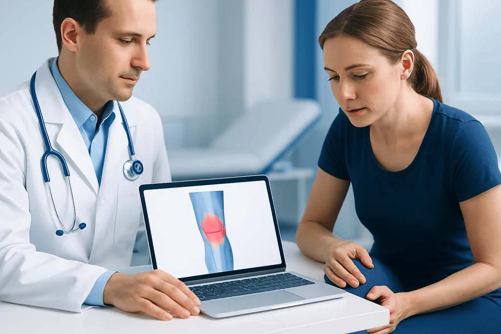
Front knee pain is commonly felt around or behind the kneecap. This pattern is often associated with patellofemoral pain syndrome, sometimes called runner’s knee, where the kneecap does not move smoothly in its groove. The pain may be worse when using stairs, squatting, kneeling, or sitting for a long time with bent knees.
Pain just below the kneecap may point to patellar tendinopathy, especially in people who jump, run, or change direction often. In children and teenagers, pain at the bony bump below the kneecap can be related to growth-related overuse, such as Osgood-Schlatter disease. In middle-aged and older adults, front knee pain can also be related to wear of the cartilage, including knee osteoarthritis.
Other clues can help separate these causes. Patellofemoral pain often causes aching or soreness without major swelling. Tendon-related pain is usually more focused and worsens with forceful activity. Arthritis is more likely to cause stiffness, grinding, or pain that gradually increases over time, especially after rest or at the end of the day.
Inner, outer, and back knee pain
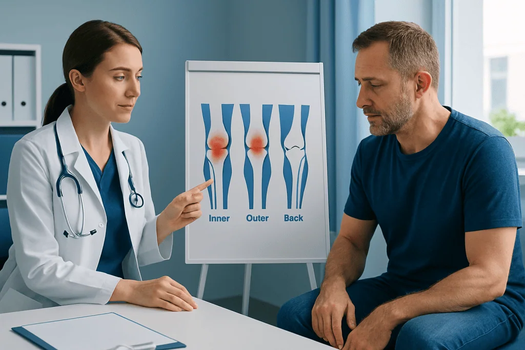
Inner knee pain, on the side closest to the other leg, may come from a medial meniscus tear, a medial collateral ligament strain, arthritis affecting one side of the joint, or irritation of the pes anserine bursa or tendons. Meniscus pain is often linked to twisting, squatting, or a catching sensation. Ligament pain is more likely after a sideways force or sports injury and may come with tenderness along the joint line.
Outer knee pain can be related to the lateral meniscus, the lateral collateral ligament, arthritis, or iliotibial band irritation. Iliotibial band syndrome often causes pain on the outside of the knee that worsens with repetitive bending, such as running downhill or cycling. Lateral meniscus problems may cause clicking, swelling, or a feeling that the knee cannot fully straighten.
Pain at the back of the knee has a different set of possibilities. It can happen with hamstring or calf tendon strain, joint swelling that tracks backward, or a Baker’s cyst, which is a fluid-filled swelling behind the knee often linked to arthritis or inflammation. In some cases, back-of-knee discomfort can also accompany injuries to deeper structures, including the cruciate ligaments, especially if there has been a twist, pop, or rapid swelling after trauma.
- Inner knee: meniscus, medial ligament, arthritis, bursitis
- Outer knee: iliotibial band, lateral meniscus, lateral ligament, arthritis
- Back of knee: Baker’s cyst, tendon strain, joint swelling, ligament injury
What triggers knee pain and who is at risk
Knee pain can begin suddenly after an injury or develop gradually from overuse, alignment problems, or age-related changes. Common triggers include twisting while the foot is planted, direct impact, sudden increases in training, repetitive kneeling, weak hip or thigh muscles, and poor movement patterns during sport or exercise. Excess body weight can also increase stress across the joint.
Some knee problems are more common in certain groups. Athletes who pivot or jump may be more prone to ligament injuries or tendon irritation. Runners and cyclists often report front or outer knee pain related to overload. Older adults are more likely to have degenerative cartilage or meniscus changes, while growing adolescents may experience pain around the kneecap or at tendon attachment points.
Not all knee pain starts in the knee itself. Pain can be influenced by the hip, ankle, lower back, or foot mechanics. Flat feet, reduced ankle mobility, and weak hip stabilizers can change how forces pass through the knee. This is one reason a full assessment may look beyond the exact painful spot.
How doctors diagnose the cause
Diagnosis usually starts with a careful history and physical examination. A clinician may ask about the exact location of pain, whether there was a pop, how quickly swelling developed, and what movements make the pain worse. During the exam, they may check the range of motion, tenderness, alignment, gait, swelling, stability, and signs of meniscus or ligament injury.
Imaging is sometimes helpful, but not always needed right away. X-rays can show fractures, alignment changes, and arthritis. Ultrasound may help assess some soft tissue problems or fluid behind the knee. MRI is often used when doctors need a closer look at cartilage, ligaments, tendons, or a suspected meniscus tear or ACL tear.
Blood tests are usually reserved for situations where infection, inflammatory arthritis, or gout is possible. If the knee is very swollen, a doctor may remove a small amount of joint fluid for testing. The goal is to identify the underlying problem accurately so treatment matches the true cause rather than just the pain location.
Treatment options based on the cause
Treatment depends on what is causing the pain and how severe it is. Many mild to moderate cases improve with activity modification, relative rest, ice, short-term support if advised, and a structured rehabilitation plan. Physical therapy is often central because it can improve strength, flexibility, balance, and movement patterns that unload the knee.
Medication may be used for symptom relief when appropriate, especially for short-term pain or inflammation, but it is usually only one part of care. In some conditions, targeted injections may be considered by a specialist. If the knee is unstable, persistently locking, or not improving with conservative care, further evaluation may be needed to discuss procedures such as knee arthroscopy or other orthopedic rehabilitation plans.
For advanced joint damage, treatment can range from bracing and exercise therapy to surgical options in selected patients. People with severe arthritis that limits daily life may be evaluated for knee replacement. The right treatment is guided by the diagnosis, age, activity goals, overall health, and whether symptoms interfere with walking, work, or sleep.
Self-care and prevention strategies
Many knee problems can be reduced or better managed with steady self-care habits. Gradually increasing exercise rather than making sudden jumps in distance, speed, or intensity can lower the risk of overload injuries. Supportive footwear, attention to running or training form, and warming up before activity may also help.
Strengthening the muscles around the knee and hip is especially important. Strong quadriceps, hamstrings, gluteal muscles, and core muscles help the leg absorb force more evenly. Flexibility and mobility work for the hips, calves, and ankles may improve movement quality and reduce strain on the knee.
Weight management can be useful for people with osteoarthritis or recurrent pain because it reduces force across the joint with everyday steps. It is also sensible to avoid pushing through sharp or worsening pain. If symptoms continue despite rest and home measures, a clinician or physiotherapist can help identify the cause and design a safe program.
When to seek medical care
Some knee symptoms need prompt medical attention. A person should seek care soon if the knee is very swollen, looks deformed, cannot bear weight, feels unstable after an injury, or is locked and cannot fully bend or straighten. Fever, redness, warmth, or feeling generally unwell together with knee pain may suggest infection or significant inflammation and should not be ignored.
Medical review is also important when pain lasts more than a few weeks, keeps returning, interrupts sleep, or prevents normal walking, work, or exercise. A gradual problem can still need treatment if stiffness, swelling, or loss of confidence in the joint is increasing over time. Early assessment may help prevent compensation patterns and longer-term limitation.
For international patients who need specialist assessment, Acibadem International’s multidisciplinary teams in JCI-accredited hospitals diagnose and treat knee conditions using imaging, rehabilitation, and orthopedic care tailored to the underlying cause. The most appropriate next step is a qualified medical evaluation, especially when symptoms are persistent, progressive, or follow trauma.
Frequently asked questions
Can a knee pain location chart diagnose the exact problem?
No. A knee pain location chart is useful for narrowing down possibilities, but it cannot confirm the exact cause by itself. Doctors usually combine pain location with symptoms, injury history, examination findings, and sometimes imaging.
What does pain on the inside of the knee usually mean?
Inner knee pain may be related to a meniscus problem, a medial collateral ligament strain, arthritis, or irritation of nearby tendons or bursae. The most likely cause depends on whether the pain followed an injury, appeared gradually, or comes with swelling or catching.
Why does the front of the knee hurt when climbing stairs?
Pain at the front of the knee with stairs often suggests irritation around the kneecap, such as patellofemoral pain syndrome. It can also happen with tendon irritation or arthritis, especially if there is stiffness or grinding.
Is pain behind the knee a sign of a blood clot?
Back-of-knee pain is more commonly caused by tendon strain, joint swelling, or a Baker’s cyst. However, sudden calf swelling, warmth, redness, or unexplained tenderness should be assessed urgently because a blood clot needs prompt medical evaluation.
When should knee pain be checked by a doctor?
A doctor should assess knee pain after a significant injury, if the knee is swollen, unstable, locked, or difficult to bear weight on. Medical review is also sensible if the pain lasts more than a few weeks, keeps returning, or limits daily activities.
Can knee pain improve without surgery?
Yes. Many causes of knee pain improve with rest, activity modification, strengthening exercises, physiotherapy, and other conservative treatments. Surgery is usually considered only when symptoms are severe, the joint is unstable or mechanically blocked, or non-surgical care has not helped enough.
References
- American Academy of Orthopaedic Surgeons
- National Institute of Arthritis and Musculoskeletal and Skin Diseases
- Mayo Clinic
- National Health Service
- American College of Rheumatology
This article is for general information only and is not a substitute for professional medical advice. Please consult a qualified doctor about your individual situation.
Joint and spine care in Turkey — expert assessment & treatment
JCI-accredited · board-certified surgeons · reply within 24h