

MRI En Español: Preparation, Procedure and Results
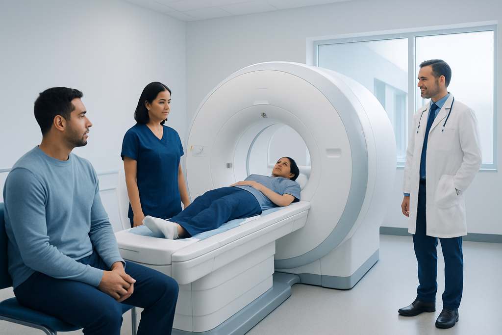
MRI does not use ionizing radiation, but its strong magnet requires careful safety screening. Preparation depends on the body area being examined and whether contrast material or sedation is planned.
Key Takeaways
- MRI does not use ionizing radiation, but its strong magnet requires careful safety screening.
- Preparation depends on the body area being examined and whether contrast material or sedation is planned.
- Most MRI scans take about 20 to 60 minutes, although complex studies can take longer.
- Patients should tell the imaging team about implants, metal fragments, pregnancy, kidney disease, allergies, and anxiety in enclosed spaces.
- A radiologist interprets the images and sends a report to the clinician who ordered the examination.
MRI en español refers to magnetic resonance imaging information for Spanish-speaking patients. An MRI is a painless imaging test that uses a powerful magnet and radio waves, not radiation, to create detailed pictures of organs, soft tissues, bones, and blood vessels.
MRI en Español: What an MRI Scan Is and How It Works
MRI en español means magnetic resonance imaging information explained for Spanish-speaking patients and families. Magnetic resonance imaging, often called MRI, is a noninvasive test that creates detailed images of structures inside the body. It can help clinicians assess the brain, spine, joints, heart, abdomen, pelvis, blood vessels, and many other areas.
An MRI scanner uses a powerful magnetic field and radiofrequency waves to arrange and detect signals from hydrogen atoms in the body. A computer turns those signals into cross-sectional images. Unlike X-rays and CT scans, MRI does not use ionizing radiation.
During the examination, the person lies still on a padded table that moves into the scanner. The machine makes repeated loud tapping, knocking, or humming sounds while pictures are acquired. The scan itself is not painful, although remaining in one position can be uncomfortable for some people.
MRI is often selected when detailed views of soft tissue are needed. For example, it may be used in the assessment of brain tumors, spinal conditions, ligament injuries, liver concerns, pelvic disorders, or blood vessel abnormalities. The referring clinician decides whether MRI is the most appropriate examination for the person’s symptoms and medical history.
Who Can Have an MRI and Important Safety Checks
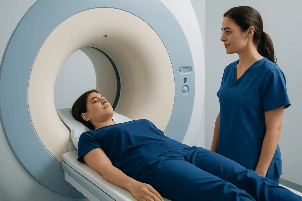
Most people can safely undergo an MRI after a standard screening process. Before the appointment, the imaging team asks about prior surgery, implanted devices, metal injuries, pregnancy, allergies, kidney health, and previous imaging examinations. These questions are essential because the MRI magnet remains powerful at all times.
Some implants are MRI-safe or MRI-conditional, meaning they can be scanned only under specified conditions. Pacemakers, defibrillators, cochlear implants, neurostimulators, aneurysm clips, infusion pumps, artificial joints, dental work, and vascular stents should always be reported. The team may need device details, manufacturer information, or additional review before proceeding.
People who have worked with metal, such as welding or machining, or who may have metal fragments in the eye, should mention this before the scan. In some situations, an X-ray may be needed to check for metal before MRI. Jewelry, watches, hearing aids, credit cards, removable dental devices, hairpins, and clothing with metallic components must be removed.
Claustrophobia, severe pain, movement disorders, and young age can make it difficult to lie still inside the scanner. The radiology team can discuss practical comfort measures, and some patients may be considered for medication or sedation when clinically appropriate. Sedation requires advance planning and may change fasting and transportation instructions.
How to Prepare for Your First MRI

For a first MRI, patients should carefully read the instructions provided by the imaging center because preparation differs by examination. Many routine MRI scans require no special diet or medication changes. However, MRI of the abdomen or pelvis, scans involving contrast, and studies performed with sedation may have additional requirements.
Patients should bring identification, referral information if needed, a current medication list, and details about implants or previous surgeries. Prior images and reports can be useful for comparison when available. It is also helpful to arrive early enough to complete the safety questionnaire without feeling rushed.
Comfortable clothing without metal is usually suitable, though the facility may provide a gown. Avoid wearing metal-containing cosmetics, temporary tattoos with metallic pigments, or accessories. The person should tell the staff if there is any chance of pregnancy, if they have kidney disease, or if they have previously had a reaction to MRI contrast material.
For individuals who feel anxious in small spaces, discussing this before the appointment is important. The team can explain the scanner layout, provide earplugs or headphones, maintain communication through an intercom, and offer a call button. In selected cases, a clinician may recommend anxiety medicine or sedation, so the patient may need someone else to drive them home.
What to Do the Night Before an MRI
The night before an MRI, patients can usually follow their normal routine unless the imaging provider has given different instructions. They should review the appointment time, confirm transport arrangements if sedation is planned, and prepare any implant identification cards or previous imaging records requested by the center.
If fasting has been advised, it is important to follow the stated time period exactly. Fasting is not necessary for every MRI, but it may be requested for certain abdominal examinations, contrast studies, or sedation. Prescription medicines should generally be taken as usual with a small sip of water unless the prescribing clinician or imaging team says otherwise.
Patients should avoid last-minute uncertainty by calling the imaging department in advance if they have a pacemaker, implanted device, kidney disease, possible pregnancy, severe claustrophobia, or questions about contrast. They should not stop important medicines independently in preparation for an MRI.
A good night’s sleep may make it easier to stay relaxed and still during the scan. People who expect discomfort from lying flat may ask their clinician whether usual pain-management medicines can be taken before the examination. Any plan should be individualized, particularly for people taking sedatives or medicines that affect alertness.
Why Do You Drink Water Before an MRI?
Some MRI examinations require drinking water beforehand, particularly certain pelvic, bladder, or abdominal studies. Water can fill the bladder or improve the visibility of nearby structures, helping the radiologist distinguish organs and tissues more clearly. The exact amount and timing depend on the specific scan.
Water instructions are not universal. For many MRI studies, including many brain, spine, and joint scans, patients do not need to drink extra water. In other situations, instructions may involve fasting rather than drinking, especially when the upper abdomen is being examined.
Patients should follow the directions supplied for their own appointment rather than relying on general advice. If the instruction is unclear or difficult to follow because of urinary symptoms, diabetes, heart failure, or another condition, they should contact the imaging center for individualized guidance.
When MRI contrast is planned, being reasonably hydrated may be encouraged in some circumstances, unless a clinician has advised fluid restriction. This does not mean that everyone should drink large amounts of water before imaging. The imaging team’s directions are the safest guide.
MRI Procedure: Step by Step and How Many Hours It Takes
After check-in, a radiology professional reviews the safety questionnaire and explains the examination. The patient removes metal items and changes clothing if necessary. If contrast is needed, a small intravenous line may be placed in the arm or hand. The team positions the body area being examined within a receiving device called a coil.
Once positioned on the table, the patient is moved into the scanner. Staff remain nearby and can communicate through an intercom. Ear protection is provided because MRI sequences produce loud sounds. The most important part of the procedure is staying as still as possible; movement can blur images and may require part of the scan to be repeated.
How many hours is the MRI procedure? Most standard MRI examinations take approximately 20 to 60 minutes. More detailed studies, scans of several body regions, specialized cardiac or vascular MRI, or examinations requiring contrast may last 60 to 90 minutes or occasionally longer. The total visit can be longer because of registration, screening, preparation, and recovery after sedation.
Contrast-enhanced imaging may use gadolinium-based contrast material to make inflammation, tumors, blood vessels, or certain organ abnormalities easier to assess. It is generally well tolerated, but the team considers kidney function and prior reactions before use. MRI findings can guide further care, including evaluation and planning for brain tumor surgery when appropriate.
After the Scan: Recovery, Benefits, Risks and Results
Most patients can return to normal activities immediately after an MRI. There is no recovery time from the magnetic field or radio waves. A person who receives sedating medicine should follow the discharge instructions, avoid driving and important decisions for the recommended period, and arrange for a responsible adult to accompany them home.
The main benefits of MRI are its detailed images and its ability to examine many soft tissues without ionizing radiation. It can provide information that may not be clearly shown by other tests. However, MRI is not always the right first test, and image findings must be interpreted alongside symptoms, examination findings, and medical history.
Risks are uncommon but include discomfort from noise or confinement, heating or movement of unsafe metal objects, and rare reactions to contrast material. Gadolinium-based contrast requires particular consideration in people with severely reduced kidney function. Patients should immediately tell staff if they experience warmth, pain, itching, breathing difficulty, dizziness, or distress during the scan.
A radiologist reviews the images and prepares a report for the clinician who ordered the MRI. Results may be available within a few days, though timing varies by facility and urgency. At Acibadem International, multidisciplinary specialists and JCI-accredited hospitals support diagnosis and treatment planning for international patients, including advanced imaging and related care such as spine surgery when indicated.
When to Seek Medical Care
An MRI appointment itself is usually planned through a clinician, but urgent medical care is needed for sudden or severe symptoms rather than waiting for routine imaging. Examples include new weakness on one side of the body, difficulty speaking, sudden loss of vision, a seizure, severe sudden headache, chest pain, severe shortness of breath, or loss of bladder or bowel control with back pain.
Patients should seek prompt medical advice if symptoms are worsening, new neurological symptoms develop, or pain follows a significant injury. The need for emergency assessment depends on the symptom pattern and overall health, not simply on whether an MRI has been scheduled.
Before the scan, patients should contact the imaging team promptly if they discover an implant, possible metal exposure, pregnancy, kidney disease, or a history of contrast reaction that was not included in their screening form. They should also report new illness or a need for sedation-related transport changes.
After an MRI with contrast, urgent assessment is appropriate for signs of a serious allergic reaction, such as trouble breathing, facial swelling, widespread hives, or fainting. Although such reactions are uncommon, timely medical evaluation is important. For nonurgent questions about the report, the ordering clinician can explain what the results mean and whether follow-up is needed.
Frequently asked questions
Does an MRI use radiation?
No. MRI uses a strong magnetic field and radiofrequency waves to create images, rather than ionizing radiation used in X-rays and CT scans. The magnetic field still makes safety screening for metal and implants very important.
Can a person eat before an MRI?
Many MRI scans do not require fasting, so a person may be able to eat normally. However, some abdominal, pelvic, contrast, or sedation-related studies have specific food and drink instructions. The instructions from the imaging center should always be followed.
Can someone with a pacemaker have an MRI?
Some people with pacemakers can have MRI under carefully controlled conditions, depending on the device and clinical circumstances. The imaging team needs complete device information before scheduling or performing the scan. A patient should never enter an MRI area without telling staff about a pacemaker or other implanted device.
Is MRI contrast safe?
Gadolinium-based MRI contrast is generally well tolerated and is used only when it may improve the diagnostic value of the examination. Reactions are uncommon, but kidney function, prior contrast reactions, and relevant health conditions should be reviewed beforehand. The care team can explain why contrast is or is not recommended.
What if a patient is claustrophobic during an MRI?
Claustrophobia is common and should be discussed before the appointment. The team may offer communication, ear protection, positioning support, and other comfort strategies. In selected cases, a clinician may recommend medication or sedation, which requires advance planning and a ride home.
When will MRI results be ready?
A radiologist reviews the scan and sends a report to the clinician who ordered it. Timing varies by center, scan complexity, and urgency, but results are often discussed within a few days. The ordering clinician is best placed to explain what the findings mean for that individual.
References
- Radiological Society of North America
- American College of Radiology
- National Institute of Biomedical Imaging and Bioengineering
- U.S. Food and Drug Administration
- National Health Service
This article is for general information only and is not a substitute for professional medical advice. Please consult a qualified doctor about your individual situation.
Joint and spine care in Turkey — expert assessment & treatment
JCI-accredited · board-certified surgeons · reply within 24h

More from the Health Library


Related Specialists

Dyt. Renan Güneş
Nutrition & Diet
Assoc. Prof. Dr. Aylin Altan Kuş
Gastrointestinal Oncology Unit Clinical Service
Dr. Metehan Bağış
Emergency Service
Dr. Ali İlbey Demirel
General Surgery