Heart Ultrasound: Preparation, Procedure and Results
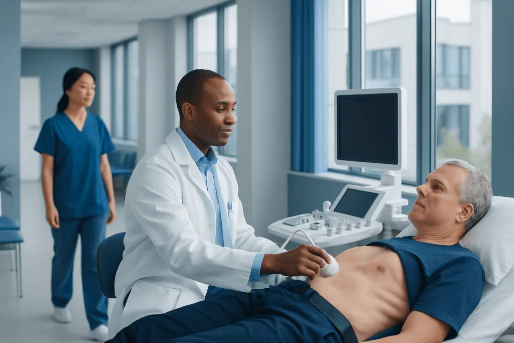
Most standard heart ultrasounds require little or no special preparation. The test is painless, does not use radiation and usually takes about 30 to 60 minutes.
Key Takeaways
- Most standard heart ultrasounds require little or no special preparation.
- The test is painless, does not use radiation and usually takes about 30 to 60 minutes.
- An echocardiogram can identify many structural and functional heart concerns, but it does not directly show coronary artery blockages.
- A cardiologist interprets the images in the context of symptoms, examination findings and other tests.
- Preparation differs for specialised tests such as transesophageal and stress echocardiograms.
A heart ultrasound, also called an echocardiogram, uses sound waves to create moving images of the heart. It is a safe, non-invasive test that helps clinicians assess heart chambers, valves, pumping strength and blood flow.
Heart Ultrasound Overview: What It Is and Why It Is Used
A heart ultrasound, commonly called an echocardiogram or echo, is an imaging test that uses high-frequency sound waves to create real-time pictures of the heart. It allows a clinician to see the heart muscle contract, watch valves open and close, and assess the direction and speed of blood flow. Unlike an X-ray or CT scan, a standard echocardiogram does not use ionising radiation.
The most common type is a transthoracic echocardiogram (TTE), performed by placing an ultrasound probe on the chest. It may be recommended for symptoms such as shortness of breath, chest discomfort, palpitations, fainting, ankle swelling or unusual fatigue. It can also be used to monitor a known heart condition, assess a heart murmur, evaluate the heart after a stroke, or check heart function before certain treatments or surgery.
Heart ultrasound findings are not interpreted in isolation. A cardiologist considers the images alongside medical history, symptoms, physical examination, blood tests and tests such as an electrocardiogram. When further assessment is needed, an echocardiogram may be part of a broader cardiology evaluation plan.
How a Heart Ultrasound Works and Who May Need One
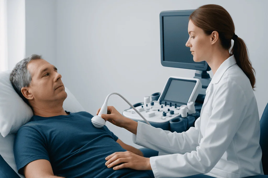
An ultrasound transducer sends sound waves into the body and receives echoes reflected from the heart and surrounding tissues. A computer converts these echoes into detailed moving images. Doppler ultrasound can also measure blood flow through the heart chambers, valves and major blood vessels, helping identify abnormal flow patterns.
A heart ultrasound may provide information about chamber size, heart-wall movement, pumping function, valve narrowing or leaking, fluid around the heart, congenital heart differences, pressure estimates within parts of the heart and some blood clots. It is often suitable for adults, children, older people and pregnant people because the standard surface examination has no radiation exposure.
Different echo techniques may be selected depending on the clinical question. A transesophageal echocardiogram (TEE) uses a small probe passed into the oesophagus to obtain closer images of certain heart structures. A stress echocardiogram records images before and after exercise or a medicine that makes the heart work harder. These specialised examinations have different preparation and monitoring requirements.
- Transthoracic echocardiogram: the routine chest-wall scan.
- Doppler echocardiogram: evaluates blood-flow speed and direction.
- Transesophageal echocardiogram: provides more detailed views from the oesophagus.
- Stress echocardiogram: assesses heart function under physical or medication-induced stress.
Preparing for a Heart Ultrasound

For a routine transthoracic heart ultrasound, people can usually eat, drink and take prescribed medicines as normal unless their clinician gives different instructions. Wearing a two-piece outfit may make changing easier, and avoiding heavy body lotion or oil on the chest on the day can help the ultrasound gel and probe make good contact with the skin.
It is helpful to bring a medication list, relevant medical records and details of previous heart tests if these are available. The person should tell the healthcare team about symptoms, a history of heart disease, previous heart surgery, allergies and any difficulty lying flat. There is normally no need to arrange transport home after a standard transthoracic echocardiogram.
Preparation is more specific for a TEE or stress echo. A person may need to avoid food and drink for several hours before a TEE because sedation and a throat procedure may be involved. For a stress test, the team may provide individual guidance about meals, caffeine, medicines and comfortable walking or exercise clothing. Instructions should be followed carefully, and medicines should not be stopped without the prescribing clinician’s advice.
What Not to Do Before a Heart Ultrasound
Before a standard chest-wall echocardiogram, there are few restrictions. People should not stop heart medicines, diabetes medicines or blood thinners on their own. They should also avoid applying thick creams, oils or powders across the chest shortly before the appointment, as these may interfere with probe contact.
For a routine test, there is generally no need to fast, avoid exercise or limit fluids unless a clinician has requested this. However, preparation can differ substantially for stress and transesophageal echocardiograms. People should not eat, drink, consume caffeine, smoke, exercise heavily or take certain medicines before these tests if their appointment instructions advise against it.
Anyone who is uncertain about their preparation should contact the testing centre before attending. This is especially important for people with diabetes, those taking medicines that affect heart rate or blood pressure, and those having a procedure involving sedation.
Heart Ultrasound Procedure: Step by Step
For a standard transthoracic echocardiogram, the person usually changes into a gown from the waist up and lies on an examination couch. Small adhesive electrodes may be placed on the chest to record the heartbeat during imaging. The sonographer or cardiac physiologist applies a small amount of clear gel to the chest and moves the handheld probe over several positions.
The person may be asked to roll onto the left side, hold their breath briefly or change position. These simple steps can improve the image quality. The probe may be pressed firmly at times, particularly between the ribs, but the examination should not be painful. The room is often dimmed so the operator can see the images clearly.
A standard study commonly takes 30 to 60 minutes, though duration varies with the type of examination and how many images are needed. Once the images are complete, the gel is wiped away and the person can usually dress and return to normal activities immediately. A TEE and stress echocardiogram involve additional steps, observation and recovery arrangements.
The main benefit of heart ultrasound is that it gives detailed functional information without surgery or radiation. Image quality can occasionally be limited by body shape, lung conditions, chest-wall anatomy or the ability to remain in a particular position. In these situations, the clinician may recommend contrast-enhanced echocardiography, TEE, cardiac MRI, CT or another test.
Heart Ultrasound Results: What They Can Show
The images are reviewed by a cardiologist or another appropriately trained clinician. The report may describe the size and shape of the heart chambers, thickness and movement of the heart walls, how effectively the heart pumps, the appearance and movement of valves, blood-flow patterns and the presence of fluid around the heart.
Results can help identify conditions including valve disease, weakened heart pumping, thickened heart muscle, enlargement of chambers, certain congenital heart conditions, inflammation or fluid around the heart, and signs that may suggest raised pressures in the lungs. If an abnormality is found, it does not always mean urgent treatment is needed; the significance depends on the finding, its severity and the person’s overall health.
How long it takes to receive results varies by centre and clinical setting. In urgent situations, a preliminary interpretation may be available the same day. For non-urgent outpatient testing, the formal report may be sent to the referring clinician within several days, who will discuss what the results mean and whether follow-up testing or treatment is appropriate.
Will a Heart Ultrasound Show a Blockage?
A standard heart ultrasound does not directly show blockages in the coronary arteries, the small blood vessels that supply the heart muscle. Coronary arteries are often too small and positioned in ways that make direct visualisation difficult with routine echocardiography.
However, an echocardiogram may show indirect signs that a section of heart muscle is not moving normally. This can sometimes occur when blood supply has been reduced or after a heart attack, but it can also have other causes. A normal echocardiogram does not exclude coronary artery disease.
When a coronary blockage is suspected, clinicians may consider tests such as an exercise test, stress echocardiogram, coronary CT angiography or invasive coronary angiography. The most suitable option depends on symptoms, risk factors, prior test results and the urgency of the situation. Sudden or persistent chest pressure, breathlessness, sweating, nausea or fainting requires urgent medical assessment.
Do You Keep Your Bra On for an Echocardiogram?
For a standard echocardiogram, the chest needs to be accessible so the probe can be placed in several positions. Most people are asked to remove clothing from the waist up and wear a gown, which usually means removing a bra. Staff should provide privacy, clear instructions and appropriate draping throughout the examination.
People can ask for a chaperone or a same-gender staff member where this is possible and preferred. They should also let the team know if they have concerns about modesty, mobility, pain, skin sensitivity or a previous difficult healthcare experience. The team can often make practical adjustments while still obtaining clinically useful images.
For some limited bedside scans, clothing arrangements may differ, but the imaging professional will explain what is needed before beginning. Comfort and dignity are important parts of the examination process.
Recovery, Risks and When to Seek Medical Care
There is no recovery period after a routine transthoracic heart ultrasound. People can eat, drive, work and resume usual activities immediately. Mild temporary skin redness from gel or probe pressure is uncommon and usually settles quickly. The test itself does not cause radiation-related risks.
TEE and stress echocardiograms have additional considerations. After sedation for a TEE, a person may need someone to take them home and should avoid driving or important decisions for the period advised by the clinical team. Stress testing is closely supervised; the team monitors for symptoms or rhythm changes and provides clear instructions after the test.
Medical care should be sought promptly for new, severe or worsening chest pain, significant shortness of breath, fainting, a very fast or irregular heartbeat with symptoms, or sudden weakness. These symptoms should not be managed by waiting for a planned imaging appointment. For non-urgent but persistent symptoms such as reduced exercise tolerance, ankle swelling or recurrent palpitations, arranging a medical review is appropriate.
Acibadem International’s multidisciplinary specialists and JCI-accredited hospitals diagnose and treat heart conditions for international patients, with care tailored to the clinical findings and individual needs.
Frequently asked questions
How long does a heart ultrasound take?
A standard transthoracic echocardiogram commonly takes around 30 to 60 minutes. The exact time depends on the images needed and whether the person needs help getting into a comfortable position. Stress and transesophageal echocardiograms generally take longer because they require additional preparation and monitoring.
How long does it take to get results back from a heart ultrasound?
In urgent hospital settings, a preliminary result may be available the same day. For planned outpatient testing, a formal report is often sent to the referring clinician within several days. The clinician will explain the result in relation to symptoms and other health information.
What not to do before a heart ultrasound?
For a routine echocardiogram, people generally do not need to fast or stop their usual medicines unless instructed otherwise. They should avoid heavy creams or oils on the chest and should not independently stop prescribed medication. Stress and transesophageal echocardiograms may have specific restrictions involving food, caffeine, exercise or medication.
Will a heart ultrasound show a blockage?
A routine heart ultrasound does not directly show blockages in the coronary arteries. It can sometimes show changes in heart-muscle movement that suggest reduced blood supply, but these changes are not specific to a blockage. Other tests may be needed when coronary artery disease is suspected.
Do you keep your bra on for an echocardiogram?
Usually, a bra is removed because the ultrasound probe needs direct access to different parts of the chest. A gown and drape are provided to support privacy during the examination. People may discuss modesty preferences or request a chaperone with the healthcare team.
Is a heart ultrasound painful or dangerous?
A standard transthoracic echocardiogram is typically painless and considered very safe. Some pressure from the probe may feel briefly uncomfortable, especially between the ribs, but it should not cause significant pain. It does not use radiation.
References
- American Heart Association
- American Society of Echocardiography
- National Heart, Lung, and Blood Institute
- Mayo Clinic
- NHS
This article is for general information only and is not a substitute for professional medical advice. Please consult a qualified doctor about your individual situation.
Ultrasound in Turkey — costs, top hospitals & a free quote
JCI-accredited · board-certified surgeons · reply within 24h
Add us as a Preferred Source to see more of our trusted health content across Google Search, AI Overviews and Discover.
Check your numbers in seconds
BMI, calories, due date, blood pressure and 30+ more clinical calculators — free, instant, doctor-reviewed ranges.

Check-up & Preventive Medicine
Comprehensive health screenings designed for early detection and prevention.
3 specialists in this unitMore from the Health Library



Related Specialists

Dr. Gökay Kurtulan
Occupational Medicine
Prof. Dr. Burak Tander
Pediatric Surgery
Dr. Ceyhun Başıoğlu
Physical Medicine & Rehabilitation
Dr. Özge Kaymaz Yılmaz
Gynecology & Obstetrics