Brain Tumor Removal: Procedure, Recovery and Results

Brain tumor removal may aim to remove a tumor completely, reduce its size, obtain tissue for diagnosis, or relieve pressure on the brain. A craniotomy is the most common approach, but some tumors can be reached through the nose or treated with other carefully selected techniques.
Key Takeaways
- Brain tumor removal may aim to remove a tumor completely, reduce its size, obtain tissue for diagnosis, or relieve pressure on the brain.
- A craniotomy is the most common approach, but some tumors can be reached through the nose or treated with other carefully selected techniques.
- Most patients spend at least one night in a high-dependency or intensive monitoring area after surgery, although the exact ICU stay varies.
- Recovery can take weeks to months and may include rehabilitation for movement, speech, thinking or daily activities.
- Surgery has important potential benefits but also risks, which should be discussed with an experienced neurosurgical team before treatment.
Brain tumor removal is a neurosurgical procedure used to remove as much of a brain tumor as safely possible, confirm its diagnosis, and reduce symptoms caused by pressure on the brain. The approach, hospital stay and recovery depend on the tumor’s location, type, size and the person’s overall health.
Overview: what brain tumor removal involves
Brain tumor removal is an operation performed by a neurosurgeon to remove all or part of a growth in or near the brain. It may be recommended to establish a precise diagnosis, ease symptoms from pressure or swelling, improve neurological function, or reduce the amount of tumor that needs further treatment. Whether complete removal is possible depends primarily on where the tumor is located and how closely it involves important brain structures, nerves or blood vessels.
Not every brain tumor requires immediate surgery. Some small, slow-growing tumors can be monitored with repeat imaging, while others may be treated with radiation therapy, medicines, chemotherapy, targeted therapy or a combination of approaches. A multidisciplinary team typically reviews brain scans, symptoms, pathology findings and personal priorities to recommend an individualized plan.
“Removal” does not always mean that every tumor cell can be safely taken out. In some situations, a planned partial removal, often called debulking, offers the best balance between reducing tumor burden and protecting speech, movement, vision, memory and other functions. Tissue removed during surgery is examined by a pathologist to identify the tumor type and guide next steps.
Who may be a candidate for surgery?
A person may be considered for brain tumor surgery when a tumor is causing seizures, headaches related to raised pressure, weakness, changes in vision, speech difficulties, balance problems or other neurological symptoms. Surgery may also be advised when imaging cannot reliably identify a tumor, when a tumor is growing, or when tissue is needed to plan radiation or drug treatment.
Before recommending an operation, the team considers the tumor’s location, size, growth pattern and likely type, as well as the person’s age, general health, medications and neurological function. MRI is central to planning, and CT, functional MRI, tractography, angiography or specialized scans may also be useful. Blood tests, anesthesia assessment and discussions about expected benefits and possible effects on daily function are part of preparation.
Some tumors sit near areas controlling language, movement, sensation, eyesight or vital functions. In these cases, surgeons may use detailed navigation, brain mapping, monitoring of nerve pathways, or an awake procedure for selected patients. These methods are intended to help maximize safe removal, not to eliminate risk completely.
- Potential goals: diagnosis, symptom relief, pressure reduction, complete removal when safe, or partial removal before further treatment.
- Reasons surgery may not be suitable: a tumor that cannot be safely reached, significant medical risk, or a situation where another treatment is more appropriate.
Brain tumor removal step by step
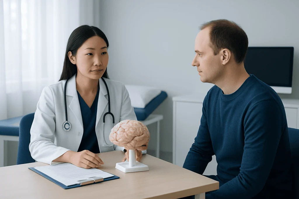
The exact brain tumor removal step by step process differs between patients. Most operations are performed under general anesthesia. For a conventional craniotomy, the head is positioned securely, hair is clipped only where needed, and the surgeon makes an incision in the scalp. A small section of skull is temporarily removed to provide access to the brain, then replaced and secured at the end of the operation.
Using an operating microscope, surgical navigation and, when appropriate, ultrasound or MRI guidance, the surgeon reaches the tumor through the safest available path. The tumor is removed in small sections while protecting normal tissue and blood vessels. In some cases, surgeons monitor speech, movement or other functions during the procedure; an awake craniotomy may be used in carefully selected patients whose tumor is near language or movement areas.
Some tumors at the base of the skull or near the pituitary gland may be approached through the nasal passages using an endoscope, avoiding a larger opening in the skull. A biopsy may be performed instead of removal when obtaining a small tissue sample is safer or more clinically useful. The removed tissue is sent for laboratory examination, and final pathology results commonly help determine whether additional treatment is needed.
After surgery, the person is transferred to a closely monitored recovery setting. Brain scans are often performed soon after the operation to assess the surgical area. The care team also checks alertness, speech, strength, pupil responses, pain, fluid balance and signs of bleeding or swelling.
Benefits and risks of brain tumor removal
The possible benefits of surgery include obtaining a definitive diagnosis, lowering pressure inside the skull, improving symptoms caused by the tumor, reducing seizures in some people, and removing or shrinking tumor tissue before other treatments. For certain tumors, complete removal can provide long-term control. For others, surgery is one part of treatment alongside radiation therapy, chemotherapy or other medicines.
All brain operations carry risks, and the individual risk profile depends on the tumor and surgical route. Possible complications include bleeding, infection, swelling, blood clots, fluid leakage, seizures, stroke, reactions to anesthesia and problems with wound healing. There can also be temporary or lasting changes in strength, sensation, balance, speech, swallowing, vision, memory, concentration or mood, especially when the tumor is close to related brain areas.
Some changes after surgery result from temporary swelling and improve as healing progresses. Others may require rehabilitation or further treatment. The neurosurgeon should explain the anticipated benefits, alternatives and relevant risks in the individual situation, including what functions may be affected and what support may be needed after discharge.
How long in ICU after brain surgery?
Many people spend the first night after a craniotomy in an intensive care unit (ICU) or a specialized high-dependency neurological unit. This allows frequent neurological examinations and close observation while the effects of anesthesia wear off. Some patients need only short-term intensive monitoring, while others stay longer because of the complexity of surgery, swelling, medical conditions or a need for additional support.
Once stable, patients are usually transferred to a regular neurosurgical ward. The total hospital stay varies widely, but the team generally looks for stable neurological findings, manageable pain, safe eating and drinking, mobility appropriate for discharge, and a clear plan for medicines, wound care and follow-up.
Family members should ask the care team what monitoring is expected and whether rehabilitation assessment will be needed before discharge. A longer ICU stay does not necessarily mean a poor long-term result; it often reflects the need for careful observation tailored to the individual operation.
What is recovery like after brain surgery?
Brain tumor removal recovery is gradual and highly individual. In the first days, tiredness, headache, scalp tenderness, facial swelling, nausea and changes in sleep are common. Some people notice temporary difficulty with concentration, word finding, balance or energy. Steroid medicines may be used for swelling, and anti-seizure medicines may be recommended for some patients, but medication choices are individualized.
Brain tumor removal surgery recovery time is often measured in weeks rather than days. Many people need several weeks before returning to lighter routines, while recovery of stamina, thinking skills or neurological function may continue for months. The operation itself, the location and type of tumor, complications, additional treatments and the person’s baseline health all influence the timeline.
Physiotherapy, occupational therapy, speech and language therapy, neuropsychological support and rehabilitation medicine can be important parts of recovery. These services help address movement, communication, swallowing, fatigue, memory, planning and return to daily activities. Follow-up appointments review wound healing, pathology results, imaging and any need for further treatment.
Driving, work, exercise, air travel and other activities should be resumed only when the treating team says they are safe. Restrictions may be particularly important after a seizure or when neurological symptoms affect reaction time, vision or coordination.
What should I do at home after a craniotomy?
At home after a craniotomy, rest should be balanced with gentle activity as advised by the medical team. Short walks and gradual increases in activity can support recovery, while heavy lifting, strenuous exercise, contact sports and activities with a fall risk are usually avoided until cleared. Keeping follow-up appointments is essential because treatment decisions may change after final pathology results are available.
The incision should be kept clean and cared for according to the discharge instructions. Patients should take prescribed medicines exactly as directed and should not stop anti-seizure medication, steroids or pain treatment without medical advice. It is helpful to have a family member or trusted person available early in recovery, particularly if fatigue, reduced mobility, memory problems or medication changes are present.
Practical strategies can make recovery easier: use a written medication schedule, prioritize sleep, eat regular nourishing meals, drink enough fluids unless advised otherwise, and break tasks into smaller steps. It may also help to keep notes of new symptoms and questions for the follow-up team. Emotional changes, uncertainty and fatigue are common after brain surgery, and psychological support can be part of comprehensive care.
Acibadem International’s multidisciplinary specialists and JCI-accredited hospitals support international patients requiring assessment, neurosurgery and coordinated follow-up for brain tumors.
Can I live a normal life with a brain tumor?
Many people with a brain tumor continue to live active and meaningful lives, although what “normal” looks like may change during treatment and recovery. Outlook depends on the tumor’s type, grade, location, molecular features, response to treatment and whether it can be fully or partly removed. Some tumors are slow growing and may be managed for many years, while others require more intensive and ongoing care.
Symptoms such as seizures, fatigue, cognitive changes or weakness may affect work, driving, relationships and independence, but rehabilitation and supportive services can help people adapt. Open discussions with the treating team about personal goals, work responsibilities and practical support allow care plans to be tailored to the individual.
Regular imaging and follow-up remain important even after a successful operation because some tumors can recur or progress. The care team can explain what monitoring is appropriate and can help patients understand which changes should prompt an earlier review.
When to seek medical care
Urgent medical assessment is needed for a first seizure, sudden severe or rapidly worsening headache, new weakness or numbness on one side, difficulty speaking, loss of consciousness, severe confusion, repeated vomiting, or sudden vision changes. These symptoms can have several causes and should not be self-diagnosed.
After surgery, patients should contact their surgical team promptly for worsening headache, fever, increasing redness or discharge from the incision, persistent vomiting, new seizures, increasing drowsiness, new neurological symptoms, or a clear watery fluid leaking from the wound or nose. Emergency services should be used for severe or rapidly developing symptoms.
Less urgent concerns, including persistent fatigue, low mood, memory difficulties or questions about returning to work and driving, should also be discussed at follow-up. Early support can improve comfort, safety and rehabilitation planning.
Frequently asked questions
How long does brain tumor removal surgery take?
The duration varies with the tumor’s size, location, blood supply and the type of operation. Some procedures take a few hours, while complex surgery may take longer. The neurosurgical team can give the most realistic estimate after reviewing imaging and the planned approach.
How long in ICU after brain surgery?
Many patients spend one night in an ICU or specialized neurological monitoring unit after surgery. Others may need a longer stay if the operation was complex or closer observation is needed. The length of stay is based on recovery needs rather than a fixed schedule.
What is recovery like after brain surgery?
Recovery often includes fatigue, headache, scalp discomfort and gradual improvement in strength, concentration and stamina. Some people return to light routines within weeks, but full recovery may take months. Rehabilitation can support speech, mobility, thinking skills and independence when needed.
What should I do at home after a craniotomy?
Follow the discharge instructions for wound care, medicines, activity and follow-up appointments. Increase activity gradually, avoid heavy exertion until cleared, and arrange support with daily tasks if needed. Contact the surgical team if symptoms worsen or the wound shows signs of infection.
Can I live a normal life with a brain tumor?
Many people with brain tumors live active lives, especially with effective treatment, monitoring and rehabilitation when needed. The outlook varies substantially by tumor type, location and response to care. A treating team can explain the likely course for an individual diagnosis.
Will a brain tumor come back after removal?
Some tumors do not return after complete removal, while others can recur or continue to grow despite treatment. Recurrence risk depends on the tumor’s biology, grade, location and extent of removal. Follow-up scans are used to monitor for changes over time.
References
- National Cancer Institute
- American Brain Tumor Association
- National Health Service
- American Association of Neurological Surgeons
This article is for general information only and is not a substitute for professional medical advice. Please consult a qualified doctor about your individual situation.
Oncology care in Turkey — second opinion and treatment plan
JCI-accredited · board-certified surgeons · reply within 24h
Add us as a Preferred Source to see more of our trusted health content across Google Search, AI Overviews and Discover.