

Mammogram Age: When to Start and How Often, by Guideline
Key Takeaways
- The US Preventive Services Task Force's 2024 update recommends mammograms every two years for all average-risk women from age 40 through 74 — ending the old age-50 default.
- Mammograms before 35 are rarely useful because young, dense breast tissue and tumors both appear white on X-ray, so ultrasound is the first tool for young women with symptoms.
- Women with a 20% or higher lifetime risk — including BRCA carriers and those with strong family histories — typically begin annual MRI around age 25 to 30, adding mammography at 30.
- About 1 in 10 screening mammograms leads to a callback, and roughly 9 in 10 of those callbacks turn out not to be cancer.
- A screening mammogram delivers about 0.4 millisieverts of radiation — comparable to roughly seven weeks of natural background exposure.
- Since September 2024, every US mammogram report must state whether your breasts are dense, a factor that both modestly raises cancer risk and can hide tumors on the image.
Most US guidelines now advise that women at average risk begin screening mammograms at age 40. The US Preventive Services Task Force recommends one every two years through age 74, while the American Cancer Society supports annual screening from 40 to 54, then every one to two years. Women at higher risk — for example, those carrying BRCA gene changes — often start earlier, sometimes around age 30, with added MRI.
Two sisters, two doctors, two different answers. One was told to book her first mammogram the month she turned 40. The other, three years older, remembers being told she could comfortably wait until 50. Neither doctor was wrong — they were simply reading from different editions of the same evolving playbook.
Breast cancer screening advice has shifted more in the past decade than almost any other area of preventive care, and the confusion is understandable. Major organizations that once disagreed by a full ten years have now largely converged, pushed by data showing breast cancer creeping into younger age groups.
So here is the current state of play: what the major guidelines actually say, why the starting age moved, why a mammogram before 35 is usually the wrong tool entirely, and — because a screening test is only as useful as your understanding of the result — what those letters and callbacks really mean.
At what age should a woman have her first mammogram?
For a woman at average risk, the answer in the United States is now 40. In April 2024, the US Preventive Services Task Force — the independent panel whose recommendations shape most insurance coverage — finalized advice that all women should begin screening mammograms at 40 and continue every other year through 74. That replaced its earlier position, which left the 40s as an individual choice and set 50 as the default starting line.
The American Cancer Society lands in nearly the same place with slightly different framing: women should have the option to start at 40, should definitely be screening annually by 45, and can shift to every one or two years at 55. The American College of Radiology goes further still, recommending annual mammograms beginning at 40 for everyone at average risk.
Notice what all three now share: 40 is the age when screening should at least be on the table, and for most women it is the age to begin. “Average risk” is doing real work in that sentence, though. It means no known BRCA1 or BRCA2 gene changes, no first-degree relative diagnosed young, no history of chest radiation in childhood or adolescence, and no prior high-risk breast biopsy result. If any of those apply, the timeline moves earlier — often substantially — which is worth a dedicated conversation with your clinician well before 40, ideally around age 25 to 30 when a formal risk assessment can be done.
Why did the starting age drop from 50 to 40?
Data, not politics. Breast cancer incidence among women in their 40s has been rising — roughly 2% per year in recent years, according to the analyses underpinning the 2024 Task Force update. When the panel re-ran its screening models with current numbers, starting at 40 instead of 50 was projected to prevent meaningfully more breast cancer deaths, on the order of one additional life saved per 1,000 women screened over a lifetime of biennial screening. Small per person; large across millions of women.
Equity weighed heavily too. Black women in the US are about 40% more likely to die of breast cancer than white women and are more often diagnosed before 50 with aggressive tumor types. Modeling suggested that beginning at 40 would narrow that mortality gap more than any other single change to the schedule.
There is also a practical truth buried in the statistics: a tumor found on a screening mammogram is, on average, smaller and less likely to have reached the lymph nodes than one found because a woman felt a lump. Earlier detection widens options and generally means less extensive treatment. Screening does not prevent breast cancer — a distinction worth keeping sharp — but decades of trial data suggest regular mammography reduces breast cancer mortality by roughly 20% among women invited to screen. Extending that benefit into the 40s is what the new starting age is designed to do.
How often should you get a mammogram — every year or every two?
Honest answer: the evidence supports both schedules, and the disagreement between guidelines reflects a genuine tradeoff rather than one side being careless.
Annual screening catches fast-growing tumors sooner and matters most in the 40s and early 50s, when breast cancers tend to grow more quickly and when premenopausal breast tissue changes rapidly. That is why the American Cancer Society specifies yearly mammograms from 45 to 54, and why the American College of Radiology recommends annual screening throughout.
Every-other-year screening, the Task Force’s choice, preserves most of the mortality benefit — modeling suggests biennial screening delivers a large share of the lives saved by annual screening — while roughly halving the downsides: callbacks, false alarms, extra imaging, and biopsies that turn out benign. Over ten years of annual screening, about half of women will experience at least one false positive; screening every two years cuts that burden substantially.
How to decide? A reasonable, evidence-grounded approach many clinicians use:
- In your 40s and early 50s, lean toward annual screening, especially if your breasts are dense or you have any family history.
- After menopause, when tumors typically grow more slowly, every two years is a well-supported rhythm.
- If a previous callback rattled you badly, or you strongly value minimizing tests, biennial screening from the start is a defensible choice — not a negligent one.
Whichever cadence you pick, consistency beats perfection. The women who benefit least are those who screen sporadically or stop after one stressful callback.
How the major guidelines compare, side by side
Seeing the recommendations next to each other makes the convergence — and the remaining differences — easy to grasp. All figures below apply to women at average risk; higher-risk women follow a different, earlier schedule covered later in this article.
| Guideline body | Start age | How often | When to stop |
|---|---|---|---|
| US Preventive Services Task Force (2024) | 40 | Every 2 years | 74 (insufficient evidence beyond) |
| American Cancer Society | Option at 40; recommended by 45 | Yearly 45–54; every 1–2 years from 55 | Continue while in good health with 10+ years life expectancy |
| American College of Radiology | 40 | Every year | Individualized by health status |
| NHS (England) | 50 (first invitation by 53) | Every 3 years | 71 (self-referral after) |
The UK’s National Health Service row is worth a pause, because it surprises many readers. Britain screens less often and starts later, and that is a deliberate national judgment about balancing benefits against false positives and overdiagnosis at the population level — not evidence that mammograms work differently across the Atlantic. It is a useful reminder that screening schedules are policy decisions built on shared science, filtered through different tolerances for the harms and costs of testing.
For an American reader, the practical synthesis is simple: no major US body now says waiting until 50 is the standard. Start the conversation at 40, settle on annual or biennial with your clinician, and put it on the calendar.
Why can't you get a mammogram before 35?
It is less a rule than a matter of physics. Mammograms are X-ray images, and on an X-ray, dense glandular breast tissue appears white. Tumors also appear white. In most women under 35, the breast is predominantly dense tissue, so radiologists describe the problem as looking for a snowball in a snowstorm. The test’s sensitivity — its ability to find what is actually there — drops enough that a “normal” result can offer false reassurance.
Three other factors reinforce the practice:
- Low prevalence. Only about 5% of breast cancers occur in women under 40, and far fewer under 35. Screening a population where disease is rare produces mostly false alarms.
- Radiation sensitivity. Younger breast tissue is more sensitive to radiation. The dose from one mammogram is small — about 0.4 millisieverts, comparable to seven weeks of natural background radiation — but starting decades of exposures early, for little diagnostic gain, tilts the math the wrong way.
- Better tools exist for this age group. When a woman under 35 finds a lump or has another concern, clinicians typically reach first for ultrasound, which distinguishes fluid-filled cysts from solid masses without radiation, adding MRI or a diagnostic mammogram when needed.
One crucial distinction: this applies to screening — testing without symptoms. A young woman with a new lump, nipple discharge, or skin change should absolutely be evaluated, whatever her age. The imaging pathway will simply be tailored, not skipped.
Who should start screening earlier than 40?
Roughly one in six women falls outside the “average risk” schedule, and for them the standard timeline can start a decade or more too late. The clearest earlier-start categories:
- Known BRCA1 or BRCA2 gene changes, or a first-degree relative who carries one. High-risk protocols typically begin annual breast MRI around age 25 to 30, with mammography added at 30.
- Chest radiation between ages 10 and 30 — most often for Hodgkin lymphoma. Screening usually starts eight to ten years after treatment or at 25, whichever comes later.
- A strong family history. A widely used clinical rule of thumb: begin screening ten years before the age at which your youngest first-degree relative was diagnosed. A mother diagnosed at 45 suggests starting around 35 — with MRI often carrying the early years, given young dense tissue.
- A calculated lifetime risk of 20% or higher on validated models that weigh family history, breast density, reproductive history, and prior biopsies. Women in this group generally qualify for annual MRI alongside mammography.
- Certain prior biopsy findings, such as atypical hyperplasia or lobular carcinoma in situ, which raise future risk and change the surveillance plan.
The most underused step here is the risk assessment itself. Professional groups recommend that every woman have her breast cancer risk formally estimated by around age 25 to 30 — a brief questionnaire-based calculation during a routine visit. Many women who qualify for earlier or supplemental screening never learn it, simply because no one ran the numbers.
What actually happens during a mammogram
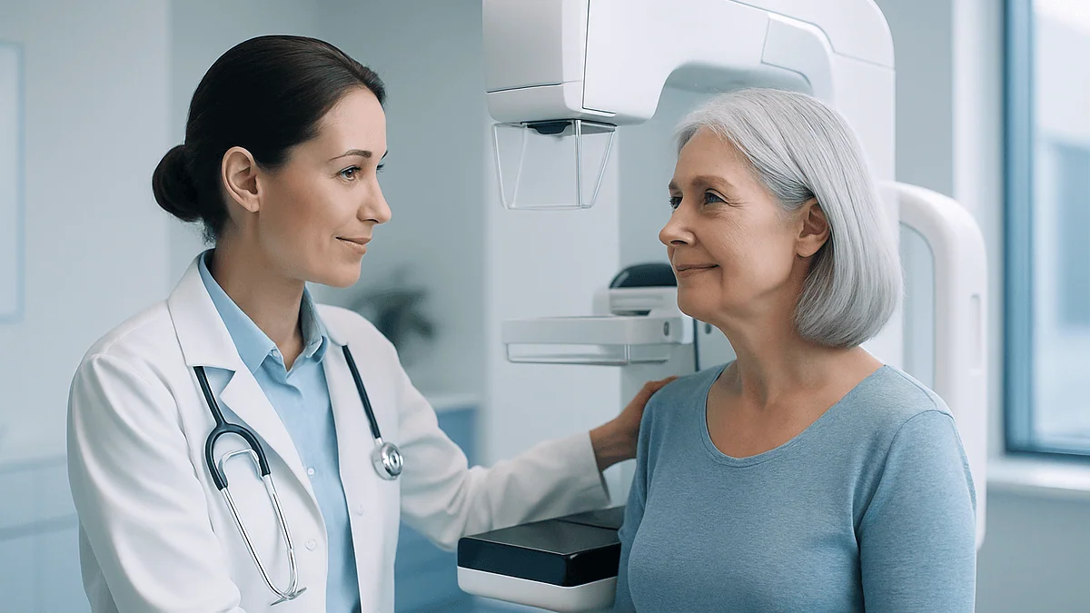
The whole appointment usually runs about 20 minutes; the imaging itself, closer to five. You will undress from the waist up, and a technologist will position one breast at a time on the machine’s platform. A clear plastic plate then compresses the breast for roughly 10 to 15 seconds per image — typically two views of each breast, top-down and angled.
The compression is the part everyone dreads, and it deserves an honest description: firm, briefly uncomfortable, occasionally painful, and over quickly. It is not gratuitous. Flattening the tissue spreads overlapping structures apart so small findings are not hidden, and a thinner target needs less radiation. Speaking of which: the total dose for a standard screening mammogram is about 0.4 millisieverts — for scale, Americans absorb around 3 millisieverts a year from natural background sources.
A few practical moves make the visit smoother:
- Skip deodorant, antiperspirant, powder, and lotion on your chest and underarms that morning — aluminum particles can mimic calcifications on the image.
- If you still menstruate, schedule for the week after your period, when breasts are least tender.
- Bring or transfer prior mammogram images if you are visiting a new facility; comparison with old films is one of the most powerful tools a radiologist has and prevents many unnecessary callbacks.
- Mention any breast symptoms when booking — a new lump changes the appointment from a screening to a diagnostic study, which involves additional views.
Results are legally required in the US to reach you within 30 days; in practice, most arrive within a week or two.
Dense breasts and 3D mammograms: what the new rules mean for you
Since September 2024, every mammogram report in the United States must tell you whether your breasts are dense — a federal requirement that turned a radiology footnote into a mainstream conversation. About 40% of women over 40 have heterogeneously dense breasts, and roughly 10% have extremely dense tissue.
Density matters twice over. Dense tissue modestly raises breast cancer risk itself, and it lowers the mammogram’s sensitivity, because dense tissue and tumors both appear white on X-ray. A woman with extremely dense breasts and a normal mammogram has genuinely useful information — but less reassurance than a woman with mostly fatty tissue and the same result.
Two responses have emerged from the evidence:
- 3D mammography (digital breast tomosynthesis) takes multiple thin image slices through the breast rather than one flat picture. Studies show it finds slightly more cancers — about one additional cancer per 1,000 women screened — and reduces callbacks, with the advantage most pronounced in dense breasts. Most US facilities now offer it, and it has effectively become the standard screening technology.
- Supplemental screening — ultrasound or MRI added to mammography — detects some cancers that mammograms miss in dense tissue. The tradeoff is real: more false positives and more benign biopsies. Guidelines stop short of recommending supplemental imaging for all dense-breasted women, so this is a genuinely individualized decision weighing your overall risk, your density category, and your tolerance for extra testing.
If your report says “dense,” do not read it as an alarm. Read it as an invitation to a ten-minute conversation about whether your screening plan should be adjusted.
How to read your mammogram results: the BI-RADS score explained
Every US mammogram report ends with a BI-RADS category — a standardized 0-to-6 scale that tells you far more than the words “normal” or “abnormal.” Decoding it takes the mystery out of the letter that arrives afterward.
- BI-RADS 0 — Incomplete. The radiologist needs more information: additional views, ultrasound, or your prior images for comparison. This is the most common reason for a callback and usually resolves benignly.
- BI-RADS 1 — Negative. Nothing to report. Continue routine screening.
- BI-RADS 2 — Benign finding. Something visible — a cyst, a calcified fibroadenoma, a stable lymph node — that is definitively not cancer. Also routine follow-up.
- BI-RADS 3 — Probably benign. The chance of cancer is under 2%. Rather than biopsy, the standard plan is a short-interval follow-up, typically at six months, to confirm stability.
- BI-RADS 4 — Suspicious. Biopsy is recommended. The category spans a wide range — from about a 2% to a 95% likelihood — and is often subdivided (4A, 4B, 4C). Most category 4 biopsies still come back benign.
- BI-RADS 5 — Highly suggestive of malignancy. At least a 95% likelihood; biopsy is needed promptly.
- BI-RADS 6 — Known, biopsy-proven cancer, used when imaging is done during treatment planning.
The single most useful thing to internalize: a 0 or a 3 is not a diagnosis, and even a 4 is a probability statement, not a verdict. The scale exists precisely so that uncertainty gets a number instead of a vague phrase.
Called back after a mammogram? Here's what the odds really say
The phone call lands, your stomach drops, and the two weeks until the follow-up appointment stretch like months. Before the worry spiral starts, the numbers deserve a hearing.
About 10% of screening mammograms in the US result in a callback for additional imaging. Of the women called back, the large majority — roughly 9 in 10 — do not have breast cancer. The callback usually means the radiologist saw overlapping tissue, an area obscured on one view, or a finding that simply needs a closer look with magnification views or ultrasound. First mammograms get called back more often than any other, for an unglamorous reason: there are no prior images to compare against, so every quirk of your normal anatomy is new information.
Even when a callback leads to a biopsy, benign results outnumber cancer. Most breast biopsies performed after screening come back showing normal tissue, cysts, or benign growths.
None of this makes the anxiety irrational — studies consistently document real, measurable distress from false positives, sometimes lasting months. It is the main cost of screening, and guideline committees weigh it seriously. Over ten years of annual mammograms, about half of women will experience at least one false positive, and 7 to 10% will undergo a benign biopsy.
Two things reduce your odds of the ordeal: screening at the same facility (or transferring prior images), and 3D mammography, which lowers callback rates. And one thing matters most of all — going to the follow-up appointment quickly rather than letting dread postpone it.
What are the warning signs of breast cancer — and when should you see a doctor?
Screening exists because early breast cancer usually causes no symptoms at all. But between mammograms, your own awareness matters, and certain changes warrant a prompt medical visit. The signs most often cited by major medical sources:
- A new lump or thickened area in the breast or underarm — the most common first sign
- A change in breast size or shape, particularly on one side
- Dimpling, puckering, or an orange-peel texture of the skin
- A nipple that newly turns inward, or a rash or scaling on the nipple
- Nipple discharge other than breast milk, especially if bloody or from one breast
- Redness, warmth, or swelling of part or all of the breast
- Persistent pain in one spot of the breast or nipple
Two honest caveats. First, most of these findings turn out to be benign — cysts, hormonal changes, infections, and harmless growths cause the same symptoms far more often than cancer does. Second, breast pain alone, especially pain in both breasts that tracks with your cycle, is rarely a cancer sign. Neither caveat is a reason to wait.
When to see a doctor: make an appointment within days — not months — for any new lump, one-sided nipple discharge, skin or nipple changes, or a breast change that persists through one full menstrual cycle. Do this regardless of your age and regardless of how recently you had a normal mammogram; a small percentage of cancers surface between screenings, and a prior normal result should never delay evaluation of a new symptom. Men, who account for about 1% of breast cancers, should apply the same rule to any chest lump.
Are there new mammogram guidelines for 2026?
Short answer: no major US guideline body has announced a new mammography recommendation taking effect in 2026, and searches suggesting otherwise are mostly chasing rumor. The current framework is the US Preventive Services Task Force recommendation finalized in April 2024 — screening every two years from 40 through 74 — alongside the American Cancer Society and American College of Radiology positions described earlier. Those remain the operative guidelines heading into 2026.
That said, the machinery of guideline revision never fully stops, and a few live questions are worth watching:
- Supplemental screening for dense breasts. The Task Force explicitly called the evidence insufficient to recommend for or against added ultrasound or MRI. Trials underway in the US and Europe may resolve this in future updates.
- Screening past 75. No randomized trial has ever included women over 74, leaving a genuine evidence gap that researchers continue to probe with observational data.
- Risk-based scheduling. Several large studies are testing whether screening intervals tailored to individual risk — rather than one calendar for everyone — outperform the current approach. Results could reshape guidelines late this decade.
A practical filter for future headlines: recommendations that change your care come from a handful of bodies — the Task Force, the American Cancer Society, the major specialty colleges — and are always published with full evidence reviews, then summarized on sites like the CDC’s. A viral post announcing “new mammogram rules” without one of those names attached deserves your skepticism. When guidance genuinely changes, your clinician’s office and your screening facility will be working from the same documents you can read yourself.
When can you stop getting mammograms?
Here the guidelines genuinely diverge, because the evidence genuinely runs out. Every major randomized trial of mammography enrolled women no older than 74, so no one can say with trial-grade certainty what screening accomplishes at 80. The Task Force therefore recommends through age 74 and formally declares the evidence “insufficient” beyond it — which is a statement about data, not a statement that screening stops working on a birthday.
The American Cancer Society takes a more functional view: continue screening as long as a woman is in good health and expected to live at least ten more years. That ten-year figure is not arbitrary. Screening finds cancers years before they would cause symptoms, so its lifesaving benefit takes roughly a decade to materialize. A woman with serious heart failure or advanced dementia is unlikely to benefit and still bears all the burdens — callbacks, biopsies, and treatment of slow-growing tumors that would never have caused harm in her remaining lifetime.
Breast cancer risk itself does not retire; incidence keeps climbing into the 70s. So the sensible question at 75 is not “Am I too old?” but “Is my overall health strong enough that finding a silent cancer now would meaningfully help me?” A vigorous 78-year-old who hikes on weekends may reasonably keep screening; her contemporary managing multiple serious illnesses may reasonably stop. Observational studies suggest healthy older women who continue screening are still diagnosed at earlier stages.
This is one of the few places in preventive medicine where “discuss it with your doctor” is not a dodge — it is literally what the evidence supports, because the deciding variables are personal ones no guideline table can capture.
Frequently asked questions
At what age should a woman have her first mammogram?
Age 40 for women at average risk, according to the 2024 US Preventive Services Task Force recommendation, with screening every two years through 74. The American Cancer Society offers the option at 40 and recommends annual screening by 45. Women at higher risk — BRCA gene changes, chest radiation in youth, or a mother or sister diagnosed young — often start earlier, sometimes around age 30, guided by a formal risk assessment.
Why can't you get a mammogram before 35?
Because it usually doesn’t work well at that age. Breast tissue in women under 35 is typically dense, and both dense tissue and tumors appear white on X-ray, sharply lowering the test’s accuracy. Breast cancer is also rare before 35, so screening produces mostly false alarms, and younger tissue is more radiation-sensitive. A young woman with a lump or other symptom should still be evaluated promptly — usually starting with ultrasound instead.
What are the 7 warning signs of breast cancer?
The most commonly cited signs are: a new lump in the breast or underarm; a change in breast size or shape; skin dimpling or an orange-peel texture; a newly inverted nipple or nipple rash; discharge other than breast milk, especially bloody or one-sided; redness or swelling of the breast; and persistent localized breast or nipple pain. Most of these turn out benign, but any of them warrants a doctor’s visit within days.
What are the new mammogram guidelines for 2026?
No major organization has announced new mammogram guidelines taking effect in 2026. The current standard remains the US Preventive Services Task Force’s 2024 recommendation: screening every two years from age 40 through 74 for average-risk women. Areas under active study — supplemental imaging for dense breasts, screening after 75, and risk-tailored intervals — could prompt future updates, which would be published with full evidence reviews and summarized on the CDC’s website.
How often should I get a mammogram after 40?
Every one or two years, depending on which guideline you and your clinician follow. The Task Force recommends every two years from 40 to 74; the American Cancer Society recommends yearly screening from 45 to 54, then every one to two years. Annual screening catches fast-growing tumors sooner but roughly doubles false positives. Many clinicians suggest yearly screening in the 40s and early 50s, then every two years after menopause.
Do I need a referral for a screening mammogram?
Often not. In much of the US, women 40 and older can self-schedule a screening mammogram directly with an accredited imaging facility, though rules vary by state and facility, and results must still be sent to a clinician who can act on them. Diagnostic mammograms — those investigating a symptom or an abnormal screening result — generally do require a clinician’s order. Call the facility ahead to confirm its policy and your insurance requirements.
How much radiation is in a mammogram, and is it safe?
A standard screening mammogram delivers about 0.4 millisieverts — roughly the amount of natural background radiation Americans absorb over seven weeks. Major health agencies consider this dose very low, and decades of evidence indicate the mortality benefit of screening from age 40 onward substantially outweighs the theoretical radiation risk. Facilities must meet federal quality and dose standards. The calculus differs for much younger women, which is one reason routine screening doesn’t start in the 20s.
What does it mean if I get called back after a mammogram?
Usually that the radiologist needs a closer look — not that you have cancer. About 10% of screening mammograms lead to callbacks for extra views or ultrasound, and roughly 9 in 10 of those women do not have breast cancer. Common reasons include overlapping tissue, an unclear area, or no prior images for comparison, which is why first mammograms are called back most often. Go to the follow-up promptly; most callbacks resolve at that visit.
When can I stop getting mammograms?
There is no universal stop age. The Task Force recommends screening through 74 and says the evidence beyond that is insufficient, because no trials included older women. The American Cancer Society advises continuing as long as you are in good health with at least a ten-year life expectancy, since screening’s benefit takes about a decade to materialize. A healthy 78-year-old may reasonably continue; a woman with serious illness may reasonably stop. Discuss it individually.
Does a normal mammogram mean I definitely don't have breast cancer?
No test is perfect, and mammograms miss roughly 1 in 8 breast cancers overall — more in women with dense breasts, where tumors can hide against dense tissue. Some cancers also emerge between screenings. A normal result is genuinely reassuring and means routine screening should continue, but it should never delay evaluation of a new lump, nipple discharge, or skin change. New symptoms warrant a doctor’s visit regardless of how recent your last normal mammogram was.
References
- CDC — Screening for Breast Cancer
- MedlinePlus — Mammography
- Cleveland Clinic — Mammogram
- NHS — Breast Screening (Mammogram)
This article is for general information only and is not a substitute for professional medical advice. Please consult a qualified doctor about your individual situation.

More from the Blog

The GGT Blood Test: The Liver Enzyme That Completes the Panel
The GGT blood test measures gamma-glutamyl transferase, an enzyme concentrated in the liver and bile ducts. Clinicians order it to help detect liver or…
The Hemolyzed Sample: Why Labs Sometimes Ask You to Come Back
A hemolyzed blood sample means red blood cells broke apart before testing, spilling their contents and skewing results such as potassium and certain enzymes.…

The Comprehensive Metabolic Panel: 14 Values, One Tube, Explained
A comprehensive metabolic panel (CMP) is a blood test that measures 14 substances to assess blood sugar, kidney function, liver health, electrolyte balance, and…

Liver Function Tests: What the Panel Really Screens
A liver function test is a blood panel that measures enzymes (ALT, AST, ALP), bilirubin, and proteins such as albumin to screen for liver…

The Lp(a) Test: The Once-in-a-Lifetime Genetic Lipid Check
The lipoprotein(a), or Lp(a), test is a blood test that measures an inherited cholesterol-carrying particle linked to higher risk of heart attack, stroke, and…

The Full Thyroid Panel: Which Tests Belong in a Complete Workup
A complete thyroid workup typically starts with TSH and free T4, adds free or total T3 when an overactive thyroid is suspected, and includes…