

3D Mammograms and Breast Ultrasound: How Modern Breast Screening Layers Up
Key Takeaways
- Combined with standard views, 3D mammography detects roughly one additional cancer per 1,000 women screened and cuts callback visits by about 15% or more compared with 2D alone.
- Compression is identical in 2D and 3D mammograms, so a 3D exam is not less painful: its comfort advantage is fewer repeat visits for false alarms.
- About 40–50% of women over 40 have dense breast tissue, and since September 2024 every US mammogram report is federally required to tell you if you do.
- Adding screening ultrasound for dense breasts finds roughly two to three extra cancers per 1,000 women, but most biopsies it triggers turn out benign.
- Ultrasound cannot replace mammography because it misses microcalcifications, often the earliest mammographic sign of highly treatable cancers.
- Screening 3D mammograms are covered without cost sharing by Medicare and most private plans, but supplemental ultrasound is often billed as diagnostic, so confirm coding before your appointment.
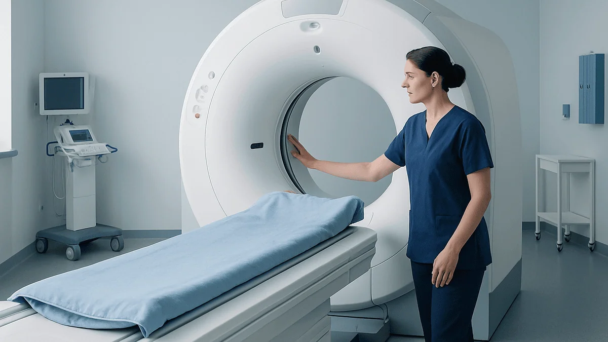
A 3D mammogram, or digital breast tomosynthesis, captures X-ray images in thin slices so radiologists can see through overlapping tissue. Studies suggest it detects roughly one additional cancer per 1,000 women screened and reduces callback visits compared with standard 2D mammography alone. Breast ultrasound adds a radiation-free look at dense tissue or a specific lump, but it supplements mammography rather than replacing it.
The letter arrives about a week after your mammogram. The result is normal, good news, but a sentence near the bottom stops you: “You have dense breast tissue, which can make cancer harder to detect.” Since September 2024, every mammography facility in the United States has been required to tell you this. Millions of women are now reading that line and wondering what, exactly, they’re supposed to do with it.
The answer usually involves layers. Modern breast screening has quietly become a stack of tools rather than a single test: a 3D mammogram as the foundation, sometimes an ultrasound on top, occasionally an MRI for those at highest risk. Each layer sees something the others miss. Each also adds cost, time, and the chance of a false alarm.
Here’s what the evidence actually shows about how these layers work together, and where the marketing gets ahead of the data.
What is a 3D mammogram, exactly?
A standard 2D mammogram takes two flat pictures of each breast, one from the top, one from the side. Everything inside the breast gets compressed into those single images, which means normal tissue can stack up and hide a small tumor, the way overlapping tree branches can hide a bird.
A 3D mammogram, known medically as digital breast tomosynthesis, solves the stacking problem mechanically. The X-ray arm sweeps in a shallow arc over the compressed breast, capturing a series of low-dose images from different angles in a few seconds. A computer then reconstructs those images into thin slices, often about a millimeter thick, that the radiologist can scroll through one at a time, like turning the pages of a book instead of trying to read them all pressed together.
That page-by-page view matters most in two situations. First, it helps reveal small cancers that overlapping tissue would otherwise camouflage. Second, it works in reverse: something that looked suspicious on a flat image often turns out to be ordinary tissue layered on top of itself, which the slices make obvious. According to Mayo Clinic, both effects show up in the data, more cancers found, fewer women called back for extra pictures.
From your side of the machine, the experience is nearly identical to a conventional mammogram. Same positioning, same compression paddle, a sweep that adds only a few seconds per view.
Is a 3D mammogram better than a regular mammogram?
Better at finding cancers and better at avoiding false alarms, yes, by modest but consistent margins. Large studies comparing tomosynthesis (usually combined with 2D images) against 2D alone have found roughly one additional invasive cancer detected per 1,000 women screened, alongside meaningful drops in recall rates. Some analyses put the callback reduction at 15% or more, which translates into thousands of women each year spared an anxious return visit.
Honesty requires a caveat, though, and it’s one the glossier articles skip. Finding more cancers at screening is not automatically the same as saving more lives. Some small, slow-growing abnormalities detected by any screening method might never have caused harm: a phenomenon researchers call overdiagnosis. What we don’t yet have is definitive proof that 3D mammography reduces deaths or advanced cancers better than 2D does. A large NIH-funded randomized trial called TMIST is following hundreds of thousands of women specifically to answer that question, with results still years away.
So the fair summary reads like this: 3D mammography is a genuine improvement in accuracy, more true positives, fewer false ones, and most major medical centers in the US now use it as their default screening tool. Whether it changes long-term outcomes is a question the evidence hasn’t finished answering. For a screening test you’ll repeat every year or two for decades, fewer false alarms alone is a real, tangible benefit.
What are the honest disadvantages of 3D mammography?
Every screening tool has a cost side of the ledger. For tomosynthesis, four items belong on it.
- Slightly more radiation, sometimes. When 3D images are taken in addition to standard 2D images, the dose roughly doubles, though it stays well under the safety limits set by the FDA. Most modern systems now sidestep this by generating a “synthesized” 2D image from the 3D data, bringing the total dose back to about the level of a conventional mammogram.
- Overdiagnosis remains possible. Finding more abnormalities includes finding some, particularly ductal carcinoma in situ, that might never have progressed. More detection can mean more biopsies and more treatment of uncertain necessity.
- Longer interpretation, occasional cost differences. A radiologist scrolls through dozens of image slices instead of four pictures. That extra reading time is one reason some insurers historically billed 3D separately, though coverage has broadened considerably (more on that below).
- No proven mortality advantage yet. As covered above, the definitive outcome data are still being gathered.
One disadvantage you can cross off the list: availability. A decade ago, tomosynthesis was a big-city amenity. Today it’s the majority of mammography units in the US, and MedlinePlus describes it as a standard screening option rather than an exotic one. If your local facility offers only 2D, that’s still a validated, effective screening test: a 2D mammogram done on schedule beats a 3D mammogram postponed.
Is a 3D mammogram less painful?
No, and it’s worth being straightforward about this, because some promotional material implies otherwise. The compression is the same. Tomosynthesis uses the same paddle, the same positioning, and roughly the same pressure as a 2D mammogram, because compression is what makes the images sharp: it spreads the tissue thin, holds it still, and reduces the radiation needed. The 3D sweep adds a few seconds per view, so if anything, each compression lasts marginally longer.
Where 3D genuinely improves the experience is downstream. Fewer false alarms means fewer callback appointments, and callbacks are where much of the discomfort of screening actually lives, both physically (repeat compressions, often firmer diagnostic views) and emotionally (days of waiting while imagining the worst). Cutting callbacks by even 15% removes a lot of cumulative squeezing and worry from the system.
If mammograms are painful for you rather than merely uncomfortable, a few evidence-backed adjustments help regardless of the technology:
- Schedule for the week after your period, when breasts tend to be least tender.
- Tell the technologist. Positioning adjustments and a brief pause can make a real difference, and compression only needs to last seconds.
- Skip caffeine that morning if you’ve noticed it makes your breasts more sensitive: the evidence here is mixed, but some women report it helps.
- An over-the-counter pain reliever beforehand is reasonable; ask your clinician or pharmacist what suits you.
Compression should feel like firm pressure, not sharp pain. A skilled technologist can usually find that line.
Dense breast tissue: why one picture isn't always enough
Breast density has nothing to do with how breasts look or feel from the outside. It refers to the ratio of glandular and fibrous tissue to fat, and it’s visible only on a mammogram. Radiologists sort density into four categories, from almost entirely fatty to extremely dense. Roughly 40 to 50 percent of American women over 40 fall into the two denser categories, according to the National Cancer Institute.
Density creates two separate problems, and the distinction matters. The first is a masking effect: dense tissue appears white on a mammogram, and so do tumors, searching for one in the other has been compared to spotting a snowball in a snowstorm. The second is independent of imaging: dense tissue itself is associated with a modestly higher risk of developing breast cancer, for reasons researchers are still working out.
Tomosynthesis helps with the masking problem, since scrolling through slices can separate a tumor from the dense tissue in front of and behind it. It helps less in extremely dense breasts, where even the slices remain overwhelmingly white. That’s the gap supplemental ultrasound was designed to fill.
Since September 2024, an FDA rule has required every US mammography report to tell you whether your breasts are dense and to note that dense tissue can hide cancers. The notification isn’t a diagnosis or an alarm: it’s a prompt to have a conversation with your clinician about whether your screening should have another layer.
Where breast ultrasound fits into the picture
Ultrasound approaches the breast with an entirely different kind of physics. Instead of X-rays, a handheld probe sends high-frequency sound waves into the tissue and listens for the echoes, building a live image from how the sound bounces back. No radiation, no compression paddle, just gel, gentle pressure, and a moving picture on a screen. Cleveland Clinic notes the exam typically takes 15 to 30 minutes.
Sound waves have a particular talent that X-rays lack: distinguishing fluid from solid. A fluid-filled cyst, extremely common and almost always benign, has a characteristic appearance on ultrasound that can settle the question on the spot, while on a mammogram the same cyst may look indistinguishable from a solid mass. This is why ultrasound is often the first tool used when a specific lump needs evaluating, and the default tool for younger patients, whose denser tissue limits mammography’s usefulness anyway.
In screening, ultrasound plays a supporting role. For women with dense breasts and a normal mammogram, adding a screening ultrasound detects, in most studies, roughly two to three additional cancers per 1,000 women, cancers the dense tissue had masked. Ultrasound also guides most needle biopsies, letting the radiologist watch the needle reach its target in real time.
The layering logic, then, is complementary rather than redundant: mammography surveys the whole breast for patterns X-rays capture best, and ultrasound investigates what sound waves characterize best.
Why ultrasound can't replace a mammogram
Given the choice between a radiation-free, compression-free scan and a mammogram, plenty of people would happily pick the ultrasound and skip the paddle forever. The evidence says that would be a mistake, for three reasons.
Microcalcifications come first. These tiny flecks of calcium, often the earliest visible sign of certain breast cancers, including some of the most treatable, show up crisply on mammograms and are largely invisible to ultrasound. A screening strategy built on ultrasound alone would systematically miss the very changes early detection is designed to catch.
Operator dependence comes second. A mammogram produces a standardized, reproducible image set; an ultrasound is only as thorough as the person or automated system sweeping the probe. Small areas can simply go unscanned, and comparing this year’s exam with last year’s is harder.
False positives come third, and the numbers are not trivial. Adding screening ultrasound to mammography in dense breasts finds those extra two to three cancers per 1,000, but it also flags many more findings that turn out to be benign after short-interval follow-ups or biopsies. In major studies, the majority of biopsies prompted by supplemental screening ultrasound found no cancer. That trade-off can be worthwhile for women with dense tissue or elevated risk; it’s a poor bargain as a wholesale mammogram replacement.
Every major medical organization, CDC included, treats ultrasound as an adjunct to mammography, not an alternative. The layers only work stacked in the right order.
2D vs. 3D vs. ultrasound at a glance
Seeing the three tools side by side makes the layering logic clearer. Note that in practice these aren’t competitors, most breast imaging centers use them in combination, choosing the mix based on your age, density, risk, and symptoms.
| Feature | 2D mammogram | 3D mammogram (tomosynthesis) | Breast ultrasound |
|---|---|---|---|
| How it sees | Two flat X-ray images per breast | X-ray sweep reconstructed into thin slices | Sound-wave echoes, live image |
| Radiation | Low dose | Similar with synthesized 2D; roughly double if 2D added separately | None |
| Compression | Yes | Yes, same as 2D | No |
| Exam time | ~10–20 minutes | ~10–20 minutes (seconds longer per view) | ~15–30 minutes |
| Strongest at | Microcalcifications, whole-breast survey | Seeing through overlapping tissue; fewer callbacks | Cyst vs. solid; dense tissue; guiding biopsies |
| Main limits | Overlap can hide or mimic tumors | Less advantage in extremely dense tissue; unproven mortality benefit vs. 2D | Misses microcalcifications; higher false-positive rate; operator-dependent |
| Typical role | Baseline screening where 3D unavailable | Primary screening at most US centers | Supplement for dense breasts; first look at lumps |
One tool surveys, one clarifies, one characterizes. The mistake is treating any single row of this table as the whole story: the strongest screening programs combine strengths deliberately rather than betting everything on one technology.
What actually happens during each exam
Knowing the choreography ahead of time takes much of the dread out of a first visit.
The mammogram. You’ll undress from the waist up and wear a gown that opens in front. Skip deodorant, powder, and lotion that morning, aluminum particles in some products can mimic calcifications on the images. The technologist positions one breast at a time on the machine’s platform; a clear plastic paddle lowers and compresses for several seconds while the image is captured. With tomosynthesis, you’ll notice the X-ray arm sweep in a small arc overhead. Two views per breast is standard, four compressions total, and the whole appointment usually runs 15 to 20 minutes. The NHS notes that any discomfort should last only as long as the compression itself: a matter of seconds each time.
The ultrasound. You’ll lie on your back, possibly with an arm raised overhead. The sonographer or radiologist applies warm gel and glides the probe across the breast, pausing to capture images and measurements. Nothing about it should hurt, though pressure over a tender area can be briefly uncomfortable. Some centers use automated whole-breast ultrasound systems for dense-breast screening, in which a larger transducer sweeps the breast systematically.
Afterward. Screening results typically arrive within days to a couple of weeks; diagnostic exams are often read the same day. If you have prior mammograms done elsewhere, arrange to have them sent ahead, comparison with old images is one of the most powerful accuracy tools a radiologist has, and it costs nothing.
Is a 3D mammogram covered by insurance?
Usually, yes, and the situation is far better than it was even five years ago, though the details reward a phone call.
Under the Affordable Care Act, most US health plans must cover screening mammograms without copays or deductibles for women 40 and older. As tomosynthesis has become the de facto standard of care, the large majority of private insurers now cover 3D screening the same way, and Medicare has covered screening tomosynthesis since 2015. A number of states have passed laws specifically requiring insurers to cover 3D at no extra cost. That said, a shrinking minority of plans still process the 3D portion as a separate billable item, which can generate a surprise charge of roughly $50 to $100. Two questions to your insurer prevent this: “Is screening digital breast tomosynthesis covered with no cost sharing under my plan?” and “Will the facility I’ve chosen bill it as part of the screening?”
Supplemental imaging is where coverage genuinely gets uneven. A screening ultrasound ordered because of dense breasts is often billed as diagnostic rather than preventive, meaning deductibles and copays can apply, although a growing list of states now mandates coverage for supplemental screening in dense breasts, and federal proposals to standardize this have been introduced repeatedly. Costs for a diagnostic workup (extra mammographic views, ultrasound, sometimes biopsy) also fall outside the free-screening guarantee.
None of this should be a reason to skip screening. Facilities routinely help patients navigate coverage, and low-cost screening programs exist in every state, including the CDC’s National Breast and Cervical Cancer Early Detection Program.
Getting called back: what the numbers really mean
The phone call, “the radiologist would like some additional images”, lands hard. It shouldn’t land quite as hard as it does, and the arithmetic explains why.
Roughly 10 in every 100 women screened with conventional mammography get called back for additional imaging; tomosynthesis lowers that number noticeably. Of those recalled, the overwhelming majority, well over 90 percent in typical screening populations, turn out not to have cancer. The finding was overlapping tissue, a cyst, a benign calcification, or simply an area that needed a clearer picture. First mammograms generate more callbacks than subsequent ones, purely because there’s no prior image for comparison.
The callback appointment itself usually involves diagnostic mammographic views (more focused, sometimes with firmer spot compression) and often an ultrasound of the specific area. Many workups end right there, with a same-day “benign, come back next year.” Some end with a recommendation for a short-interval follow-up in six months: a category radiologists use when a finding is almost certainly benign but worth watching. A minority proceed to needle biopsy, and even then, most biopsies come back benign.
None of this means callbacks are trivial; the anxiety is real and well documented, and it’s a legitimate cost of screening that honest medicine should acknowledge rather than wave away. It’s also precisely the cost that 3D mammography most reliably reduces. If you’re weighing where tomosynthesis earns its keep, this is the line item: fewer of those phone calls, fewer of those long weekends of waiting.
Who should consider adding ultrasound, or MRI, to screening?
Layering makes sense when your baseline layer has a known blind spot or your baseline risk is elevated. Three groups come up most often in the evidence.
- Women with dense breasts and average risk. This is the classic supplemental-ultrasound scenario: the mammogram is normal, but density limits how much that normal result can reassure. Adding ultrasound finds some masked cancers at the price of more false positives. Professional guidelines stop short of recommending it universally, which makes it a genuine shared decision between you and your clinician, one worth having rather than defaulting either way.
- Women at high lifetime risk. For those with certain inherited gene variants, a strong family history, or prior chest radiation in youth, major guidelines recommend annual breast MRI in addition to mammography, not instead of it. MRI is the most sensitive breast imaging tool available, and for this group its higher false-positive rate is an accepted trade.
- Anyone with a new symptom. A palpable lump gets diagnostic imaging tailored to age and the finding itself, often ultrasound first, usually with mammography, regardless of when the last screening happened or what it showed.
Risk calculators (your clinician may mention names like Tyrer-Cuzick or the NCI’s Breast Cancer Risk Assessment Tool) can put a number on lifetime risk and clarify which tier you’re in. The point of all this stratification is efficiency: aim the extra layers, with their extra false alarms, at the people whose odds justify them.
When to see a doctor, don't wait for your next screening
Screening is for people without symptoms. The moment you have one, the calendar becomes irrelevant: a change you can see or feel deserves evaluation now, even if your last mammogram was three months ago and pristine. Mammograms miss a meaningful minority of cancers, particularly in dense tissue, and a recent normal result should never talk you out of getting a new finding checked.
Contact your clinician promptly if you notice any of the following, in either breast or armpit:
- A new lump or area of thickening that feels different from the surrounding tissue
- Nipple discharge that occurs without squeezing, especially if bloody or from one breast only
- A nipple that has newly turned inward or changed shape
- Skin changes, dimpling or puckering (sometimes compared to an orange peel), redness, scaling, or warmth that doesn’t resolve
- A change in the size or contour of one breast
- Persistent pain in one specific spot that doesn’t track with your menstrual cycle
Two reassurances belong alongside that list. Most of these findings turn out to be benign, cysts, fibroadenomas, hormonal changes, and infections vastly outnumber cancers. And breast pain by itself, especially when it affects both breasts and ebbs with your cycle, is rarely a sign of cancer. But “probably benign” is a conclusion for a clinician with imaging in hand, not a bet to make alone at home. Men, take note too: male breast cancer is rare but real, and a lump behind the nipple warrants the same prompt evaluation.
When should you start, and how often should you go?
For women at average risk, the current US Preventive Services Task Force recommendation, updated in 2024, is to begin screening mammography at age 40 and repeat it every other year through age 74: a notable shift from earlier guidance that started routine screening at 50. Other respected bodies, including the American Cancer Society, support annual screening options starting in the 40s. The differences reflect honest disagreement about how to weigh earlier detection against more cumulative false positives, not a dispute about whether screening works.
What the guidelines agree on matters more than where they diverge: screening should start no later than your 40s, it should happen on a regular schedule rather than sporadically, and the interval, annual versus every two years, is a legitimate conversation to have with your clinician based on your density, family history, and preferences. Consistency beats perfection here; a mammogram every two years, every time, outperforms annual intentions that slip into three-year gaps.
Higher-risk women follow a different playbook, often starting earlier, sometimes a decade before the age at which a first-degree relative was diagnosed, and adding MRI. If breast or ovarian cancer runs in your family, especially at young ages, raise it with your clinician before you turn 40 rather than after.
And whichever schedule you land on, try to return to the same facility, or have prior images transferred. A radiologist comparing this year’s slices against last year’s catches subtle changes that no single exam, however advanced, can reveal on its own. That comparison is the quietest layer in the stack, and one of the most powerful.
Frequently asked questions
Is a 3D mammogram better than a regular mammogram?
By modest, consistent margins, yes. Studies show tomosynthesis finds about one additional cancer per 1,000 women screened and reduces false-alarm callbacks compared with 2D alone, because thin image slices see through overlapping tissue. What hasn’t been proven yet is whether it saves more lives than 2D; a large NIH trial is studying exactly that. A 2D mammogram done on schedule remains a validated, effective test.
What are the disadvantages of 3D mammography?
The main drawbacks are a slightly higher radiation dose when 3D images are added to separate 2D views (though still well under FDA limits, and largely eliminated by synthesized 2D software), the possibility of detecting slow-growing abnormalities that might never have caused harm, occasional extra insurance charges, and the fact that a survival advantage over 2D mammography hasn’t yet been demonstrated in completed trials.
Is a 3D mammogram covered by insurance?
Usually. Medicare has covered screening tomosynthesis since 2015, and most private plans now cover it without cost sharing under the Affordable Care Act’s preventive-care rules, with many states mandating it explicitly. A minority of plans still bill the 3D portion separately, which can add roughly $50–$100. Call your insurer beforehand and ask whether screening digital breast tomosynthesis is covered with no cost sharing.
Is a 3D mammogram less painful than a regular one?
No. The compression, the uncomfortable part, is identical, and the 3D sweep adds a few seconds per view. The real comfort benefit is indirect: fewer false-positive callbacks mean fewer repeat compressions and less anxious waiting. To reduce discomfort with any mammogram, schedule the week after your period, tell the technologist if positioning hurts, and remember each compression lasts only seconds.
Does a 3D mammogram expose me to more radiation?
Sometimes slightly, and it stays within FDA safety limits either way. When 3D images are captured alongside separate 2D images, the dose roughly doubles. Most modern systems avoid this by generating a synthesized 2D image from the 3D data, bringing total exposure back to about the level of a conventional mammogram, comparable to a few weeks of natural background radiation.
Can a breast ultrasound replace a mammogram?
No. Ultrasound misses microcalcifications, the tiny calcium flecks that are often the earliest visible sign of certain treatable cancers, and it produces more false positives when used for whole-breast screening. Its strengths are different: distinguishing fluid-filled cysts from solid masses, examining dense tissue, evaluating specific lumps, and guiding biopsies. Every major medical organization treats it as a supplement to mammography, not a substitute.
What does dense breast tissue on my results letter mean?
It means your breasts contain more glandular and fibrous tissue than fat, something visible only on a mammogram, affecting roughly 40–50% of women over 40. Dense tissue can mask tumors on X-ray images and is itself linked to modestly higher breast cancer risk. The letter, required by FDA rule since September 2024, is a prompt to discuss whether supplemental screening such as ultrasound makes sense for you.
How long does a 3D mammogram take?
About 15 to 20 minutes for the full appointment, essentially the same as a 2D mammogram. Each of the four compressions lasts only seconds, with the 3D sweep adding a few extra seconds per view. Skip deodorant, powder, and lotion that morning, since particles in some products can mimic calcifications on the images, and bring or transfer any prior mammograms for comparison.
Should I panic if I get called back after a mammogram?
No, callbacks are common and usually resolve benignly. Around 10% of women screened with 2D mammography are recalled (fewer with 3D), and well over 90% of those recalled do not have cancer. Most callbacks involve extra mammographic views and sometimes an ultrasound, often with same-day reassurance. First mammograms trigger more callbacks simply because there are no prior images to compare against.
At what age should I start getting mammograms?
Current US Preventive Services Task Force guidance, updated in 2024, recommends starting at age 40 and screening every other year through 74 for women at average risk; some organizations support annual screening. If you have a strong family history, known gene variants, or other risk factors, talk with your clinician well before 40, high-risk screening often starts earlier and adds MRI to mammography.
References
This article is for general information only and is not a substitute for professional medical advice. Please consult a qualified doctor about your individual situation.

More from the Blog

APOE4: What Carrying the Alzheimer’s Risk Gene Means, and Does Not Mean
APOE4 is the most common gene variant linked to late-onset Alzheimer's disease. One copy roughly doubles to triples lifetime risk compared with the most…

Vitamin D Test: What Level Is Actually Deficient and Who Needs Testing
A vitamin D test measures 25-hydroxyvitamin D in blood. Most US laboratories and the NIH call a level below 12 ng/mL (30 nmol/L) deficient…
Preventive Care Checklist: The Screenings and Vaccines Recommended by Age
Preventive care is the set of screenings, vaccines and counseling that find or prevent disease before symptoms appear. For most adults that means blood…

Whole Genome Sequencing and Polygenic Risk Scores: What Consumer Genomics Can Predict
Whole genome sequencing reads nearly all 3 billion letters of a person's DNA and can reliably identify rare single-gene variants linked to conditions such…

Protein in Urine: What a Urinalysis Finding Means for Your Kidneys
Protein in urine, called proteinuria, means the kidneys' filters are letting protein such as albumin leak through. A single positive result on a urinalysis…
Lynch Syndrome: The Inherited Cancer Risk Worth Knowing About
Lynch syndrome is an inherited condition, caused by a change in one of the DNA mismatch repair genes, that raises lifetime risk of colorectal,…