

Skin Cancer Screening and Mole Mapping: What a Proper Skin Check Involves
Key Takeaways
- Localized melanoma has a five-year relative survival around 99 percent, but that figure falls to roughly 35 percent once it has spread to distant sites, millimeters and months genuinely matter.
- The 'E' in ABCDE, evolving, is the most sensitive warning sign; a small changing spot outranks a large stable one, and the 'ugly duckling' mole that doesn't match its neighbors deserves equal attention.
- Mole mapping is standardized total-body photography whose two proven benefits are catching change earlier and sparing stable moles from unnecessary biopsies: it helps most in people with 50-plus or atypical moles.
- Dermoscopy, the lighted 10x scope pressed against the skin, improves melanoma detection over the naked eye while reducing biopsies of benign spots, but only in trained hands.
- In people with darker skin tones, melanoma most often appears on palms, soles, and under nails, sites unrelated to sun exposure and routinely missed in both self-checks and clinical exams.
- A sore that hasn't healed within three to four weeks, or a new dark streak under a nail without injury, warrants a medical exam even if it causes no pain at all.
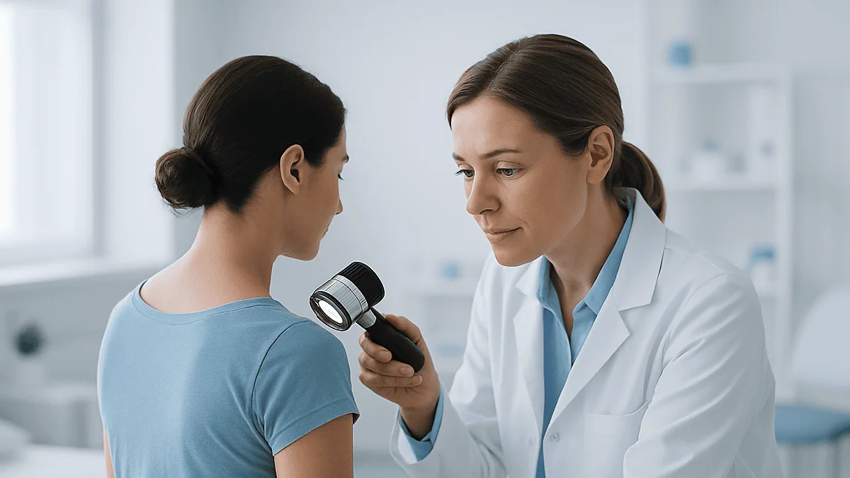
A skin cancer screening is a head-to-toe visual exam in which a clinician checks your skin, often using a handheld magnifier called a dermatoscope, looking for suspicious moles or growths. Mole mapping adds standardized photographs so subtle changes can be tracked over months and years. Most exams take 10 to 20 minutes, and any concerning spot is typically biopsied to confirm what it is.
The person who first notices a melanoma is often not a doctor. It’s a hairdresser who spots a dark, lopsided patch behind an ear, a spouse who asks about a mole on a shoulder blade, or someone catching an unfamiliar spot in the mirror after a shower. Skin is the one organ we can examine with nothing more than good light and honest attention, which makes it both the easiest cancer site to watch and the easiest to ignore.
Yet most adults have never had a professional skin exam, and many aren’t sure what one involves. Do you have to undress completely? Does mole mapping mean lasers and machines? What exactly is the doctor looking for?
Here’s a candid walk-through of the whole process, what happens in the room, what the photographs are for, what early skin cancer actually looks like, and where the scientific evidence is stronger (and weaker) than the marketing suggests.
Why most skin cancers are found by looking, not by lab tests
There is no blood test for skin cancer. No scan, no swab, no annual panel. Detection still comes down to eyes, yours, a partner’s, a clinician’s, noticing that something on the skin is new, growing, or simply doesn’t belong. According to the Cleveland Clinic, skin cancer is the most common cancer in the United States, with roughly one in five Americans developing it in their lifetime, and the vast majority of cases begin as something visible.
That visibility is the whole opportunity. A melanoma caught while it’s still confined to the top layers of skin is a fundamentally different disease than one that has traveled to lymph nodes. The difference between those two scenarios can be a matter of millimeters of depth, and often a matter of months in noticing.
Here’s the part worth being opinionated about: the single most valuable skill in skin cancer detection isn’t owned by any clinic. It’s your own familiarity with your own skin. A dermatologist meeting you for the first time doesn’t know whether that brown spot on your calf appeared last spring or has been there since childhood. You do. Professional exams, dermoscopy, and mole mapping are all powerful tools, but they work best layered on top of a patient who pays attention between visits. Everything below builds on that foundation.
How do I get screened for skin cancer?
The path is simpler than most people expect. You can start with your primary care clinician, who can examine any spot that worries you and perform a general skin check during a routine visit. If something looks ambiguous, or if you have significant risk factors, you’ll usually be referred to a dermatologist for a full-body exam. In many places you can also book directly with a dermatology practice; some communities offer free screening events, often in spring before sun season.
When you call, ask specifically for a “full-body skin exam” or “skin cancer screening” rather than a general appointment: it changes how the visit is scheduled and how much time is reserved. MedlinePlus notes that the exam itself is noninvasive: no needles, no fasting, no preparation beyond showing up with clean, product-free skin.
A few practical steps make the visit more useful:
- Remove nail polish beforehand, melanoma can appear under fingernails and toenails as a dark streak.
- Skip makeup and leave your hair loose or easy to part, since the scalp and face need checking too.
- Photograph or jot down any spots you’ve wondered about, including ones on your back you’ve only glimpsed.
- Bring your family history: a first-degree relative with melanoma meaningfully changes your risk profile.
If you’ve never had a baseline exam and you’re an adult with fair skin, a history of sunburns, or many moles, booking one is a reasonable act of self-knowledge, not hypochondria.
What happens during a full-body skin exam, step by step
You’ll be asked to change into a gown, typically keeping your underwear on. A chaperone can be present if you’d like one, say so at booking. The clinician then works systematically from scalp to soles, and a thorough exam really does mean thorough: the part lines of your hair, behind the ears, inside the mouth if warranted, under the breasts, between fingers and toes, the soles of the feet, and under the nails.
Those overlooked zones matter more than they seem. Melanomas in people with darker skin tones disproportionately appear on the palms, soles, and nail beds, sites that have nothing to do with sunbathing. In men, melanoma most often shows up on the trunk, especially the back; in women, on the legs. Both are areas people rarely inspect themselves.
Expect the whole exam to take 10 to 20 minutes if nothing needs closer study. The clinician may stretch the skin, use bright raking light, or press a dermatoscope against individual spots. You’ll likely hear the word “benign” a lot, seborrheic keratoses, cherry angiomas, and ordinary moles make up the overwhelming majority of what’s found.
Two things you should do during the exam: point out any spot that is new, changing, itching, or bleeding, even if it seems trivial, and ask the clinician to teach you what your normal moles look like. That second request turns a passive check into a lesson you’ll use every month afterward.
Dermoscopy: what that little lighted scope actually does
At some point the clinician will likely press a device the size of a small flashlight against a mole. That’s a dermatoscope: a magnifier, usually 10x, with polarized light that lets the examiner see through the skin’s surface glare into structures beneath: pigment networks, blood vessel patterns, dots and globules invisible to the naked eye.
Why it matters: the naked eye is decent at flagging obviously abnormal moles but mediocre at telling an unusual-but-harmless mole from an early melanoma. Dermoscopy narrows that gap. Reviews of the technique consistently find that trained users detect more melanomas and perform fewer unnecessary biopsies of benign spots than examiners using eyes alone. It’s one of the rare tools that improves both sensitivity and specificity at once.
The examination is painless and takes seconds per spot. Some clinicians apply a drop of gel or alcohol for contact dermoscopy; others use non-contact polarized scopes. Neither version involves radiation or discomfort.
A worthwhile caveat: dermoscopy is only as good as the person holding it. The skill takes formal training and case volume to develop, which is one reason a dermatologist’s full-body exam and a quick glance during an unrelated appointment are not equivalent experiences. If a spot has genuinely worried you, an exam that includes dermoscopic evaluation is worth seeking out. It’s also the technology that makes mole mapping, the next step up, more than just photography.
Mole mapping explained: photography, not fortune-telling
Strip away the futuristic name and mole mapping is systematic photography. In its full form, often called total body photography, you stand in a series of standardized poses while high-resolution images capture essentially your entire skin surface. Individual moles of interest may also get close-up dermoscopic photos. The images are stored, and at future visits (or via automated comparison software in newer systems) your skin is checked against its own baseline.
The logic is elegant: melanoma announces itself through change. A mole that looks mildly atypical but has been identical for five years is behaving like a mole. A spot that wasn’t in last year’s photos, or one that has grown two millimeters, is behaving suspiciously, even if it doesn’t yet look dramatic. Photography converts a clinician’s fallible memory (and yours) into an objective record.
Mole mapping earns its keep in two ways. First, it catches new and changing lesions earlier than unaided exams. Second, and this is underappreciated, it prevents biopsies. When a slightly odd mole can be proven unchanged against a dated photograph, it can be safely watched instead of cut.
What mole mapping is not: a diagnosis, a guarantee, or a substitute for examination. Fast-growing melanomas, particularly the nodular type, can appear and progress between annual photo sessions. The photos are a comparison tool; the judgment still comes from the human reading them and from you reporting changes between visits.
Who genuinely benefits from mole mapping, and who can skip it
Not everyone needs their skin photographed. For a person with a handful of ordinary moles and no significant risk factors, standard periodic exams and self-checks do the job. Mole mapping’s value concentrates in people whose skin is genuinely hard to monitor by memory alone.
Dermatologists most often recommend baseline photography for:
- People with many moles, commonly 50 to 100 or more, where no examiner could plausibly remember each one.
- Those with multiple atypical (dysplastic) moles, which are both harder to assess and markers of elevated melanoma risk.
- Anyone with a personal history of melanoma, whose risk of a second primary melanoma is substantially higher than the general population’s.
- People with a strong family history of melanoma, especially in first-degree relatives.
- Patients on long-term immunosuppression, such as organ transplant recipients, whose skin cancer risk rises steeply over time.
For these groups, photography changes clinical decisions: it flags true change early and spares stable moles from the scalpel. For everyone else, it’s optional, reasonable if it gives you peace of mind and you understand its limits, unnecessary if it doesn’t.
One honest note on cost and access: total body photography isn’t universally covered by insurance and availability varies. If formal mapping isn’t accessible to you, well-lit smartphone photos of your own moles, taken with a ruler or coin for scale and repeated every few months, capture much of the same benefit at home. Consistency matters more than equipment.
What does skin cancer look like at the start?
Early skin cancer is usually undramatic: that’s precisely why it gets missed. Forget the textbook images of large black lesions; those are late findings. At the beginning, each of the three main types tends to look like something forgettable.
Early basal cell carcinoma, the most common type, often appears as a small pearly or translucent bump, sometimes with fine visible blood vessels, or as a flat pink patch. People routinely mistake it for a pimple that never resolves or a nick that keeps re-crusting. On darker skin, it may look brown or glossy black.
Early squamous cell carcinoma typically starts as a rough, scaly, persistent patch or a firm red nodule, often on sun-exposed skin, ears, lips, hands, scalp. It can be tender. Its precursor, actinic keratosis, feels like fine sandpaper before it looks like much of anything.
Early melanoma most often looks like a mole with something slightly off: one edge blurrier than the rest, a second shade of brown creeping in, a diameter that’s grown past its neighbors. The Mayo Clinic notes it can arise in a longstanding mole or, more often, as an entirely new spot, and in people of color, frequently on palms, soles, or under nails as a dark streak.
The common thread across all three: persistence and change. Normal skin blemishes heal, fade, or stay identical. Skin cancers don’t.
The ABCDE rule, and the "ugly duckling" that outranks it
The ABCDE checklist remains the most useful mnemonic for melanoma, and it’s worth knowing cold:
- A: Asymmetry: one half of the spot doesn’t match the other.
- B: Border: edges that are ragged, notched, or blurred rather than smooth.
- C: Color: multiple shades in one lesion, browns, black, tan, sometimes red, white, or blue.
- D: Diameter: larger than about 6 millimeters, roughly a pencil eraser, though melanomas can be smaller when found early.
- E: Evolving: any change in size, shape, color, elevation, or new symptoms like itching or bleeding.
If you memorize only one letter, make it E. Evolution is the most sensitive signal on the list; a small, single-colored spot that is visibly changing deserves more concern than a large, stable one that has looked the same for a decade.
Then there’s the concept dermatologists lean on that never made it into the acronym: the ugly duckling sign. Most people’s moles resemble one another: a personal “signature” pattern of size and shade. A melanoma frequently breaks that pattern. It’s the one spot that’s darker, larger, or simply different from all its siblings. When scanning your own skin, don’t judge each mole in isolation; judge the crowd and look for the outlier.
Neither rule diagnoses anything. Both exist to answer one question: does this spot deserve a professional’s eyes? When in doubt, the answer is yes.
What are the 7 warning signs of skin cancer?
Search engines love the number seven, but here’s the honest framing first: there is no official medical list titled “the 7 warning signs of skin cancer.” What circulates online is a distillation of genuine red flags documented by sources like the NHS and Mayo Clinic. Gathered into seven, they look like this:
- A new spot appearing in adulthood, especially after age 40, when new moles become less common.
- A changing spotgrowing, darkening, developing new colors, or rising above the skin.
- A sore that won’t heal within three to four weeks, or one that heals and reopens in the same place.
- Bleeding, oozing, or crusting from a mole or bump without a clear injury.
- Persistent itch, tenderness, or pain in a single defined spot, sensation where there was none.
- A shiny, pearly, or translucent bump, sometimes with a rolled edge or a dip in the center, that slowly enlarges.
- Pigment spreading from a mole’s border into surrounding skin, or a dark streak under a nail that isn’t from trauma.
Notice what the list does not require: no dramatic size, no black color, no pain. Several early skin cancers are pink, small, and painless. Notice also what unites the seven, persistence over weeks and deviation from your normal. A spot triggering even one of these signs warrants an exam, not because it’s probably cancer (most flagged spots aren’t), but because the ones that are reward speed.
The three main skin cancers, compared
“Skin cancer” is really a family of diseases with very different personalities. The two most common types, basal cell and squamous cell carcinoma, arise from keratinocytes and grow slowly enough that they’re often called “non-melanoma” skin cancers as a group. Melanoma, arising from pigment cells, is far less common but drives the large majority of skin cancer deaths because of its capacity to spread.
| Basal cell carcinoma | Squamous cell carcinoma | Melanoma | |
|---|---|---|---|
| Share of skin cancers | Roughly 8 in 10 | Most of the rest | About 1 in 100, but most deaths |
| Typical early look | Pearly bump; flat pink patch; sore that recurs | Rough scaly patch; firm red nodule; may be tender | Irregular mole; new dark spot; nail streak |
| Common locations | Face, nose, ears, neck | Ears, lips, hands, scalp, forearms | Back (men), legs (women); palms, soles, nails in darker skin |
| Growth pace | Slow, months to years | Moderate, can invade locally | Variable; nodular type can advance in weeks |
| Spread risk | Very rarely spreads | Small but real risk if neglected | Significant once deep, depth drives prognosis |
The practical takeaway from the table: basal cell carcinomas almost never threaten life but do destroy local tissue if ignored, squamous cell carcinomas deserve prompt attention especially on the ears and lips, and melanoma is the reason “watch and wait” is the wrong strategy for any changing pigmented spot.
Is skin cancer serious if caught early?
Serious enough to act on, and far more manageable than most cancers when found soon: that’s the fair two-part answer. The numbers behind it are striking. For melanoma diagnosed while still localized to the skin, U.S. cancer registry data show a five-year relative survival around 99 percent. Once melanoma reaches regional lymph nodes, that figure drops into the 70s; with distant spread, to roughly 35 percent. Few diseases in medicine show such a steep gradient between early and late detection.
For basal and squamous cell carcinomas, the early-detection story is less about survival and more about what removal involves. Caught small, most are treated with a minor outpatient procedure and a modest scar. Left for years, a basal cell carcinoma on the nose or eyelid can erode into cartilage and require reconstructive surgery, not usually life-threatening, but genuinely disfiguring. Squamous cell carcinoma, particularly on the ear or lip or in immunosuppressed patients, carries a real though small risk of spreading if neglected.
Two honest qualifiers belong here. First, statistics describe populations, not individuals; your situation depends on the specific tumor’s type, depth, and biology. Second, “early” is measured in melanoma by millimeters of depth, which is why a delay of a few months can matter more than it would in slower cancers. The message isn’t fear: it’s that vigilance has an unusually high payoff for this particular disease.
How to do a monthly self-check at home
A self-exam takes about ten minutes and needs three tools: a full-length mirror, a hand mirror, and good light. Once a month is the rhythm most sources suggest, frequent enough to catch change, spaced enough that you’ll actually notice it.
Work in the same order every time so nothing gets skipped:
- Face, ears, and neck, including the rims and backs of the ears.
- Scalp, parting hair section by section: a comb or a partner’s help makes this feasible.
- Arms and hands: tops, palms, between fingers, and under fingernails.
- Torso front and back, using the hand mirror for your back and the backs of your shoulders; women should lift the breasts to check beneath.
- Legs all around, then feet, tops, soles, between toes, and under toenails.
- Buttocks and genital area, which do develop skin cancers despite never seeing sun.
Photograph anything you want to track, with a coin beside it for scale, and date the photo. Comparing images taken three months apart beats trusting memory every time. Note new spots, anything matching the ABCDEs, and any “ugly duckling” that stands out from your usual pattern.
For people with darker skin, add deliberate attention to palms, soles, and nails: the sites where melanoma most often appears and where diagnosis is most often delayed. The historical pattern of later-stage diagnosis in Black patients is partly a story of nobody, including clinicians, looking in the right places.
What the screening evidence honestly shows, and what it doesn't
Here’s the part most articles skip. In 2023, the U.S. Preventive Services Task Force reviewed the research and concluded that current evidence is insufficient to weigh the benefits and harms of routine full-body screening exams in asymptomatic adults without symptoms or elevated risk. The CDC likewise notes there is no standard population-wide screening recommendation for skin cancer the way there is for breast or colon cancer.
Read that carefully, because it’s widely misunderstood. “Insufficient evidence” does not mean skin exams don’t work. It means no large randomized trial has yet proven that screening every symptom-free adult reduces skin cancer deaths at the population level, and such trials are genuinely hard to run for a cancer this visible and this often self-detected. The potential downside researchers weigh isn’t the exam itself, which is harmless, but downstream effects: biopsies of benign spots, scarring, anxiety, and possible overdiagnosis of lesions that would never have caused trouble.
What the evidence does support is a risk-stratified approach. People with a personal or family history of melanoma, many or atypical moles, fair sun-damaged skin, a history of blistering sunburns or tanning bed use, or immunosuppression have clearly elevated risk, and regular professional exams for these groups are standard dermatologic practice. Prompt evaluation of any spot that is new, changing, or symptomatic is universally recommended: that’s diagnosis, not screening, and its value isn’t in dispute.
In plain terms: if you’re low-risk with unremarkable skin, an annual exam is a reasonable choice rather than a proven mandate. If you carry risk factors, make it a habit.
When to see a doctor
Some skin findings justify an appointment within days to weeks rather than at your next routine check. Make the call if you notice:
- A mole or spot that has changed in size, shape, or color over weeks to months.
- A new pigmented spot appearing in adulthood, particularly one growing or darker than your other moles.
- A sore, pimple-like bump, or scaly patch that hasn’t healed after three to four weeks.
- Any spot that bleeds, oozes, crusts, itches persistently, or becomes tender without injury.
- A new dark streak under a fingernail or toenail not explained by a recent bang or bruise.
- Pigment spreading from a mole into the surrounding skin, or a mole developing a blurry, notched border.
The NHS uses a similar threshold, moles that change, plus sores that fail to heal within about four weeks, as the trigger for prompt evaluation. None of these findings means you have cancer; most turn out benign. They mean the question deserves a trained answer rather than months of self-monitoring.
Seek care more urgently if a skin lesion is growing visibly week to week, bleeding repeatedly, or accompanied by a new lump in the nearby lymph node areas of the neck, armpit, or groin. And if you’ve previously had any skin cancer, a lower threshold applies to everything above: report changes early, keep your follow-up schedule, and treat your skin history as the risk factor it is. Nobody at a dermatology office ever considers a changing mole a wasted appointment.
What happens if the doctor finds something suspicious
First, breathe: being asked to biopsy a spot is not a diagnosis. It’s how the question gets answered, and most biopsied lesions come back benign.
The procedure itself is minor. After numbing the area with a local anesthetic injection, the pinch of the needle is genuinely the worst part, the clinician removes some or all of the spot. A shave biopsy takes a thin surface layer; a punch biopsy removes a small cylinder a few millimeters across; an excisional biopsy takes the entire lesion with a rim of normal skin, often preferred for suspected melanoma so a pathologist can measure its full depth. Most biopsies take under fifteen minutes, and you drive yourself home.
The sample goes to a pathologist, who examines it under a microscope. Results typically arrive within one to two weeks: an admittedly long-feeling wait. The report will name the diagnosis and, for melanoma, describe its thickness, which is the single most important factor guiding what happens next.
If the result shows a skin cancer, your care team will discuss options appropriate to its type, depth, and location; for early lesions, that conversation usually centers on removal procedures done in outpatient settings. If it’s benign, you’ll still have gained something: a documented answer, a baseline for that area of skin, and, worth insisting on, a habit. Ask when your next full skin exam should be, put it on the calendar, and keep doing your monthly checks between visits. Screening isn’t a single event; it’s a rhythm.
Frequently asked questions
How do I get screened for skin cancer?
Book a full-body skin exam with a dermatologist, or ask your primary care clinician to check your skin and refer you if needed. The exam is visual, no needles or preparation, and takes about 10 to 20 minutes. When scheduling, specifically request a skin cancer screening so enough time is reserved. Arrive without makeup or nail polish, and bring notes on any spots that are new, changing, itching, or bleeding.
What does skin cancer look like at the start?
Usually something small and unimpressive: a pearly bump mistaken for a pimple, a rough scaly patch that feels like sandpaper, a sore that keeps re-crusting, or a mole with one blurry edge or a new shade of brown. Early melanoma can also appear as an entirely new dark spot, or a streak under a nail. The unifying clues are persistence beyond a few weeks and visible change, normal blemishes heal; skin cancers don’t.
What are the 7 warning signs of skin cancer?
There’s no official list of seven, but the red flags documented by major medical sources are: a new spot in adulthood; a changing mole; a sore that won’t heal within three to four weeks; bleeding or crusting without injury; persistent itch or tenderness in one spot; a shiny pearly bump that slowly enlarges; and pigment spreading beyond a mole’s border or appearing as a nail streak. Any single one justifies a professional exam.
Is skin cancer serious if caught early?
Early detection changes the picture dramatically. Melanoma found while confined to the skin carries a five-year relative survival around 99 percent in U.S. registry data, versus roughly 35 percent after distant spread. Basal and squamous cell carcinomas caught small are typically handled with minor outpatient procedures, while neglected ones can invade surrounding tissue. Early skin cancer still requires proper treatment and follow-up, but the outlook at that stage is among the best in oncology.
How often should I have a professional skin check?
It depends on your risk. There’s no universal schedule for low-risk adults: the evidence for routine screening of symptom-free people is officially rated insufficient, though many choose an annual exam. If you have a personal or family history of melanoma, many or atypical moles, fair sun-damaged skin, or take immunosuppressive medicines, regular exams every six to twelve months are standard practice. Ask your dermatologist to set your interval, and do monthly self-checks regardless.
What does mole mapping involve, and does it hurt?
Nothing painful at all, mole mapping is photography. You stand in a series of standardized poses while high-resolution images document your skin surface, and individual moles may get magnified dermoscopic photos. The session typically takes 20 to 40 minutes. At future visits, your skin is compared against these baseline images so new or changing spots stand out. It involves no radiation, no contrast, and no discomfort beyond standing still in a gown.
Can a doctor tell if a mole is cancerous just by looking?
Not with certainty. Visual exam and dermoscopy let a trained clinician sort spots into ‘clearly benign,’ ‘clearly suspicious,’ and ‘uncertain’ with impressive accuracy, but the definitive answer always comes from a biopsy examined by a pathologist under a microscope. That’s why being sent for a biopsy isn’t a diagnosis: it’s the standard way to answer the question, and most biopsied spots turn out to be benign.
Do people with dark skin need skin cancer screening?
Yes. Skin cancer is less common in darker skin but historically diagnosed at later stages, partly because it appears in places nobody checks: palms, soles, under nails, and inside the mouth. Melanoma in people of color frequently arises at these non-sun-exposed sites. Self-exams should deliberately include the hands, feet, and nails, and any new dark streak under a nail or non-healing sore deserves prompt professional evaluation regardless of skin tone.
Are smartphone mole-checking apps reliable?
Not reliable enough to rule out cancer. Studies of algorithm-based apps have found variable and sometimes poor accuracy, with real risk of falsely reassuring users about dangerous lesions. Where phones genuinely help is documentation: dated, well-lit photos of your moles, taken with a coin for scale and repeated every few months, create a change record that makes both self-checks and clinical visits more effective. Use the camera; be skeptical of the verdict.
What should I do to prepare for a skin cancer screening?
Arrive with bare, product-free skin: no makeup, and no nail polish, since melanoma can appear under nails. Wear hair loose or easy to part so your scalp can be examined. Beforehand, note or photograph any spots that are new, changing, itching, or bleeding, including hard-to-see areas like your back. Bring your family history of skin cancer and a list of past sunburns, tanning bed use, or previous skin biopsies if you’ve had them.
References
- MedlinePlus: Skin Cancer Screening
- NHS: Melanoma Skin Cancer
- CDC: About Skin Cancer
- Cleveland Clinic: Skin Cancer
This article is for general information only and is not a substitute for professional medical advice. Please consult a qualified doctor about your individual situation.

More from the Blog

The Sleep Study: What a Night of Polysomnography Actually Records
A sleep study, or polysomnography, is an overnight test that records brain waves, eye movements, muscle tone, heart rhythm, breathing effort, airflow, blood oxygen,…

Malaria Tablets and Vaccines: What Exists and Who Needs Them
Yes. Since 2021 the World Health Organization has recommended two malaria vaccines, but only for young children living in parts of Africa with moderate…

DNA and Paternity Testing During Pregnancy: What Is Possible
Yes. A paternity DNA test can be done during pregnancy using a blood sample from the pregnant person, which carries small fragments of the…

Retinal Scans: What an Eye Photo Reveals About Diabetes, Blood Pressure and the Brain
A retinal scan is a photograph or light-based scan of the back of the eye. It can show early blood-vessel damage from diabetes and…

The BRCA Gene Test: Who Should Consider It and What Results Change
The BRCA gene test is a blood or saliva test that looks for inherited changes in the BRCA1 and BRCA2 genes, which can substantially…
Prostate MRI: How It Changed Prostate Cancer Diagnosis
Prostate MRI, a detailed magnetic scan of the prostate gland, is now widely used before biopsy when a PSA test or examination raises concern.…