Skin Cancer Treatment
Skin cancer care includes diagnosis, staging and personalized treatment for melanoma and non-melanoma skin cancers, using surgery, systemic therapy or radiotherapy when needed.
Quick answer
Skin cancer is the uncontrolled growth of abnormal cells in the skin, most often triggered by ultraviolet damage. The main types are basal cell carcinoma, squamous cell carcinoma and melanoma. Treatment usually starts with a biopsy to confirm the diagnosis, followed by surgical removal; radiotherapy or systemic drug therapy may be added for higher-risk or advanced disease.
What Is Skin Cancer?
Skin cancer is the uncontrolled growth of abnormal cells in the skin, most often caused by DNA damage from ultraviolet radiation in sunlight or tanning devices. It develops when damaged skin cells begin to multiply without the normal controls that keep tissue growth in check, forming a lesion that can invade deeper layers and, in some types, spread to lymph nodes or distant organs. It can affect anyone, though people with fair skin, a history of sunburn, many moles, previous skin cancer or a weakened immune system carry a higher risk.
Skin cancer is not one disease. It includes several cancers with different behaviours, risks and treatment pathways. Some grow slowly and respond very well to a single precise surgical procedure. Others, especially melanoma and certain high-risk squamous cell carcinomas, require careful staging and coordinated care from several specialists. The most important first step is understanding exactly what type of skin cancer you have, how deep or advanced it is, and which treatment plan offers the best balance between cancer control, safety, function and cosmetic outcome. That understanding comes from pathology and staging, not from appearance alone.
A skin cancer diagnosis raises predictable questions. Has it spread? Will surgery change my appearance? Will I need radiotherapy or drug treatment? How quickly do I need to decide? At Acibadem, skin cancer care is organised around accurate diagnosis, individualised treatment planning and multidisciplinary decision-making when the case requires it. Dermatology, plastic and reconstructive surgery, surgical oncology, medical oncology, radiation oncology, pathology, radiology and nuclear medicine specialists may all be involved, depending on the diagnosis.
This page explains what skin cancer care covers: how the disease is recognised, how it is diagnosed and staged, how surgery, radiotherapy and systemic therapy are used, what recovery typically involves, and which factors genuinely influence outcomes. It is written to help you ask better questions, whoever ends up treating you.
Types of Skin Cancer
Types of skin cancer differ fundamentally in how they grow, how likely they are to spread, and how they are treated. Knowing which type you have matters more than almost anything else in planning care, because a strategy that is appropriate for one type can be entirely wrong for another.
Basal cell carcinoma
Basal cell carcinoma is the most common form of skin cancer. It usually grows slowly and very rarely spreads to distant organs, but it should not be dismissed for that reason: if neglected, it can invade deeper tissues, cartilage or bone and become considerably harder to remove, particularly on the face. Basal cell carcinomas take several forms, superficial, nodular and infiltrative among them, and the infiltrative and recurrent variants require more careful surgical planning because their edges extend further than the visible lesion suggests.
Squamous cell carcinoma
Squamous cell carcinoma is the second most common type and can behave more aggressively than basal cell carcinoma. Most are low risk when found early, but the risk rises when a tumour is large, deep or recurrent, when it sits on a high-risk site such as the lip or ear, when it involves nerves, or when the patient has a weakened immune system, for example, organ transplant recipients on long-term immune-suppressing medication. High-risk squamous cell carcinoma can spread to lymph nodes, which is why staging matters in these cases.
Melanoma
Melanoma is less common than basal and squamous cell cancers but has a markedly greater tendency to spread if it is not diagnosed and treated early. It arises from the pigment-producing cells of the skin and can develop within an existing mole or as a new dark lesion. Because its behaviour is strongly linked to tumour thickness, melanoma is the most time-sensitive of the common skin cancers. Its evaluation and treatment follow a distinct pathway, described in detail on our melanoma skin cancer page.
Merkel cell carcinoma and rarer skin cancers
Merkel cell carcinoma is an uncommon but aggressive skin cancer that often requires multidisciplinary treatment combining surgery, radiotherapy and, in some cases, systemic therapy. Other rare skin malignancies, including certain adnexal tumours and cutaneous lymphomas, also fall within comprehensive skin cancer care. For these diagnoses, expert pathology review is particularly important, because rare tumours are the ones most often misclassified at first assessment.
Skin Cancer Symptoms and Warning Signs
Skin cancer symptoms are usually visible rather than felt, which makes the skin unusual among organs: you can inspect it yourself. Some lesions are obvious, while others are subtle and easy to overlook. A new spot that does not heal, a changing mole, a bleeding bump, a rough scaly patch, a dark streak under a nail, or a sore on the lip or ear all deserve assessment by a qualified physician. Evaluation is especially important if a lesion is changing in size, colour, shape or texture, or if it becomes painful, itchy, crusted or ulcerated. Change over time is the single most useful warning sign.
What does skin cancer look like?
What does skin cancer look like? It depends on the type, and there is no single appearance. Basal cell carcinoma often appears as a pearly or waxy bump, sometimes with visible small blood vessels, or as a flat, pale, scar-like patch; it may crust, bleed with minor trauma and fail to heal. Squamous cell carcinoma typically looks like a firm red nodule or a rough, scaly, crusted patch that may ulcerate. Melanoma usually appears as a dark lesion with irregular features: the widely used ABCDE checklist points to Asymmetry, irregular Borders, uneven Colour, a Diameter larger than a pencil eraser, and Evolution or change over time. Amelanotic melanoma, which lacks the usual dark pigment, is a reminder that appearance alone is never conclusive.
Early stage skin cancer: what to watch for
Early stage skin cancer is often small, flat and unremarkable, which is exactly why it is missed. An early basal cell carcinoma may look like a small shiny spot or a patch of dry skin that keeps returning. An early squamous cell carcinoma may resemble a stubborn rough patch. Early melanoma may be a mole that has begun to change subtly in shade or outline. The practical lesson is that persistence matters as much as appearance: a lesion that has been present and slowly changing for weeks or months warrants a professional look, even if it seems trivial. Early lesions are the ones treated with the smallest procedures and the simplest recoveries.
Skin cancer pictures: how useful are they?
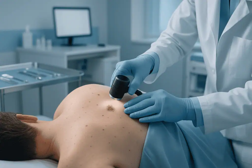
Skin cancer pictures found online can help you understand the range of appearances, but they cannot diagnose your lesion, and they should not reassure you. Photograph collections tend to show classic, well-developed examples, while real lesions, especially early ones, are often subtler. Pictures also cannot show the features a dermatologist assesses with dermoscopy, a magnified, illuminated examination that reveals structures invisible to the naked eye. Use pictures for awareness; use a physician for assessment.
Melanoma pictures and the limits of self-diagnosis
Melanoma pictures typically illustrate the ABCDE features: asymmetric shape, ragged borders, mixed browns and blacks, larger diameter and documented change. They are useful for training your eye during self-examination, but melanoma can also be small, uniform in colour or entirely non-pigmented, and benign moles can look alarming. Comparing your own skin to photographs is a starting point for vigilance, not a substitute for dermoscopy and, where indicated, biopsy.
Does skin cancer itch?
Does skin cancer itch? Sometimes. Basal cell and squamous cell carcinomas can itch, sting or feel tender, and some melanomas itch as they grow. However, many skin cancers cause no sensation at all, and most itchy skin lesions are benign, eczema, insect bites and dry skin itch far more often than cancer does. Itch is therefore neither a reliable warning sign nor a reassurance. What matters is the combination: a persistent lesion that itches, bleeds, crusts or changes deserves assessment regardless of how it feels.
What cancer can cause itchy skin?
Several cancers can be associated with itching. Skin cancers themselves sometimes itch locally, at the site of the lesion. Persistent, generalised itching without a visible rash is a different phenomenon: it is usually caused by benign conditions such as dry skin, allergy, liver or kidney problems or thyroid changes, but in a small number of cases it accompanies internal disease, including some blood cancers such as lymphoma. This is a question of context rather than alarm: a physician evaluating unexplained itching will consider the whole clinical picture, not a single symptom.
Who May Need Skin Cancer Evaluation, and How Diagnosis Works
Patients who may need skin cancer care fall into several groups: those with a newly diagnosed biopsy result, those with a suspicious lesion that has not yet been biopsied, and those seeking a second opinion after being advised to have surgery, radiotherapy or systemic therapy. Some patients come for treatment after a cancer has returned following previous removal. Others need multidisciplinary review because the cancer sits close to the eye, nose, mouth, ear, scalp, hand or genital area, where cancer control and reconstruction both require careful planning.
How is skin cancer diagnosed?
Skin cancer is diagnosed by biopsy, no examination, photograph or scan replaces tissue under a microscope. The pathway usually begins with a clinical examination of the skin and, when appropriate, dermoscopic assessment. Dermoscopy allows the physician to view patterns and structures not visible to the naked eye, helping to distinguish benign lesions from suspicious ones and to decide which lesions genuinely need sampling. If cancer is suspected, a biopsy is performed. The method, excisional biopsy, punch biopsy, shave biopsy or incisional biopsy, depends on the lesion and its location. Pathology then confirms the diagnosis and provides the details that drive treatment: cancer type, depth, ulceration, margin status, nerve involvement and other risk features. In skin cancer, a few millimetres of depth or a single positive margin can change the entire plan, which is why a reliable pathology report is worth more than a fast one.
How is skin cancer staged?
Staging determines whether the cancer is confined to the skin or has spread, and it dictates strategy. For most small basal cell carcinomas, no staging beyond examination is needed. For melanoma and higher-risk squamous cell carcinomas, additional steps may include imaging studies, lymph node ultrasound or sentinel lymph node biopsy: a procedure that samples the first lymph node draining the tumour to check for microscopic spread. Imaging may involve ultrasound, computed tomography, magnetic resonance imaging, or positron emission tomography combined with computed tomography; brain imaging is considered for selected melanoma patients. In advanced melanoma and selected other cancers, molecular testing of the tumour can identify specific mutations that influence drug treatment choices. Accurate staging matters because early-stage disease and advanced disease require very different strategies, and treating one as the other serves nobody.
Conditions and Indications Addressed
Comprehensive skin cancer care addresses both common and complex skin malignancies, with the pathway adapted to tumour type, stage and patient priorities. Indications may include first-time treatment, recurrence management, staging, reconstruction, or coordination of drug therapy and radiotherapy.
- Basal cell carcinoma: including superficial, nodular, infiltrative or recurrent tumours, especially in cosmetically or functionally sensitive areas.
- Squamous cell carcinoma: including low-risk lesions, high-risk tumours, recurrent disease and cancers with nerve involvement or lymph node spread.
- Melanoma: from early thin melanoma to thicker tumours, nodal disease or metastatic melanoma requiring systemic therapy.
- Merkel cell carcinoma: a less common but aggressive skin cancer that often requires multidisciplinary treatment.
- Skin cancers in high-risk locations: including the eyelids, nose, lips, ears, scalp, hands, feet and genital area.
- Recurrent skin cancer: cancers returning after prior surgery, radiotherapy or other treatment.
- Skin cancers in immunosuppressed patients: including transplant recipients and patients receiving long-term immune-suppressing medication prescribed by their own physicians.
- Advanced or metastatic skin cancer: disease that has spread to lymph nodes, soft tissue, bone, lung, liver, brain or other organs.
Not every patient needs every treatment modality: this point deserves emphasis. A small, low-risk basal cell carcinoma may be managed with a single localised procedure, while melanoma may require wider excision, nodal evaluation and oncology review. The value of a coordinated programme is that each patient’s plan is based on the biology of the cancer rather than a single standard approach applied to everyone.
How Skin Cancer Treatment Is Performed
Preparation and treatment planning
The process begins with a detailed medical review. Patients are usually asked to provide pathology reports, biopsy slides or blocks if available, prior operation notes, imaging studies, photographs of the lesion, medication lists and relevant medical history. Original biopsy material matters more than patients expect: when the slides or tissue blocks themselves are available, the pathology team can re-examine the tissue directly rather than relying only on the written report, which is where subtle classification differences are caught.
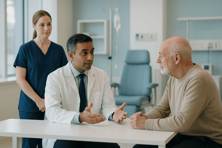
During the initial consultation, the physician examines the lesion and surrounding skin, checks regional lymph nodes when relevant, and reviews the pathology. If the diagnosis is uncertain, a second pathology review may be requested: a routine safeguard rather than a criticism of the original laboratory. When staging is necessary, the imaging described above is arranged, and for some melanoma patients sentinel lymph node biopsy is planned alongside the main operation. Only when type, depth and extent are clear does treatment selection begin in earnest.
Surgery for localised skin cancer
Surgery is the most common treatment for localised skin cancer, and for many patients it is the only treatment needed. A typical surgical pathway follows these steps:
- Planning: the surgeon maps the tumour and marks a margin of healthy-appearing tissue around it, guided by tumour type, size, depth and international protocols. For melanoma, the margin is determined chiefly by tumour thickness.
- Anaesthesia: many excisions are performed under local anaesthesia as outpatient procedures; larger operations, lymph node procedures or complex reconstruction may require sedation or general anaesthesia.
- Excision: the tumour is removed together with the planned margin. For selected high-risk non-melanoma cancers, particularly where tissue preservation matters, such as on the face, margin-controlled techniques may be used, in which the tissue edges are examined systematically before the wound is closed.
- Lymph node procedures when indicated: sentinel lymph node biopsy may be performed in the same session for appropriate melanoma cases, to establish whether the cancer has begun to travel through the lymphatic system.
- Reconstruction: closure ranges from a simple layered repair to local flaps, skin grafts or more complex reconstructive techniques, chosen to preserve function and achieve the best reasonable cosmetic result.
- Pathology review: the removed specimen is examined to confirm the diagnosis and assess whether the margins are clear. This report determines whether treatment is complete or whether a further step is advisable.
Reconstruction deserves particular attention for cancers near the eyelid, lip, nose, ear or hand, where small differences in tissue alignment affect daily life. The aim is never simply to close a wound; it is to close it in a way that protects sight, speech, eating, hearing and grip while keeping cancer clearance uncompromised. When those two aims pull against each other, cancer safety comes first, and the reconstructive plan adapts around it.
Radiotherapy when needed
Radiotherapy uses carefully planned radiation beams to damage cancer cells while limiting exposure to surrounding healthy tissue. It may serve as the primary treatment when surgery is not medically appropriate, or when an operation would cause unacceptable functional or cosmetic consequences. It may also be recommended after surgery for selected high-risk squamous cell carcinomas, for cancers with nerve involvement, for positive margins that cannot be further excised, or in certain lymph node situations.
Before radiotherapy begins, the patient undergoes simulation and planning: imaging defines the treatment area, and the radiation oncology team calculates the dose and delivery pattern. Treatment is typically delivered over multiple sessions, with the exact schedule depending on tumour type, location, treatment intent and the patient’s overall condition. Side effects may include skin redness, dryness, peeling, fatigue or localised discomfort; these are expected, monitored and managed throughout the course rather than left to the patient to endure alone.
Systemic therapy for advanced or high-risk disease
Systemic therapy treats cancer throughout the body and is used mainly for advanced melanoma, metastatic disease, unresectable tumours or selected high-risk situations. Immunotherapy helps the immune system recognise and attack cancer cells and has become central to advanced melanoma care. Targeted therapy may be used for cancers carrying specific molecular changes identified through tumour testing. Chemotherapy plays a smaller role in melanoma than it once did, but it retains a place in selected skin cancers and treatment sequences. These decisions sit within the broader framework described on our oncology and cancer treatment page.
Choosing systemic therapy requires careful evaluation of disease extent, molecular findings, autoimmune conditions, organ function, prior treatments and the patient’s own preferences. Treatment is monitored with clinical examinations, blood tests and imaging at appropriate intervals. Side effects vary by medication and may affect the skin, bowel, liver, lungs, endocrine system or other organs; recognising and managing them early is an integral part of safe cancer care, not an afterthought.
How long does treatment take?
The time required varies widely, and it is worth setting expectations honestly. A small surgical excision may be an outpatient procedure lasting less than an hour. Complex surgery with reconstruction or lymph node procedures takes longer and may involve a short hospital stay. Radiotherapy usually means repeated visits over days or weeks. Systemic therapy is delivered in cycles or scheduled intervals, and the overall course depends on cancer type, response and tolerability. After minor surgery, many patients return to light daily activities quickly, with wound care instructions and a temporary restriction on strenuous exercise until healing is secure. Stitches may be removed after several days to two weeks depending on the area treated. More complex reconstruction needs closer follow-up and a more cautious recovery.
Why Acting Early Matters
Early treatment generally means smaller procedures, simpler reconstruction and a greater chance of controlling the cancer before it spreads. Basal cell carcinoma rarely spreads to distant organs, but it invades deeper tissues over time and becomes harder to remove. Squamous cell carcinoma can reach lymph nodes or other sites, especially when high-risk features are present. Melanoma is particularly time-sensitive because the risk of spread rises as the tumour grows thicker and more biologically aggressive. Delay can turn an outpatient excision into a complex operation, radiotherapy or systemic therapy, and it can narrow reconstructive options, especially on the face, scalp, hands or feet.
Is skin cancer deadly?
Skin cancer can be deadly, but the risk varies enormously by type and stage, and most cases are treated successfully when found early. Basal cell carcinoma very rarely causes death, though neglected tumours can be locally destructive. Squamous cell carcinoma is more dangerous when it carries high-risk features or spreads to lymph nodes. Melanoma accounts for the greatest share of skin cancer deaths despite being less common, precisely because of its tendency to spread, and it is also the type where early detection changes the picture most dramatically. The honest summary: the same word, “skin cancer”, covers conditions ranging from routinely treatable local lesions to serious systemic disease, and only diagnosis and staging tell you which one you are dealing with.
Acting early does not mean rushing into treatment before you understand the diagnosis. It means obtaining a reliable pathology report, completing the necessary staging, and moving forward with an evidence-based plan in a timely way. A few extra days spent confirming pathology is time well spent; months spent watching a growing lesion is not.
Potential Benefits of Skin Cancer Treatment
The benefits of treatment depend on the type and stage of cancer, but timely, well-planned care offers several concrete advantages.
| Benefit | What It Means for You |
|---|---|
| Local cancer control | Removing or treating the visible cancer and the surrounding at-risk tissue reduces the chance of continued growth in the treated area. |
| Lower risk of spread in appropriate cases | For melanoma and high-risk squamous cell carcinoma, accurate staging and timely treatment help identify and address disease before it advances further. |
| Preservation of function | Careful planning near the eyes, lips, nose, ears, fingers or joints protects daily activities such as sight, speech, eating and grip as far as possible. |
| Considered cosmetic planning | When surgery is required, reconstructive techniques help minimise visible change while keeping cancer clearance uncompromised. |
| Personalised treatment choices | Patients with advanced disease receive therapy selected according to tumour type, stage, molecular findings and overall health, not a one-size plan. |
| Structured follow-up | Ongoing surveillance helps detect recurrence, new skin cancers or treatment-related issues at an earlier, more manageable stage. |
Recovery Timeline After Skin Cancer Treatment
Recovery varies with the treatment performed, the size and location of the cancer, and your general health, but the following timeline reflects common expectations.
| Time Period | What Patients Can Expect |
|---|---|
| Day 1 | After minor surgery, patients usually go home the same day with a dressing and wound care instructions. After more complex surgery, observation or a hospital stay may be needed. |
| First week | Swelling, bruising, tightness or mild discomfort around the treated area is common. Patients protect the wound, avoid heavy activity and attend scheduled checks. |
| First month | Most surgical wounds become stronger and more comfortable. Stitches have usually been removed, and pathology results are reviewed to confirm whether further treatment is needed. |
| During radiotherapy or systemic therapy | Patients attend planned sessions or treatment cycles. The care team monitors skin reactions, fatigue, laboratory results and any immune-related or medication-related side effects. |
| Longer term | Scars continue to mature for months. Follow-up visits focus on recurrence surveillance, skin checks, sun protection guidance and early detection of new lesions. |
Factors That Influence Outcomes
Skin cancer outcomes depend on many factors, and no responsible physician predicts an individual result before a complete evaluation. Several features, however, consistently shape the plan. The type of cancer is one of the strongest determinants: basal cell carcinoma behaves differently from melanoma, and a superficial tumour differs from one that has invaded deeply or reached the lymph nodes.
Stage at diagnosis is equally central. Tumours found while small and localised usually need less extensive treatment. For melanoma, thickness, ulceration, mitotic activity, lymph node involvement and distant spread are the key prognostic details. For squamous cell carcinoma, tumour size, depth, location, perineural invasion, immune status and prior recurrence shape the risk. Margin status after surgery indicates whether the cancer has been fully removed or whether a further step should be considered.
Location matters more than patients often expect. A small cancer on the cheek may be straightforward, while a similar-sized tumour on the eyelid, nose, ear canal, lip or finger demands more intricate planning. Prior treatment also affects outcomes: recurrent tumours may sit within scar tissue, altered anatomy or a biologically more aggressive lesion. The patient’s immune system, age, other medical conditions and current medications influence both healing and treatment tolerance.
The quality of diagnosis and coordination makes a measurable practical difference. Accurate pathology review, appropriate imaging, thoughtful surgical margins, careful reconstruction and timely oncology input keep treatment aligned with the actual risk of the disease, neither undertreating a dangerous tumour nor overtreating an indolent one. For advanced melanoma and selected other cancers, access to immunotherapy, targeted therapy, molecular testing and experienced side-effect management are important components of care.
Finally, your own participation counts. Following wound care instructions, attending follow-up visits, reporting new symptoms, protecting your skin from ultraviolet exposure and performing regular self-examinations all support long-term surveillance. Anyone treated for skin cancer carries an increased likelihood of developing further skin cancers over time, so follow-up is never only about the treated lesion: it is about monitoring the skin as a whole, for years.
How Skin Cancer Care Is Organised at Acibadem
Patients facing skin cancer usually need more than a procedure: they need a reliable diagnosis, a clear treatment sequence, experienced physicians and coordinated scheduling. At Acibadem, the medical pathway is shaped by the complexity of the case rather than a fixed template. A patient with a straightforward basal cell carcinoma may be managed by dermatology and surgical specialists with pathology confirmation. A patient with melanoma, high-risk squamous cell carcinoma, recurrent disease or metastatic cancer may be discussed by a multidisciplinary tumour board, where the relevant physicians review pathology, imaging, surgical options, radiotherapy indications and systemic therapy choices together. This structure supports treatment plans based on international, evidence-based protocols while still adapting to the individual in front of the team.
Technology serves the strategy rather than replacing it. Dermoscopic evaluation helps identify suspicious lesions. High-quality pathology supports precise classification and risk assessment. Cross-sectional and functional imaging stage advanced disease. Modern radiotherapy planning focuses treatment on the intended area while limiting exposure to nearby tissue. In the operating room, microsurgical and reconstructive techniques support repair after tumour removal in delicate locations. In oncology units, systemic treatments are administered with structured monitoring and side-effect management.
The breadth of specialties, dermatology, plastic and reconstructive surgery, surgical oncology, medical oncology, radiation oncology, radiology, nuclear medicine and pathology, allows patients to move between disciplines when their condition requires it. That flexibility matters most when decisions are genuinely finely balanced: whether a melanoma patient needs sentinel lymph node biopsy, whether a high-risk squamous cell carcinoma should receive postoperative radiotherapy, or whether advanced melanoma should begin with immunotherapy or targeted therapy.
Personalised planning is central because the best option is not always the most extensive one. For some patients, the safest plan is a focused outpatient procedure. For others, it is surgery followed by radiotherapy, or systemic therapy with scheduled imaging. The aim is care that is medically appropriate, technically sound and respectful of the patient’s priorities, including recovery needs, appearance, work responsibilities and family support.
Life After Skin Cancer Treatment
A skin cancer diagnosis deserves careful attention, but it does not always mean a long or complicated treatment journey. Many skin cancers are highly treatable when addressed early, and even complex cases can usually be managed with a structured plan and the right combination of specialists. For most patients, the sensible sequence is the same: confirm the diagnosis with reliable pathology, complete any staging the case requires, understand all suitable treatment options, including the reasoning behind each, and then proceed without unnecessary delay.
After treatment, life shifts from active treatment to vigilance. Scheduled follow-up appointments track the treated site and screen the rest of the skin, since new, unrelated skin cancers are more likely once you have had one. Between visits, regular self-examination, ideally with photographs to document change, helps you notice new or evolving lesions early. Consistent sun protection reduces further ultraviolet damage: shade during peak hours, protective clothing and broad-spectrum sunscreen become permanent habits rather than seasonal ones. Scars continue to soften and fade for many months after surgery, and any lingering questions about appearance, sensation or function belong on the agenda of your follow-up visits, where they can be assessed properly. Skin cancer is one of the few cancers you can watch for yourself; patients who learn what their own skin normally looks like give their doctors the earliest possible warning when something changes.
Preparation
- Preparation starts with dermatologic examination, biopsy review and imaging if spread is suspected. Patients should share all medications, allergies and previous skin cancer history. Blood tests or anesthesia assessment may be required before surgery or systemic treatment.
Aftercare
- Aftercare depends on the treatment type and may include wound care, pain control and follow-up pathology review. Patients should protect the skin from sun exposure and attend regular skin checks. Additional oncology treatments may be planned if the cancer is advanced or high risk.
Frequently Asked Questions
What affects the cost of skin cancer treatment?
The final cost depends on the diagnosis, stage, tumor location, pathology tests, imaging, surgical complexity, reconstruction needs, radiotherapy or systemic therapy, hospital stay and follow-up requirements. A personalised quote can be prepared after medical records are reviewed.
How can I get a personalised quote from Acibadem?
You can request a free consultation and share biopsy reports, pathology results, imaging, clinical photos if appropriate and previous treatment notes. The specialist team can then recommend the likely pathway and provide a tailored cost estimate.
Is surgery always included in the treatment package?
Not always. Some patients need biopsy and excision only, while others may require reconstruction, lymph node assessment, radiotherapy or systemic therapy. Package content depends on the confirmed treatment plan.
Will pathology and staging tests change the quote?
They can. Skin cancer care often depends on detailed pathology and staging. If additional tests show that the cancer is more complex or requires a different treatment approach, the plan and cost estimate may change.
Are travel and language services included?
International patient services may help with appointment coordination, interpreter support, airport transfers and accommodation guidance. The exact inclusions should be confirmed during the free consultation because they vary by package and clinical need.
Medically reviewed by the Acıbadem International Medical Board August 30, 2026
See our medical review board →
Update history
- PublishedJune 8, 2026
- Medical review approvedAugust 30, 2026
- Last content updateSeptember 8, 2026
References3
- Skin Cancer medlineplus.gov
- Skin Cancer (Including Melanoma): Patient Version cancer.gov
- Skin cancer (non-melanoma) nhs.uk
Trusted care for international patients
From the Acibadem Blog
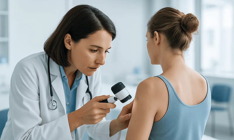
When Is Radiotherapy Used for Skin Cancer Instead of Surgery? How the Choice Is Made
Radiotherapy is usually chosen instead of surgery for skin cancer when an operation would be difficult, risky, or cosmetically costly: for example, in people…
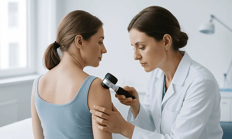
Life After Skin Cancer Treatment: Sun Habits, Skin Self-Checks and Regular Reviews
Sun protection after skin cancer means daily habits rather than a single product: seeking shade when the UV index is 3 or higher, wearing…
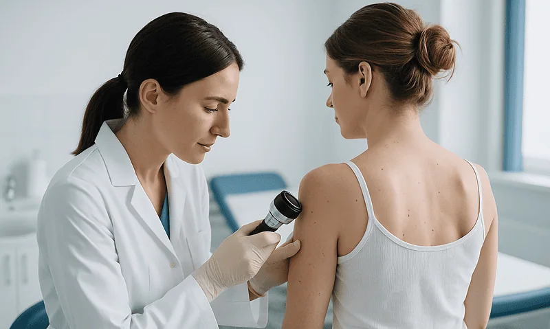
Healing After Skin Cancer Surgery: Dressings, Scars and When Normal Activity Resumes
Skin cancer surgery healing usually takes several weeks, with most stitched wounds closing within one to three weeks and Mohs wounds settling over roughly…